Autoimmune Bullous Diseases Dr Abdullah ALAKEEL Assistant Professor

Autoimmune Bullous Diseases Dr. Abdullah ALAKEEL Assistant Professor & Consultant Department of Dermatology KKUH
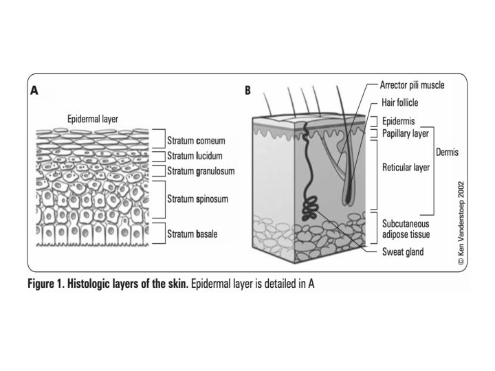

• Circumscribed skin lesions containing fluid (If the size ≤ 5 mm = vesicle If the size > 5 mm = bulla)

• Classification Of Vesiculobullous Diseases:

Autoimmune bullous diseases • A- Loss of intraepidermal adhesion: Pemphigus group : • 1 - Pemphigus vulgaris (PV) with subtypes • a- Classic • b- Pemphigus vegetans 2 - Pemphigus foliaceus with subtypes: a- Classic - Fogo selvagum - Pemphigus erythematosus ( Senear-Usher) b- Paraneoplastic pemphigus c- Drug induced pemphigus d- Ig. A pemphigus

Autoimmune bullous diseases B- Loss of subepidermal adhesion : 1 - Pemphigoid a- Bullous pemphigoid b- Pemphigoid gestationis c- Cicatricial pemphigoid 2 - Linear Ig. A disease - of childhood - Adult form 3 - Epidermolysis bullosa acquisita 4 - Dermatitis herpetiformis

Autoimmune bullous diseases • Pemphigus Group : • A group of disorders with loss of intraepidermal adhesion due to autoantibodies directed against proteins of the desmosomal complex that hold keratinocytes together. The desmosome is a complex structure, with many of its components targets for autoantibodies.
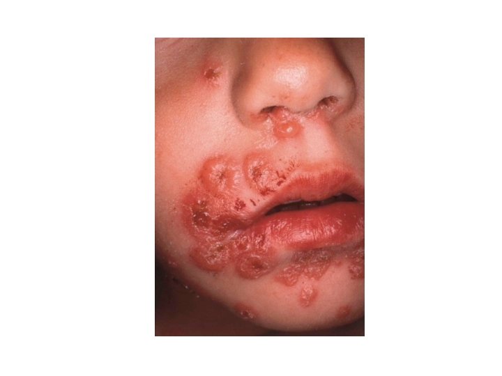
: • Definition : severe, potentially fatal disease with intraepidermal")
PV • Pemphigus vulgaris (PV): • Definition : severe, potentially fatal disease with intraepidermal blister formation on skin and mucosa caused by autoantibodies against desmogleins. • Epidemiology : 0. 1 -0. 5/ 100000 yearly, most patients middle aged.

PV • Pathogenesis: • - Genetic predisposition: HLA-DRQ 402 - DQ 0505 • - Antibodies against desmoglein 3 (Dsg 3) and later desmoglein 1 (Dsg 1 ). The bound antibodies activate proteases that damage the desmosome, leading to acantholysis. • - Serum antibody titer usually correlates with severity of disease and course.
 are more likely to cause")
PV - Agents containing sulfhydryl groups (penicillamine, captopril, piroxicam) are more likely to cause PV. - Those without sulfhydryl groups tend to cause PV ( beta-blockers, cephalosporins, penicillins, & rifampicin). - Note : drugs from either group can cause either type of pemphigus.

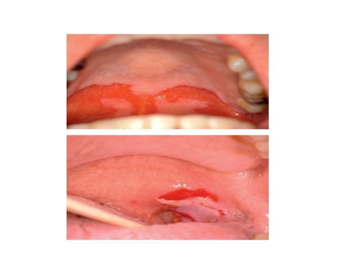
• Clinical features: PV • Sites: oral mucosa, scalp , face, mechanically stressed areas, nail fold, intertriginous areas. • Bliters are NOT stable, epidermis falls apart, erosions & crusts are common • Oral involvement: 70%, anti-Dsg 3 (Dsg 3 is the main desmoglein on mucosa) • Additional localized disease; scalp • Note: always check scalp when confronted with unexplained oral erosions.

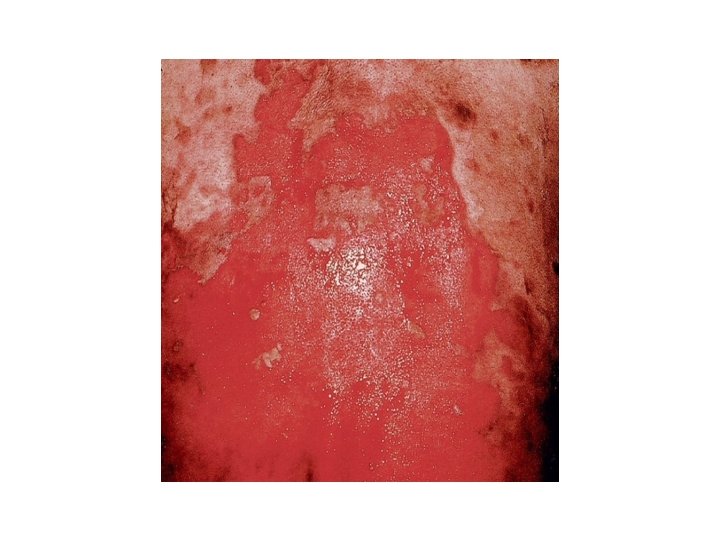
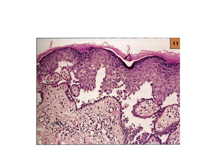
PV • Generalized disease due to development of antibodies against Dsg 1 which is present in skin along with Dsg 3. • Pruritus is uncommon. • Histology: acantholysis, retention of basal layer keratinocytes (tombstone effect), mild dermal perivascular infiltrates.
, less")
PV • • Diagnostic approach: Clinical evaluation Nikolsky sign Pseudo-Nikolsky sign (Asboe-Hansen sign), less specific
, C 3")
PV • Histology: • DIF: perilesional shows deposition of Ig. G (100%), C 3 (80%) • Indirect IF • ELISA: to identify anti-Dsg 3, 1

PV • Differential diagnosis: • When skin is involved: • Bullous impetigo, dyskeratotic acanthoytic disorders ( Hailey-Hailey, Grover disease) • When oral mucosa is involved: • Denture intolerance • Erosive candidiasis • Chronic recurrent aphthae • Erythema multiforme • Erosive lichen planus • Herpetic ginigivitis

PV • Therapy : • 1 - Systemic corticoisteroids • The main cause of morbidity & mortality in patients is CS side effects, have to combine with steroid-sparing agent, check for osteoporosis and latent TB • A- combination pulse therapy : prednisolone 1 g + cyclophosphamide 7. 5 -10 mg/kg every 3 -4 weeks • With cyclophosphamide in interval 1 -2 mg/kg daily.

PV • Prednisolone- azathioprine therapy • Alternative immunosuppressive agents: cyclosporine, mycophenolate mofetil, chlorambucil • Topical measures: local anesthetic gels • Therapy resistant course: IVIG

 • Clinical")
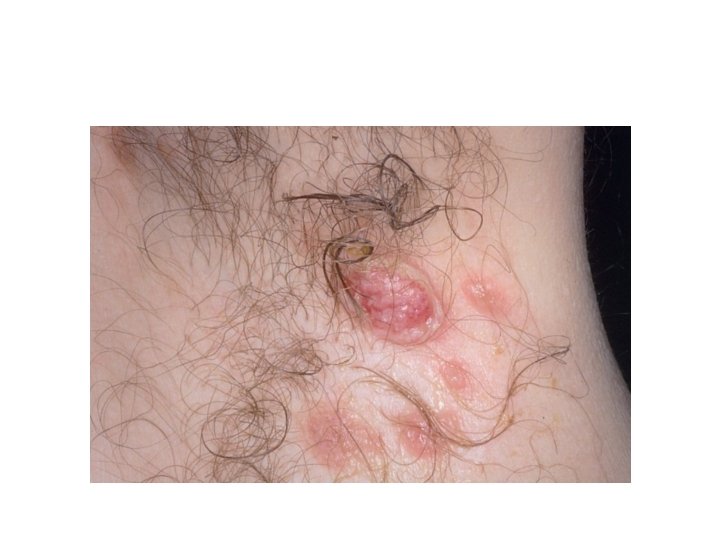
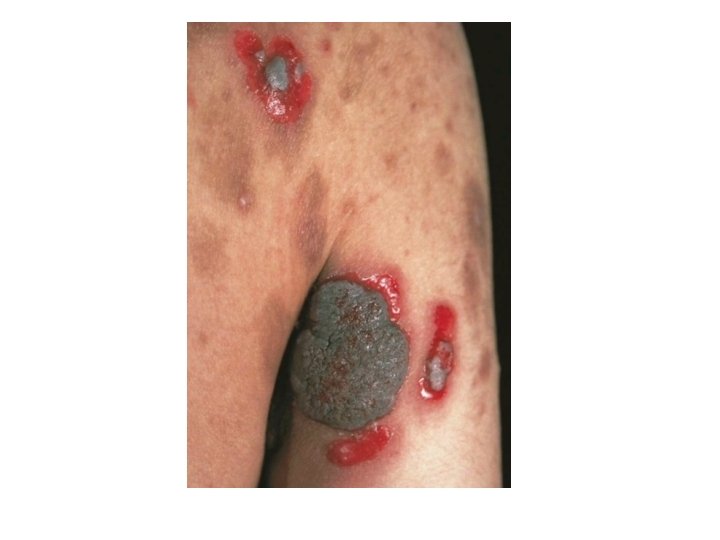
Pemphigus vegetans • Unusual variant of PV with hyperkeratotic verruciform reaction (vegetans) • Clinical features : • Originally typical PV, then development of white macerated plaques in involved areas • (pyodermite végétante) : limited to intertriginous areas, starts as pustules that evolve into vegetating lesions.

Pemphigus vegetans • Diagnostic approach : as for PV • DDx: if mild and localized can be confused with Hailey-Hailey • Therapy : see PV

Pemphigus Foliaceus • Form of pemphigus with superficial blisters caused by anti-Dsg 1 • Pathogenesis: • Anti-Dsg 1, the main desmoglein on the upper epidermis. • More often drug induced than PV, usually sulfhydryl groups : captopril, penicillamine, peroxicam • Maybe caused by sunburn or paraneoplastic sign

PF • Clinical features: • Scalp, face, chest and back, can progress to involve large areas with diffuse scale and erosions. • Diagnostic approach: • • • Clinical Biopsy ? Not helpful DIF: superficial deposition of Ig. G ELISA: reveals Ig. G antibodies against Dsg 1 Medication history Therapy: same approach as PV, but usually more responsive to therapy. Dapsone maybe helpful

Pemphigus eryhthematosus • Uncommon feature of pemphigus foliaceus with additional features of lupus erythematosus. • More likely to be triggered by sunlight or medications than other forms of PF

Ig. A Pemphigus • Pustular acantholytic dermatosis with intercellular Ig. A deposition in epidermis. • Can be associated with gammopathy • Clinical features: • Subcorneal pustular dermatosis (Sneddon. Wilkinson disease): broad, annular erythematous patches with peripheral flaccid pustules and central crusting, favours flexures and trunk, never mouth, pruritic
: clinically similar, sunflower lesions •")
Ig. A Pemphigus • Intradermal neutrophilic dermatosis (Huff syndrome): clinically similar, sunflower lesions • Diagnostic approach: • DIF; showing Ig. A directed against keratinocytes • Therapy : most cases are responsive to dapsone, if not , corticosteroids & other immunosuppressive agents

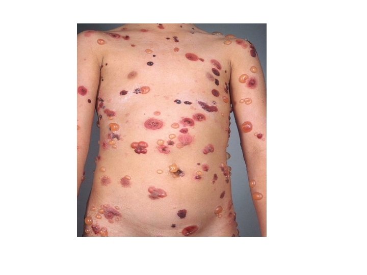
Paraneoplastic Pemphigus • Most often associated with lymphoma, leukemia, thymoma, Castleman tumor. • Not with SCC or adenocarcinoms • Clinical: severe persistent painful stomatitis extending from lips to pharynx, larynx and esophagus, conjunctival involvement may lead to blindness. • Cutaneous changes are polymorphic

Paraneoplastic Pemphigus • Note: if patient is sick and has lesions resembling erythema multiforme, lichen planus and a blistering disease , be highly suspicious of paraneoplastic pemphigus. • Histology is rarely helpful. • Therapy : treat the underlying tumour , prognosis correlates with the response • No consensus on what immunosuppressive regimen. Good success with ant-CD 20 (rituximab)
: • Subepidermal blistering disease caused by autoantibodies to")
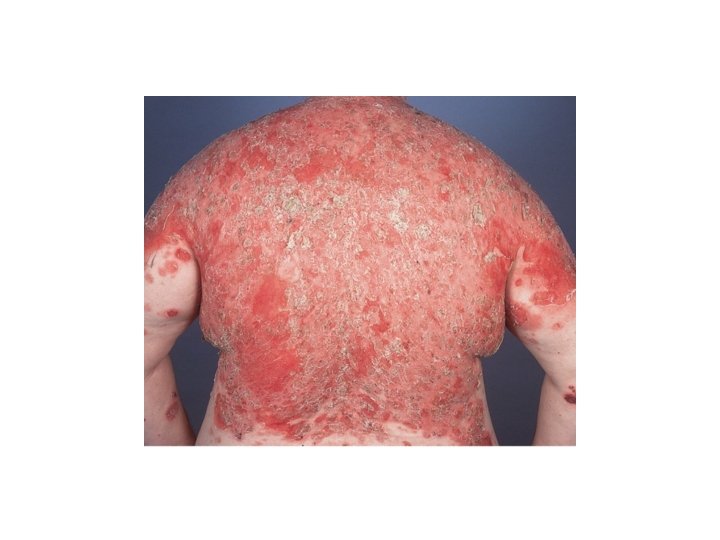
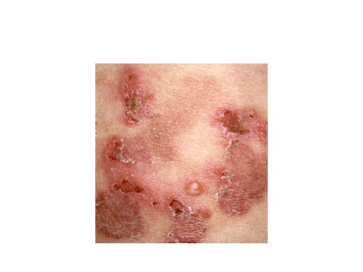
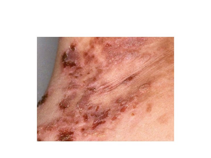
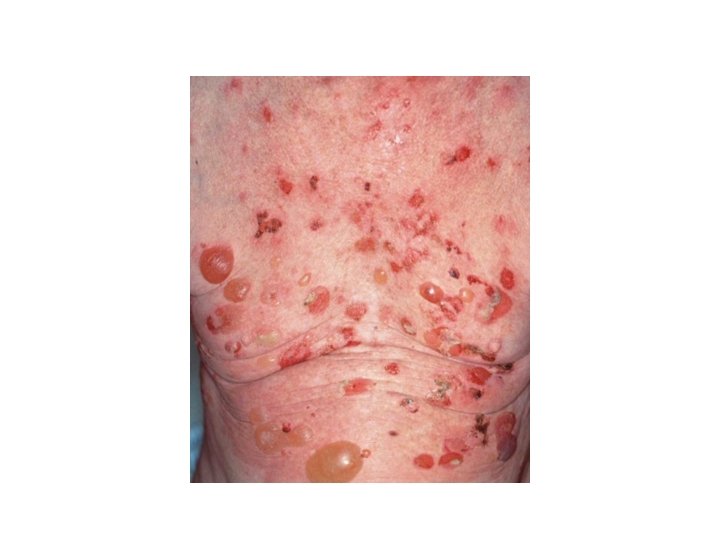
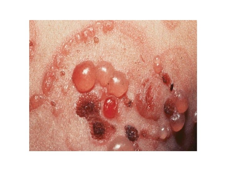
Pemphigoid Group • Bullous pemphigoid (BP): • Subepidermal blistering disease caused by autoantibodies to components of hemidesmosomes in the basement membrane zone (BMZ). • Most common autoiummune bullous disease, 1/100000, favours elderly male<female.

BP • Pathogenesis : • Autoantibodies directed against 2 hemidesmosomal proteins: • - BP 230 • - BP 180 • BP 180 is most likely to be more involved in the initial immune response, since it is transmembrane.
, sunlight, and")
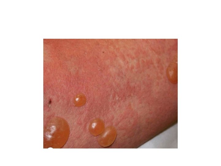
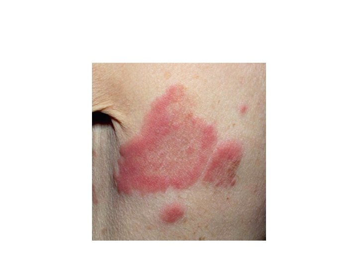
BP • Less common causes include drugs ( benzodiazepine, furosemide, penicillin, sulfasalazine), sunlight, and ionizing radiation. • Clinical : • before blisters develop, pruritus, urticarial lesions may be present, blisters tend to develop in these areas. • Note: always keep BP in mind when confronted with an elderly patient with persistent « urticaria » . • Blisters are stable and tense. • Oral mucosal involvement in <20%.

BP • Histology : • Prebullous lesions: presence of unexpected eosinophils is • • • a good clue. Later subepidermal blister formation. Diagnostic approach: Labs: elevated ESR, eosinophils, and increased Ig. E in 60%. DIF: best taken from erythematous area at periphery, not blister itself; band of Ig. G & C 3 along BMZ. Indirect IF: using Na. CL split skin ELISA: identifies ab against both BP 230 & 180 in 60 -80% of pts.

BP • • Therapy: Steroids ? Methotrexate 15 -20 mg weekly Mostly widely used steroid-sparing agents, azathiporine & mycophenolate mofetil.

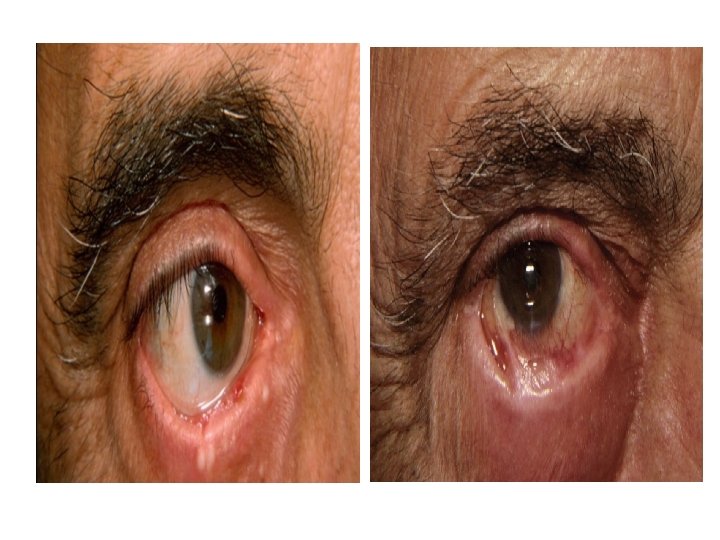
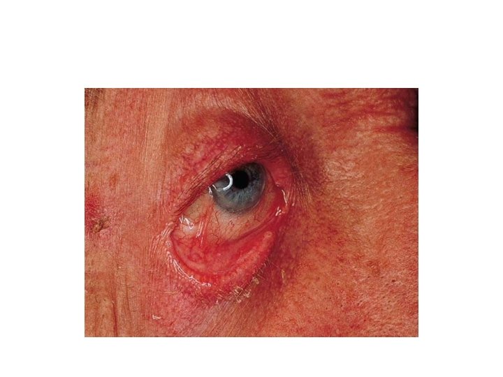

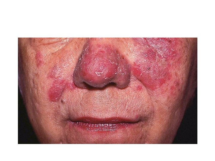
Cicatricial Pemphigoid • Chronic subepidermal blistering disease favoring mucus membrane especially mouth and eyes. • Patients > 65 years. Women > men

Cicatricial Pemphigoid • Clinical features: • Conjunctiva affected in 75% of cases. Starts unilaterally , within 2 years usually bilateral. Adhesions, ectropion, corneal damage • Oral mucosa: much less painful than PV. • Esophagus and larynx can develop strictures, requiring surgery. • Genitalia: narrowing of vaginal orifice; adhesions between glans & foreskin. • Skin: only involved in 25%

Cicatricial Pemphigoid Diagnostic approach : DIF Indirect IF Therapy: Ocular: topical or systemic corticosteroids, OPH consultation • Mucosal: topicals • Widespread: Pred + azathoiprine or pred+ cyclophosphamide pulse therapy • IVIG • • •

Pemohigoid Gestationis • Synonym : herpes gestationis • Form of BP occuring during pregnancy. • Occur in 1/10000 -40000 pregnancies. • No maternal risk, no increase in birth defects but complications of pregnancy in 15 -30% with fetal death rate. • Pathogenesis: • mothers often HLA-88, -DR 3, -DR 4, father often HLADR 2. possible that mothers are sensitized against placental antigens. Target antigens are BP 180

Pemohigoid Gestationis • Erythematous urticarial plaques, alone or with papules, vesicles, blisters in sub-epidermal area, erosions. • Intense pruritus. • Sites: abdomen, proximal extremities. • Rarely appears postpartum, resolve within 3 months. Occasionally recur with menses or ingestion of OCP, tends to be worse in next pregnancy. • The ab cross the placenta, the newborn can have blisters for a few weeks.

Pemohigoid Gestationis • Diagnostic approach: Labs : eosinophilia, DIF, Indirect IF • Management: • Systemic Potent steroids: For blisters, avoid the systemic in the 1 st trimester (topicals) • Skin care : to prevent infections • Anti-histamines: For pruritis.
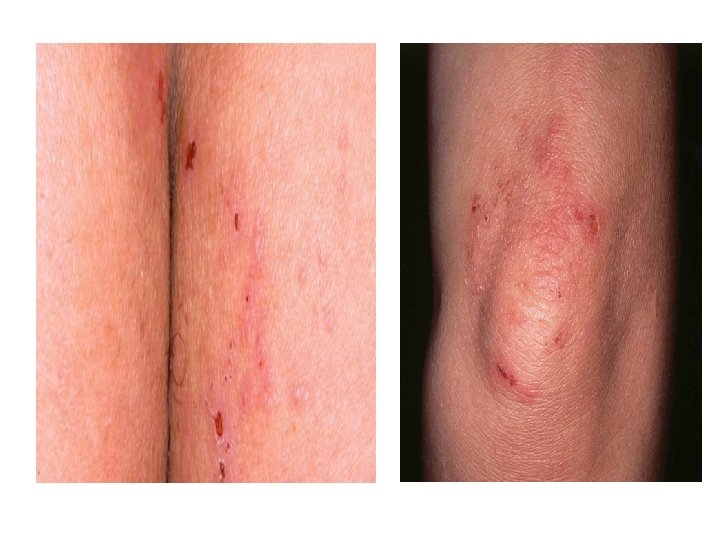
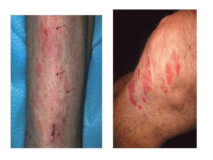

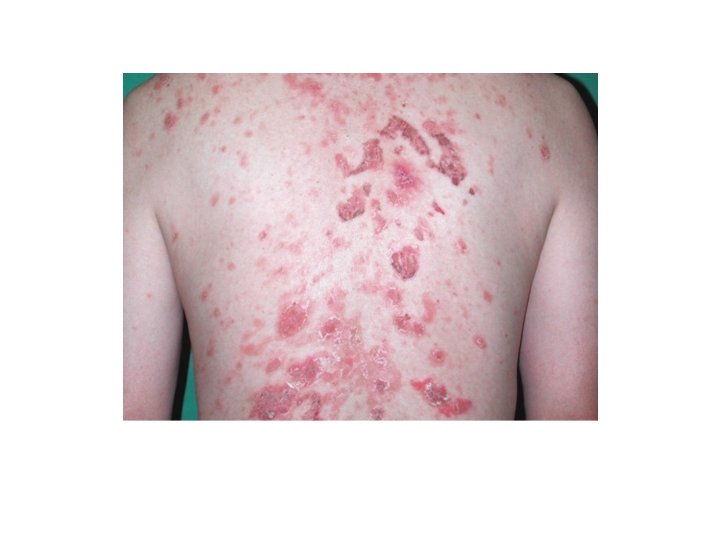
Dermatitis Herpetiformis • Pruritic vesicular disease caused by Ig. A ab directed against epidermal transglutaminase & presenting with granular pattern in papillary dermis. • M>F 1: 2, disease of young adults. • DH & gluten-sensitive enteropathy are closely related (i. e. celiac disease).

Dermatitis Herpetiformis • Grouped papules/vesicles/urticarial wheals on an erythematous base, associated with intense pruritus, burning, stinging, excoriations. • sites: extensor surfaces of elbows/knees, sacrum, buttocks, scalp. • Spontaneous remissions may occur, but disease often lifelong.

Dermatitis Herpetiformis • Histology: • Neutrophilic microabcesses in the papillary dermis are the hallmark • Approach: • Skin biopsy • DIF: granular deposits of Ig. A in dermal papillae • Indirect IF: • ELISA: identifies Ig. A ab against transglutaminase in at least 80% • Jejunal biopsy: flattening of villi

Dermatitis Herpetiformis • Therapy: • Gluten free diet. • Dapsone: amazingly effective


Linear Ig. A disease • Subepidermal blistering disease caused by deposits of Ig. A along BMZ. • Maybe identical to DH but without GI involvement , or resemble BP. • Over 50% have mucosal involvement • Approach: • DIF • OPH, and to exclude celiac (jejunal biopsy. . ) • Therapy: CS, Dapsone

Linear Ig. A disease • Childhood type: • Before 5 years of age and resolves spontaneously • Large tense blisters arranged in a rosette fashion, predilection of abdomen, groin , axillae and face mucosal disease very common 90% approx. GI disease extremely rare.

Linear Ig. A disease • • • Diagnostic approach: DIF: Ig. A deposits IIF: Ig. A Therapy: Dapsone , sulfapyridine If CI or failure: CS

Summary

Summary

Summary

• Thank you !
- Slides: 71