
Authors University of Michigan Medical School Department of



- Slides: 56
Author(s): University of Michigan Medical School, Department of Cell and Developmental Biology License: Unless otherwise noted, the content of this course material is licensed under a Creative Commons Attribution Noncommercial Share Alike 3. 0 License. http: //creativecommons. org/licenses/by-nc-sa/3. 0/ We have reviewed this material in accordance with U. S. Copyright Law and have tried to maximize your ability to use, share, and adapt it. The citation key on the following slide provides information about how you may share and adapt this material. Copyright holders of content included in this material should contact open. michigan@umich. edu with any questions, corrections, or clarification regarding the use of content. For more information about how to cite these materials visit http: //open. umich. edu/education/about/terms-of-use. Any medical information in this material is intended to inform and educate and is not a tool for self-diagnosis or a replacement for medical evaluation, advice, diagnosis or treatment by a healthcare professional. Please speak to your physician if you have questions about your medical condition. Viewer discretion is advised: Some medical content is graphic and may not be suitable for all viewers.
Citation Key for more information see: http: //open. umich. edu/wiki/Citation. Policy Use + Share + Adapt { Content the copyright holder, author, or law permits you to use, share and adapt. } Public Domain – Government: Works that are produced by the U. S. Government. (17 USC § 105) Public Domain – Expired: Works that are no longer protected due to an expired copyright term. Public Domain – Self Dedicated: Works that a copyright holder has dedicated to the public domain. Creative Commons – Zero Waiver Creative Commons – Attribution License Creative Commons – Attribution Share Alike License Creative Commons – Attribution Noncommercial Share Alike License GNU – Free Documentation License Make Your Own Assessment { Content Open. Michigan believes can be used, shared, and adapted because it is ineligible for copyright. } Public Domain – Ineligible: Works that are ineligible for copyright protection in the U. S. (17 USC § 102(b)) *laws in your jurisdiction may differ { Content Open. Michigan has used under a Fair Use determination. } Fair Use: Use of works that is determined to be Fair consistent with the U. S. Copyright Act. (17 USC § 107) *laws in your jurisdiction may differ Our determination DOES NOT mean that all uses of this 3 rd-party content are Fair Uses and we DO NOT guarantee that your use of the content is Fair. To use this content you should do your own independent analysis to determine whether or not your use will be Fair.
Cells and Tissues Sequence Medical Histology Integumentary System, Glands/mammary gland Fall, 2008 Cell and Developmental Biology
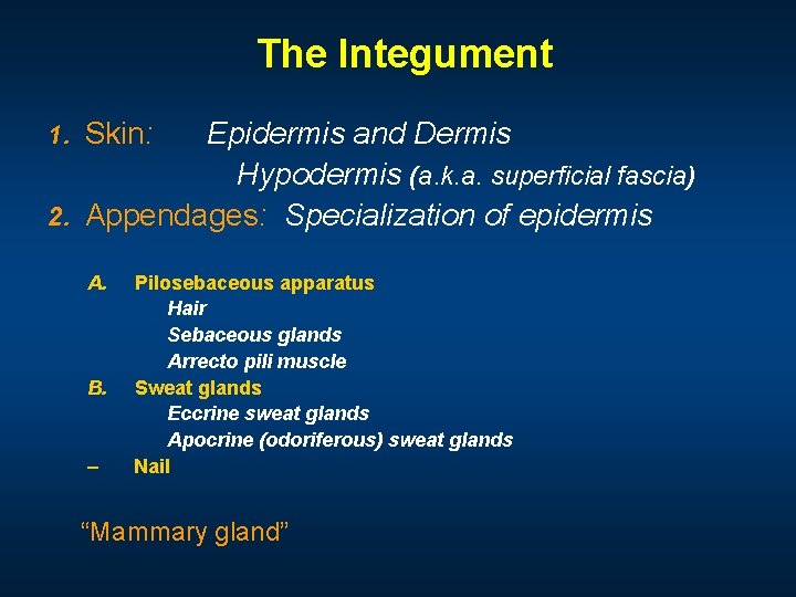
The Integument 1. Skin: Epidermis and Dermis Hypodermis (a. k. a. superficial fascia) 2. Appendages: Specialization of epidermis A. B. – Pilosebaceous apparatus Hair Sebaceous glands Arrecto pili muscle Sweat glands Eccrine sweat glands Apocrine (odoriferous) sweat glands Nail “Mammary gland”
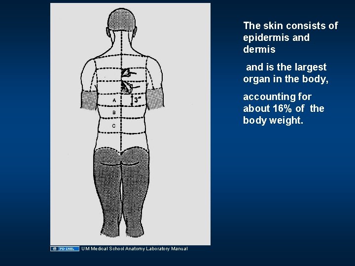
The skin consists of epidermis and is the largest organ in the body, accounting for about 16% of the body weight. UM Medical School Anatomy Laboratory Manual
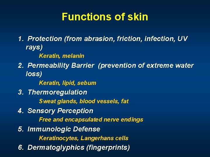
Functions of skin 1. Protection (from abrasion, friction, infection, UV rays) Keratin, melanin 2. Permeability Barrier (prevention of extreme water loss) Keratin, lipid, sebum 3. Thermoregulation Sweat glands, blood vessels, fat 4. Sensory Perception Free and encapsulated nerve endings 5. Immunologic Defense Keratinocytes, Langerhans cells 6. Dermatoglyphics (fingerprints)
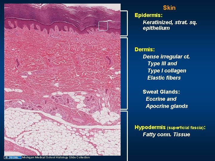
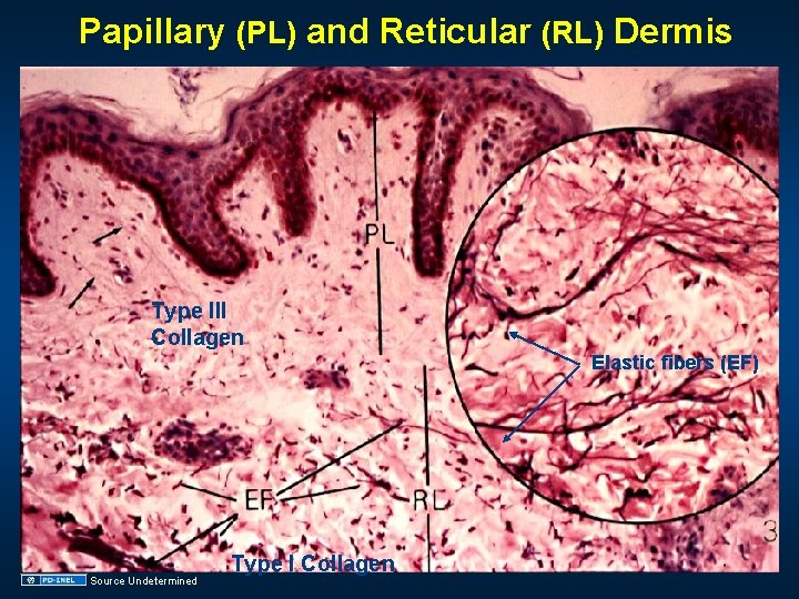
Skin Epidermis: Keratinized, strat. sq. epithelium Dermis: Dense irregular ct. Type III and Type I collagen Elastic fibers Sweat Glands: Eccrine and Apocrine glands Hypodermis (superficial fascia): Fatty conn. Tissue Michigan Medical School Histology Slide Collection
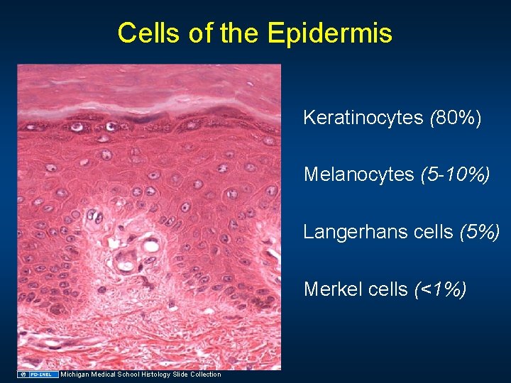
Cells of the Epidermis Keratinocytes (80%) Melanocytes (5 -10%) Langerhans cells (5%) Merkel cells (<1%) Michigan Medical School Histology Slide Collection
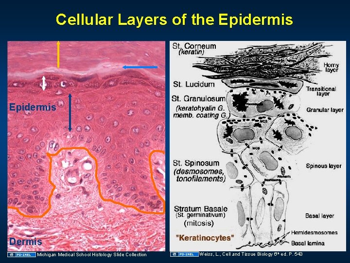
Cellular Layers of the Epidermis Dermis Michigan Medical School Histology Slide Collection Weiss, L. , Cell and Tissue Biology 6 th ed. P. 543
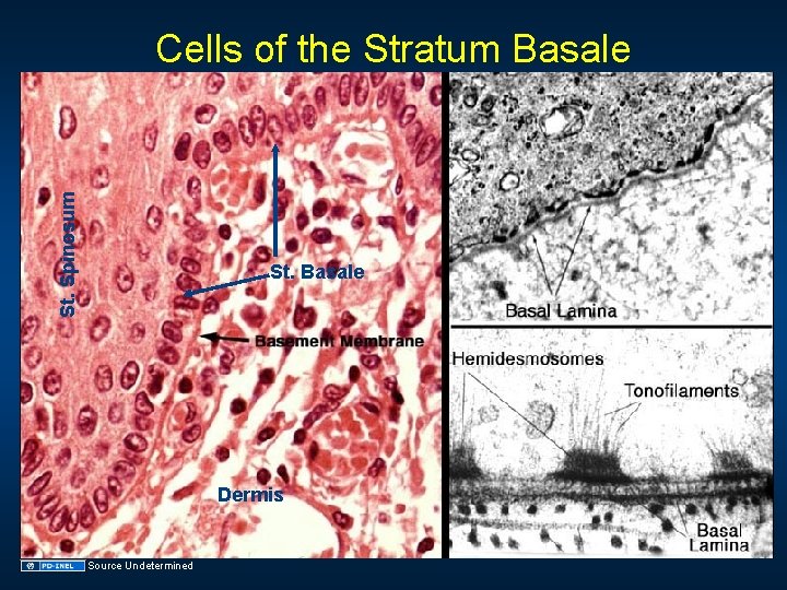
St. Spinosum Cells of the Stratum Basale St. Basale Dermis Source Undetermined
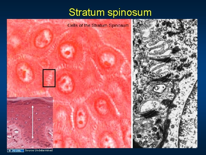
Stratum spinosum Source Undetermined
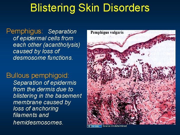
Blistering Skin Disorders Pemphigus: Separation of epidermal cells from each other (acantholysis) caused by loss of desmosome functions. Bullous pemphigoid: Separation of epidermis from the dermis due to blistering in the basement membrane caused by loss of anchoring filaments and hemidesmosomes. Source Undetermined
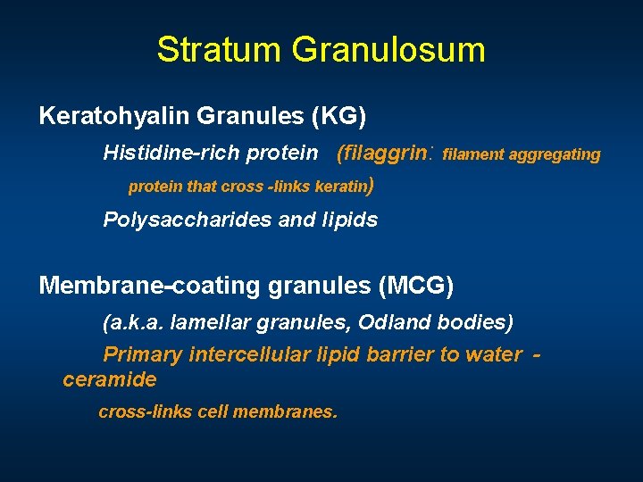
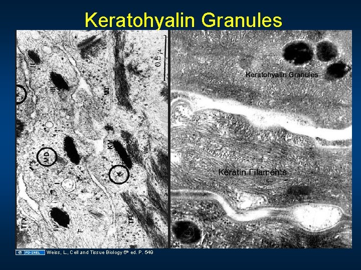
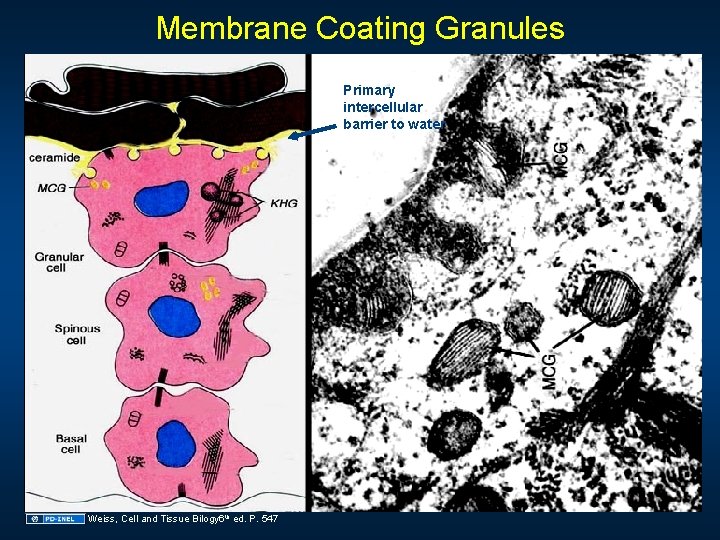
Stratum Granulosum Keratohyalin Granules (KG) Histidine-rich protein (filaggrin: protein that cross -links keratin ) filament aggregating Polysaccharides and lipids Membrane-coating granules (MCG) (a. k. a. lamellar granules, Odland bodies) Primary intercellular lipid barrier to water ceramide cross-links cell membranes.
Keratohyalin Granules Weiss, L. , Cell and Tissue Biology 6 th ed. P. 549
Membrane Coating Granules Primary intercellular barrier to water Weiss, Cell and Tissue Bilogy 6 th ed. P. 547
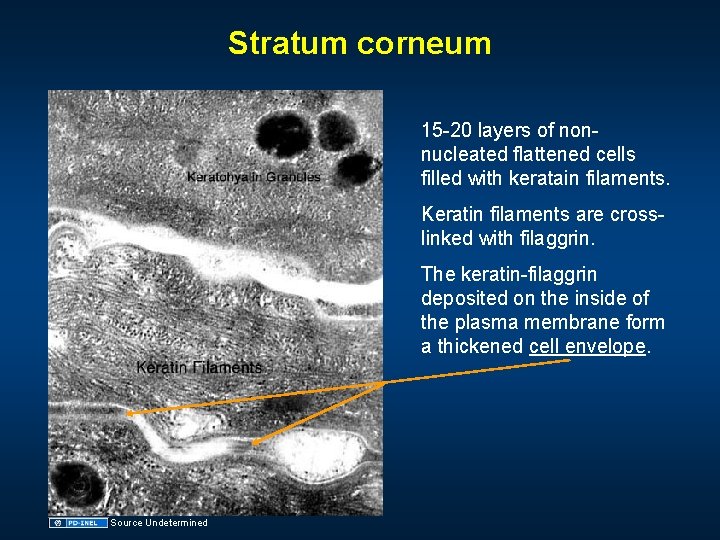
Stratum corneum 15 -20 layers of nonnucleated flattened cells filled with keratain filaments. Keratin filaments are crosslinked with filaggrin. The keratin-filaggrin deposited on the inside of the plasma membrane form a thickened cell envelope. Source Undetermined
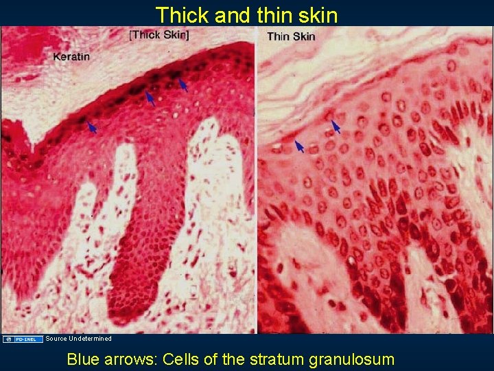
Thick and thin skin Source Undetermined Blue arrows: Cells of the stratum granulosum
Melanocytes Skin color Red blood cells in the dermal vascular beds. Carotenes from exogenous foods stored in fatty tissues. Hemoglobin and bilirubin (endogenous degradation products). Melanin (pigment produced by melanocytes)
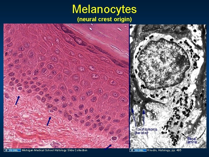
Melanocytes (neural crest origin) Michigan Medical School Histology Slide Collection Rhodin, Histology, pp. 485
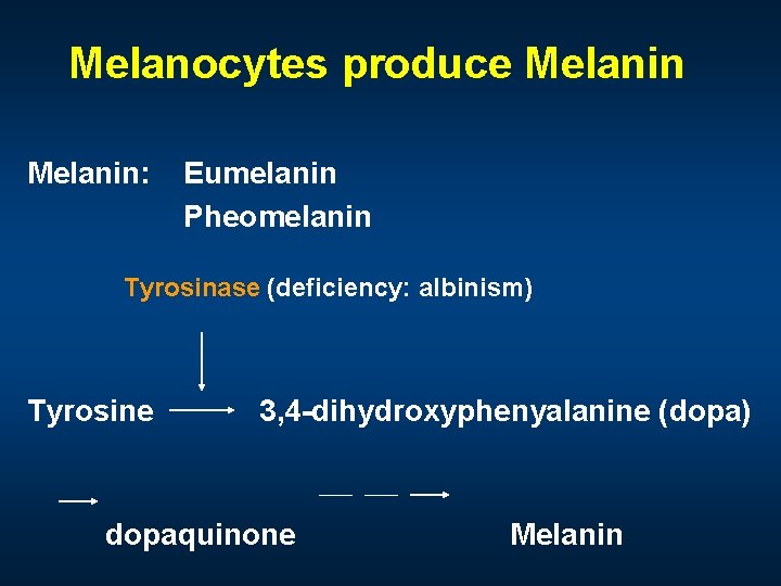
Melanocytes produce Melanin: Eumelanin Pheomelanin Tyrosinase (deficiency: albinism) Tyrosine 3, 4 -dihydroxyphenyalanine (dopa) dopaquinone Melanin
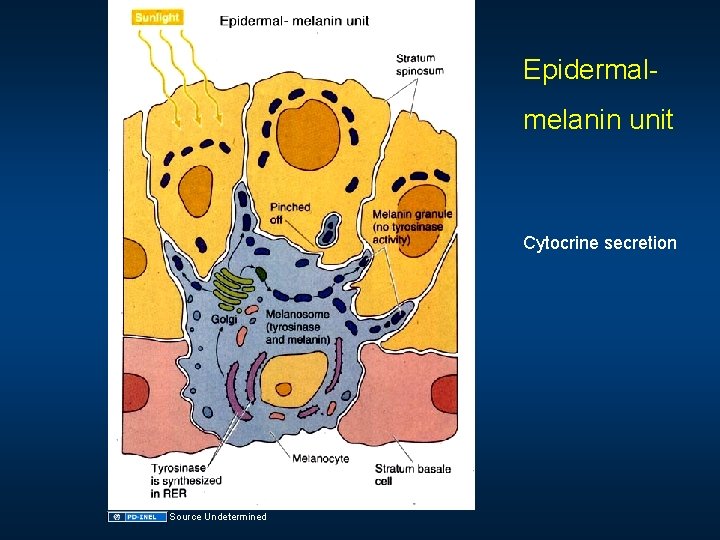
Epidermalmelanin unit Cytocrine secretion Source Undetermined
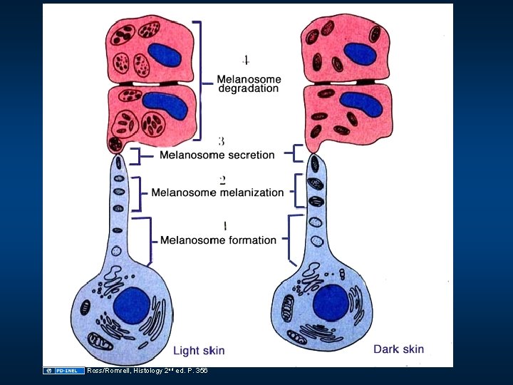
Ross/Romrell, Histology 2 nd ed. P. 356
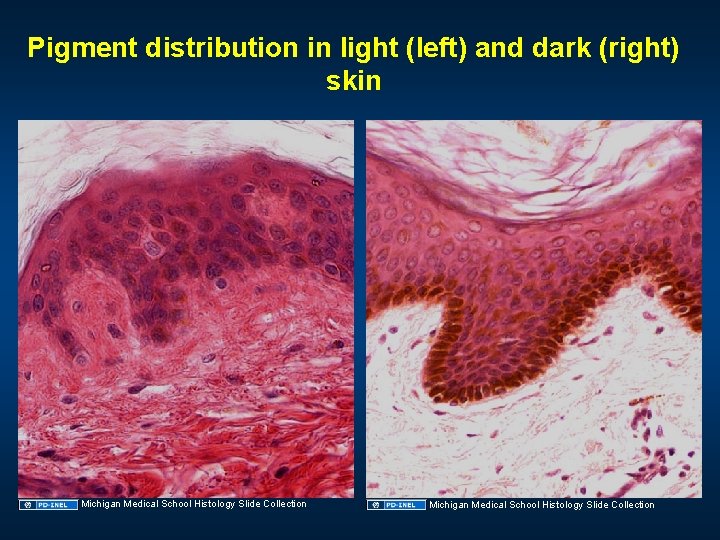
Pigment distribution in light (left) and dark (right) skin Michigan Medical School Histology Slide Collection
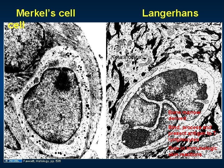
Merkel’s cell Langerhans Bone marrow derived Bind, process and present antigen to Tlymphocytes Role in immunologic skin reactions. Fawcett, Histology, pp. 536
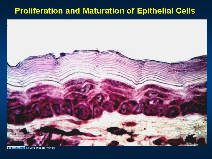
Proliferation and Maturation of Epithelial Cells Source Undetermined
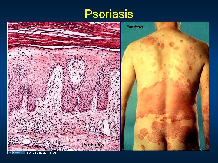
Psoriasis Source Undetermined
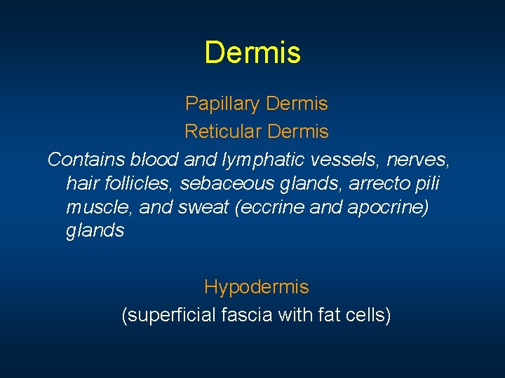
Dermis Papillary Dermis Reticular Dermis Contains blood and lymphatic vessels, nerves, hair follicles, sebaceous glands, arrecto pili muscle, and sweat (eccrine and apocrine) glands Hypodermis (superficial fascia with fat cells)
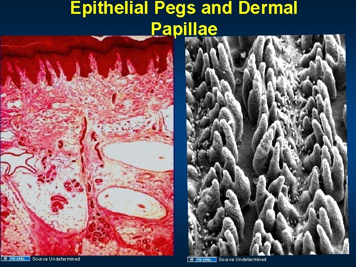
Epithelial Pegs and Dermal Papillae Source Undetermined
Papillary (PL) and Reticular (RL) Dermis Type III Collagen Elastic fibers (EF) Source Undetermined Type I Collagen
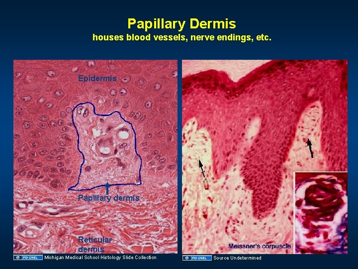
Papillary Dermis houses blood vessels, nerve endings, etc. Epidermis Papillary dermis Reticular dermis Michigan Medical School Histology Slide Collection Source Undetermined
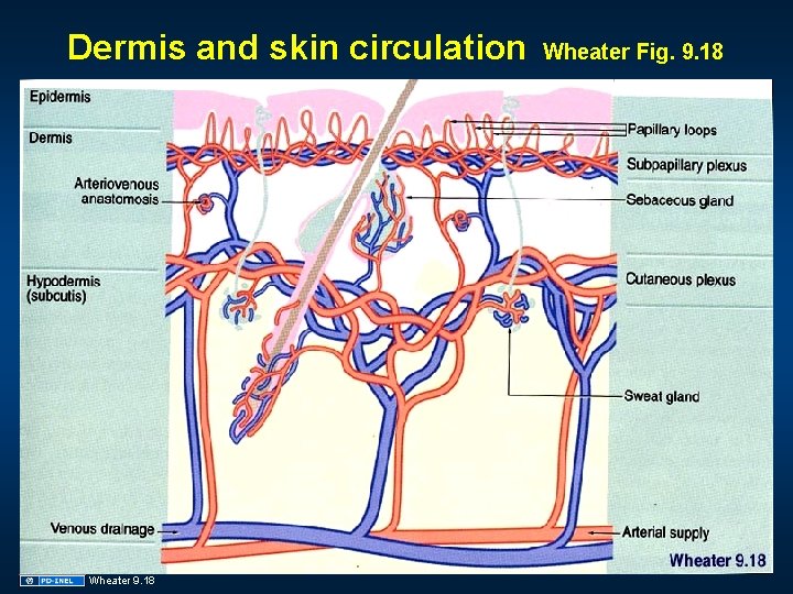
Dermis and skin circulation Wheater 9. 18 Wheater Fig. 9. 18
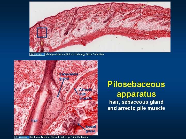
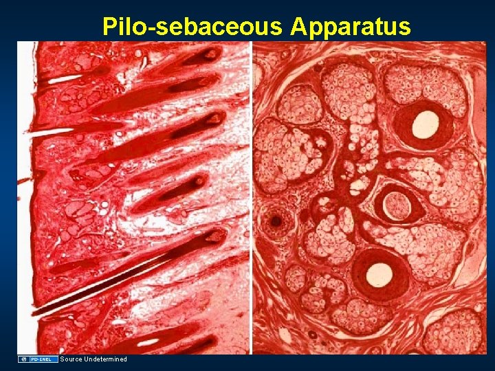
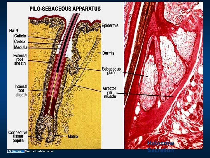
Michigan Medical School Histology Slide Collection Sebaceous gland Arrecto pile muscle hair Swaet gland Michigan Medical School Histology Slide Collection Pilosebaceous apparatus hair, sebaceous gland arrecto pile muscle
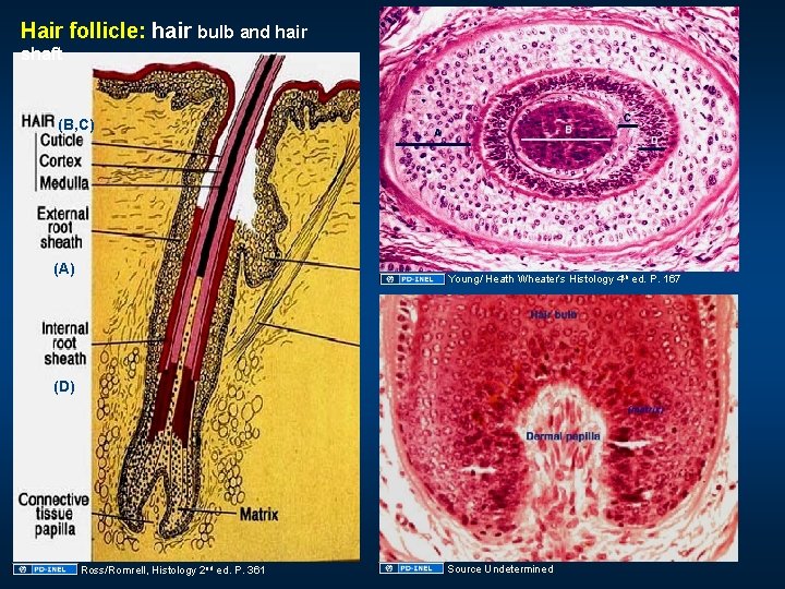
Hair follicle: hair bulb and hair shaft (B, C) (A) Young/ Heath Wheater’s Histology 4 th ed. P. 167 (D) Ross/Romrell, Histology 2 nd ed. P. 361 Source Undetermined
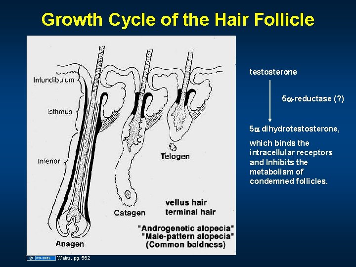
Growth Cycle of the Hair Follicle testosterone 5 a-reductase (? ) 5 a dihydrotestosterone, which binds the intracellular receptors and Inhibits the metabolism of condemned follicles. Weiss, pg. 562
Pilo-sebaceous Apparatus Source Undetermined
Source Undetermined Holocrine Secretion
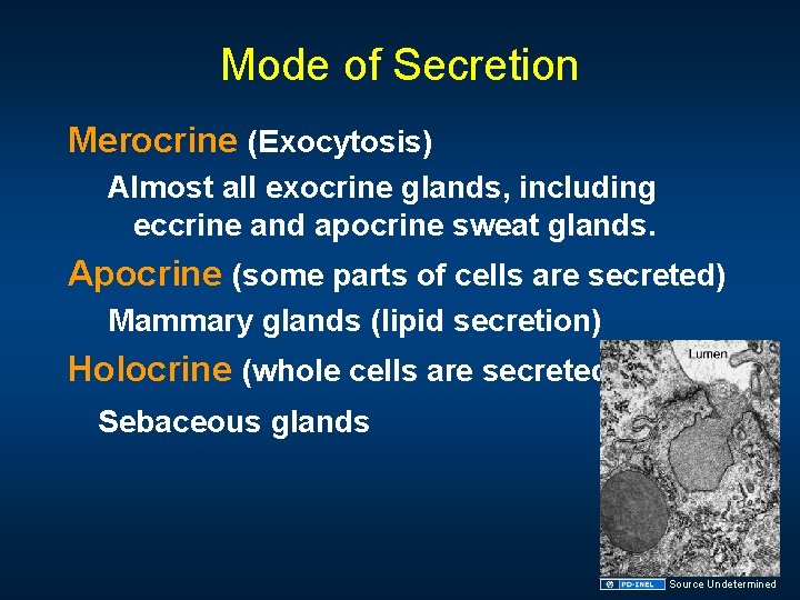
Mode of Secretion Merocrine (Exocytosis) Almost all exocrine glands, including eccrine and apocrine sweat glands. Apocrine (some parts of cells are secreted) Mammary glands (lipid secretion) Holocrine (whole cells are secreted) Sebaceous glands Source Undetermined
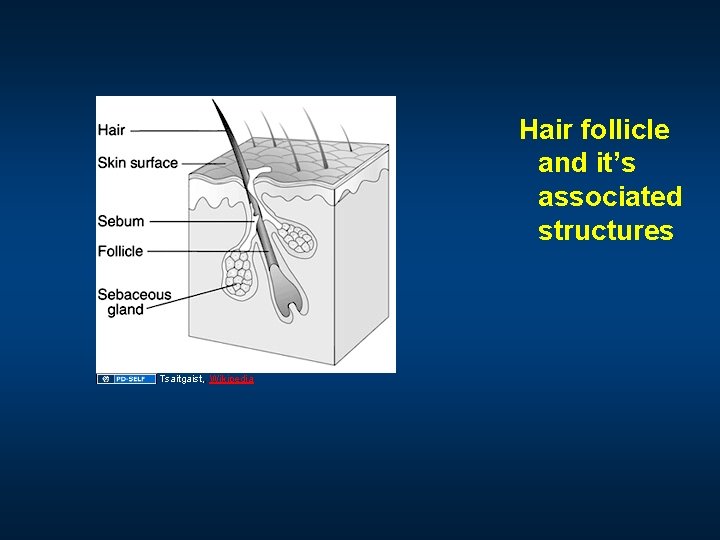
Hair follicle and it’s associated structures Tsaitgaist, , Wikipedia
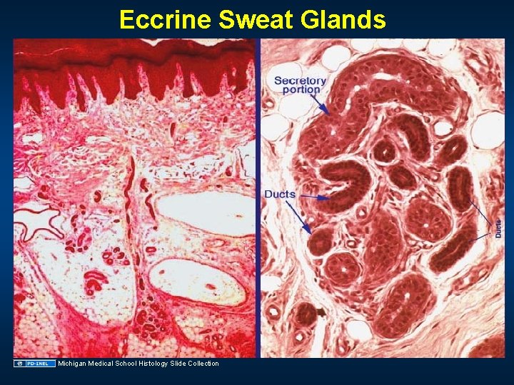
Eccrine Sweat Glands Michigan Medical School Histology Slide Collection
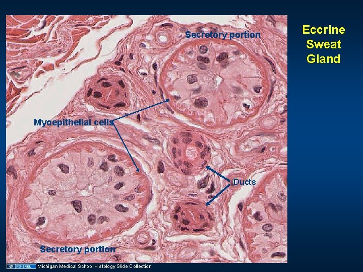
Secretory portion Myoepithelial cells Ducts Secretory portion Michigan Medical School Histology Slide Collection Eccrine Sweat Gland
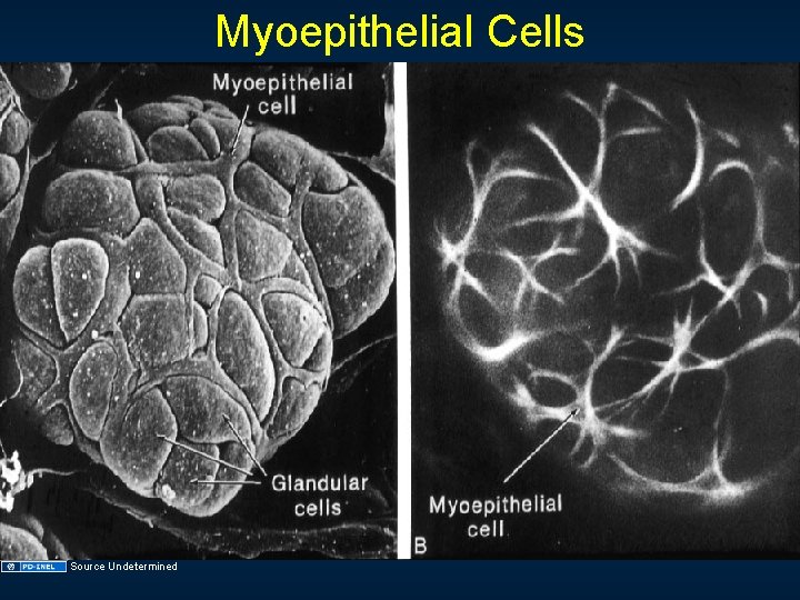
Myoepithelial Cells Source Undetermined
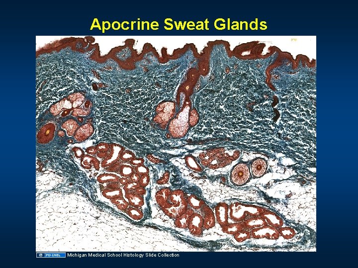
Apocrine Sweat Glands Michigan Medical School Histology Slide Collection
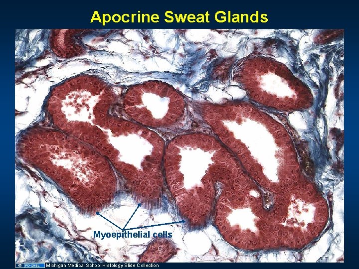
Apocrine Sweat Glands Myoepithelial cells Michigan Medical School Histology Slide Collection
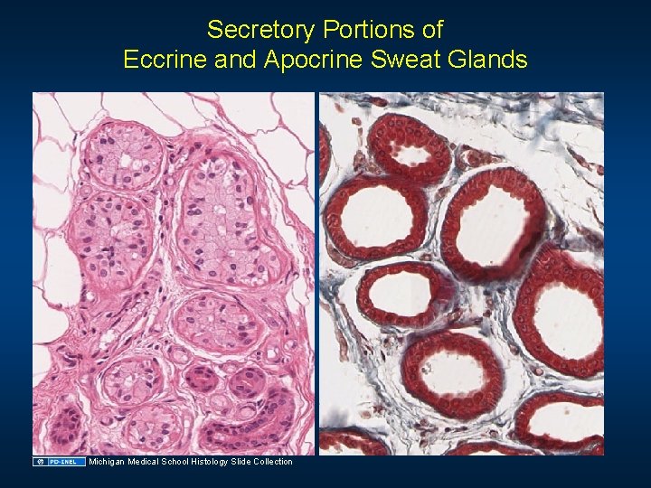
Secretory Portions of Eccrine and Apocrine Sweat Glands Michigan Medical School Histology Slide Collection
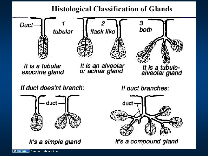
Source Undetermined
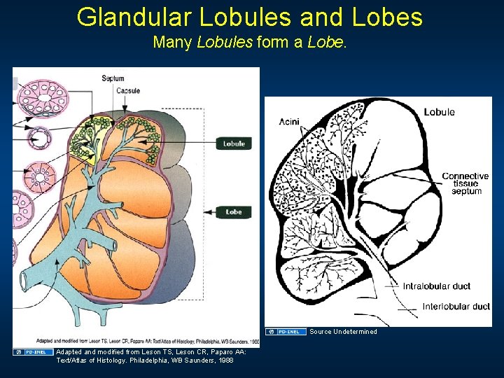
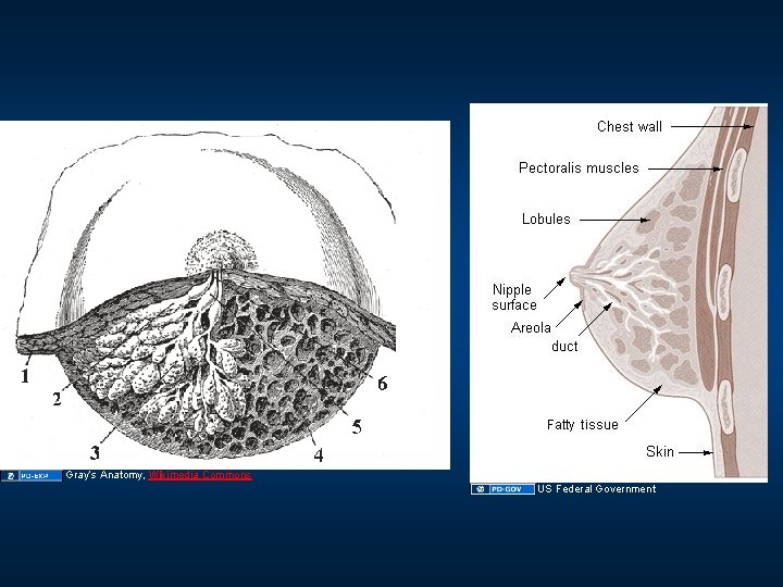
Glandular Lobules and Lobes Many Lobules form a Lobe. Source Undetermined Adapted and modified from Leson TS, Leson CR, Paparo AA: Text/Atlas of Histology. Philadelphia, WB Saunders, 1988
Gray’s Anatomy, Wikimedia Commons US Federal Government
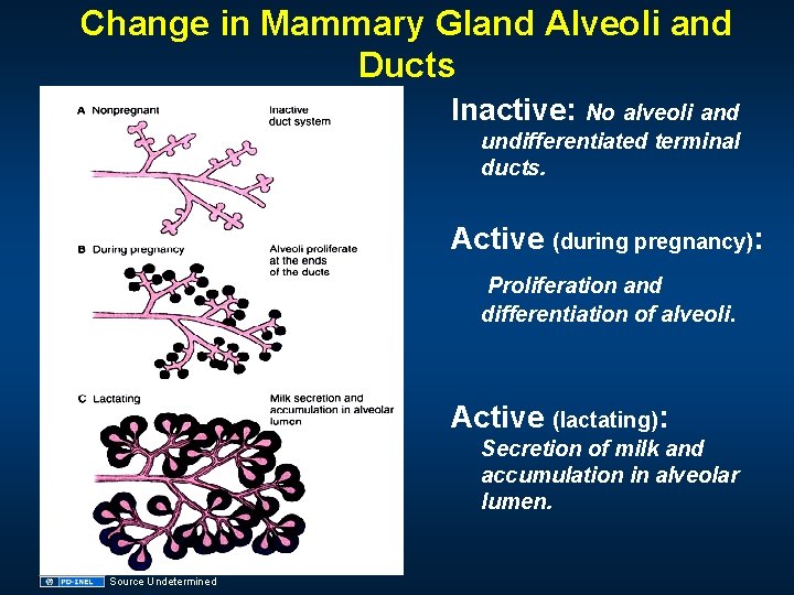
Change in Mammary Gland Alveoli and Ducts Inactive: No alveoli and undifferentiated terminal ducts. Active (during pregnancy): Proliferation and differentiation of alveoli. Active (lactating): Secretion of milk and accumulation in alveolar lumen. Source Undetermined
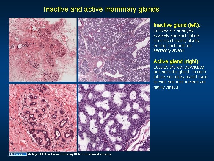
Inactive and active mammary glands Inactive gland (left): Lobules are arranged sparsely and each lobule consists of mainly bluntly ending ducts with no secretory alveoli. Active gland (right): Lobules are well developed and pack the gland. In each lobule, secretory alveoli have formed and their lumens are highly dilated. Michigan Medical School Histology Slide Collection (all images)
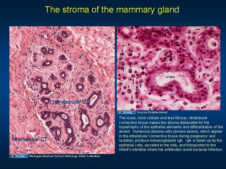
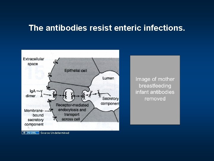
The stroma of the mammary gland Lactiferous duct Intralobular CT Lobule Source Undetermined Interlobular CT Michigan Medical School Histology Slide Collection The loose, more cellular and less fibrous, intralobular connective tissue makes the stroma distensible for the hypertrophy of the epithelial elements and differentiation of the alveoli. Numerous plasma cells (arrows above), which appear in the intralobular connective tissue during pregnancy and lactation, produce immunoglobulin Ig. A is taken up by the epithelial cells, secreted in the milk, and transported to the infant’s intestine where the antibodies resist bacterial infection.
The antibodies resist enteric infections. Source Undetermined
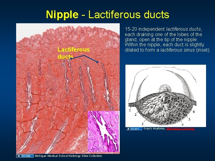
Nipple - Lactiferous ducts 15 -20 independent lactiferous ducts, each draining one of the lobes of the gland, open at the tip of the nipple. Within the nipple, each duct is slightly dilated to form a lactiferous sinus (inset). Gray’s Anatomy, Wikimedia Commons Michigan Medical School Histology Slide Collection
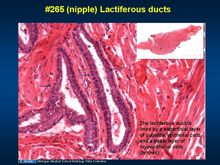
#265 (nipple) Lactiferous ducts The lactiferous duct is lined by a superficial layer of cuboidal epithelial cells and a basal layer of myoepithelial cells (arrows). Michigan Medical School Histology Slide Collection
Learning Objectives • • Be able to identify principal layers of the skin (epidermis, dermis and hypodermis) at the light microscope level and know the major functions of each layer. Be able to identify the strata of the epidermis in thick and thin skin and know the major cellular events that take place in each layer in the process of keratinization. Be able to identify the cells in different layer of the epidermis at the electron microscope level by recognizing characteristic organelles and structures present in each layer. Be able to recognize melanocytes and know the process of pigment formation in the skin. Be able to identify eccrine and apocrine sweat glands at the light microscope level and distinguish ductal and secretory portions. Be able to identify the components of the pilosebacous apparatus and know the structural relationship between each component and the epidermis. Be able to identify the mammary gland, by recognizing its structural components (lactiferous ducts, alveoli, lobules, the stromal connective tissue), and know the histological differences in active and inactive glands.
Additional Source Information for more information see: http: //open. umich. edu/wiki/Citation. Policy Slide 5: UM Medical School Anatomy Laboratory Manual Slide 7: Michigan Medical School Histology Slide Collection Slide 8: Michigan Medical School Histology Slide Collection Slide 9: Michigan Medical School Histology Slide Collection ; Source Undetermined Slide 10: Source Undetermined Slide 11: Source Undetermined Slide 12: Source Undetermined Slide 14: Source Undetermined Slide 15: Source Undetermined Slide 16: Source Undetermined Slide 17: Source Undetermined Slide 19: Michigan Medical School Histology Slide Collection ; Source Undetermined; Slide 21: Source Undetermined Slide 22: Source Undetermined; Slide 23: Michigan Medical School Histology Slide Collection ; Source Undetermined Slide 24: Source Undetermined Slide 25: Source Undetermined Slide 26: Source Undetermined Slide 28: Source Undetermined Slide 29: Source Undetermined Slide 30: Michigan Medical School Histology Slide Collection ; Source Undetermined Slide 31: Wheater 9. 18 Slide 32: Michigan Medical School Histology Slide Collection Slide 33: Sources Undetermined Slide 34: Weiss, pg. 562 Slide 35: Source Undetermined
Slide 36: Source Undetermined Slide 37: Source Undetermined Slide 38: Tsaitgaist, Wikipeda, http: //en. wikipedia. org/wiki/File: Hair_follicle-en. svg Slide 39: Michigan Medical School Histology Slide Collection Slide 40: Michigan Medical School Histology Slide Collection Slide 41: Source Undetermined Slide 42: Michigan Medical School Histology Slide Collection Slide 43: Michigan Medical School Histology Slide Collection Slide 44: Michigan Medical School Histology Slide Collection ; Source Undetermined Slide 45: Source Undetermined Slide 46: Adapted and modified from Leson TS, Leson CR, Paparo AA: Text/Atlas of Histology. Philadelphia, WB Saunders, 1988; Source Undetermined Slide 47: Gray’s Anatomy, Wikimedia Commons, http: //commons. wikimedia. org/wiki/File: Dissected_lactating_breast_gray 1172. png; U. S. Federal Government Slide 48: Source Undetermined Slide 49: Michigan Medical School Histology Slide Collection Slide 50: Michigan Medical School Histology Slide Collection ; Source Undetermined Slide 51: Source Undetermined; Slide 52: Gray’s Anatomy, Wikimedia Commons, http: //commons. wikimedia. org/wiki/File: Dissected_lactating_breast_gray 1172. png; Michigan Medical School Histology Slide Collection Slide 53: Michigan Medical School Histology Slide Collection