Atopic Dermatitis v Dry skin pruritus erythematous papules


")
, drugs, chemicals, food")
 v Intensely pruritic, bright-red")
; herpes simplex virus")
; drugs (e. g.")


 v Varicella-zoster virus v Macules (2– 3 mm) evolving into papules, then")


: Janeway lesions")


 or erythema.")
 v Paramyxovirus v First disease v Discrete lesions that become confluent as")

 v Togavirus v Third disease v Spreads from hairline downward, clearing")
 v S. aureus, phage group II v Diffuse")

 v Diffuse")
 v Second disease")


: The generalized rash: part I &")
- Slides: 41
Atopic Dermatitis v Dry skin; pruritus; erythematous papules; excoriations; scaling; lichenification; accentuation of skin lines; v Personal or family history of atopy
Contact Dermatitis v Erythema; edema; vesicles; bullae in linear or geometric pattern; common causes include cosmetics, topical medications, metal, latex, poison ivy, textiles, dyes, sunscreens, cement, food, benzocaine, neomycin v linear or geometric pattern and distribution of lesions
Cutaneous Reaction to Arthropod Bite v Urticarial papules and plaques v Outdoor exposure (usually) and distribution of lesions where insects are likely to bite
Cutaneous Small-Vessel Vasculitis v Infections (including group A Streptococcus, viral hepatitis), drugs, chemicals, food allergens, idiopathic causes. v Palpable purpuric lesions appearing in crops on legs or other dependent areas; may become vesicular or ulcerative; usually resolve over 3– 4 weeks. v Occurs in a wide spectrum of diseases, including connective tissue disease, cryoglobulinemia, malignancy, Henoch. Schönlein purpura (HSP); more common in children. v Fever, malaise, arthralgias, myalgia.
Exanthematous Drug-induced Eruption v Drugs (antibiotics, anticonvulsants, diuretics, etc. ) v Intensely pruritic, bright-red macules and papules, symmetric on trunk and extremities; may become confluent.
Erythema Multiforme v Drug intake (i. e. , sulfa, phenytoin, penicillin); herpes simplex virus or Mycoplasma pneumoniae infection, etc. v Target lesions (central erythema surrounded by area of clearing and another rim of erythema) up to 2 cm; symmetric on knees, elbows, palms, soles; may become diffuse; may involve mucosal surfaces; lifethreatening in maximal form (Stevens -Johnson syndrome).
Erythema Nodosum v Infections (e. g. , streptococcal, fungal, mycobacterial, yersinial); drugs (e. g. , sulfas, penicillins, oral contraceptives); sarcoidosis; idiopathic causes v Large, violaceous, nonulcerative, subcutaneous nodules; exquisitely tender; usually on lower legs but also on upper extremities.
Exfoliative Erythroderma v Underlying psoriasis, eczema, drug eruption, mycosis fungoides v Usually occurs in adults over age 50; more common in men. v Fever, chills (i. e. , difficulty with thermoregulation); lymphadenopathy.
Folliculitis v Bacterial, viral or fungal v Multiple small pustules localized to hair follicles on any body surface v Hair follicle at center of each lesion v Hot tub rash: pseudomona
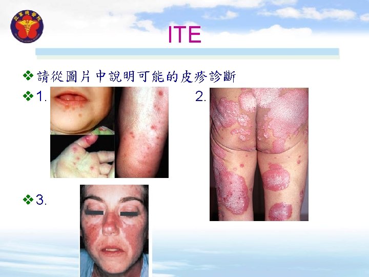
Hand-Foot-and-Mouth Disease v Coxsackievirus A 16 most common cause v Tender vesicles, erosions in mouth; 0. 25 -cm papules on hands and feet with rim of erythema evolving into tender vesicles. v Transient fever.
Miliaria Rubra/Crystallina v Erythematous non-follicular papules/vesicles associated with heat exposure or fever; lesions on back, trunk, neck, or occluded areas v History of heat exposure and distribution of lesions
Plaque Psoriasis v Thick, sharply demarcated, round or oval, erythematous plaques with thick silvery white scale; lesions on extensor surfaces, elbows, knees, scalp, central trunk, umbilicus, genitalia, lower back, or gluteal cleft v Positive Auspitz sign (removal of scale produces bleeding points) and Koebner phenomenon
Scabies v Discrete, small burrows, vesicles, papules, and pinpoint erosions on fingers, finger webs, wrists, elbows, knees, groin, buttocks, penis, scrotum, axillae, belt line, ankles, and feet v Distribution of lesions, intense pruritus, and positive mineral oil mount
Seborrheic Dermatitis v Erythematous patches with greasy scale; lesions behind ears or on scalp and scalp margins, external ear canals, base of eyelashes, eyebrows, nasolabial folds, central chest, axillae, inframammary folds, groin, and umbilicus.
Systemic Lupus Erythematosus v Autoimmune disease v Macular and papular erythema, often in sun-exposed areas; discoid lupus lesions (local atrophy, scale, pigmentary changes); periungual telangiectasis; malar rash; vasculitis sometimes causing urticaria, palpable purpura; oral erosions in some cases. v Arthritis; cardiac, pulmonary, renal, hematologic, and vasculitic disease.
Tinea Corporis v Flat, red, scaly lesions progressing to annular lesions with central clearing or brown discoloration; keys to diagnosis are annular lesions with central clearing and positive KOH preparation.
Urticaria v Discrete and confluent, raised, edematous, round or oval, waxing and waning lesions with large variation in size v Erythematous border (flare) and pale center (wheal) v History of drug, food, or substance exposure
Varicella (Chickenpox) v Varicella-zoster virus v Macules (2– 3 mm) evolving into papules, then vesicles (sometimes umbilicated), on an erythematous base ("dewdrops on a rose petal"); pustules then forming and crusting; lesions appearing in crops; may involve scalp, mouth; intensely pruritic. v Malaise; generally mild disease in healthy children; more severe disease with complications in adults and immunocompromised children.
Uncommon causes of generalized skin rash
Acute Meningococcemia v Neisseria meningitidis v Initially pink maculopapular lesions evolving into petechiae; petechiae rapidly becoming numerous, sometimes enlarging and becoming vesicular; trunk, extremities most commonly involved; may appear on face, hands, feet; may include purpura fulminans reflecting disseminated intravascular coagulation. v Hypotension, meningitis.
Bacterial Endocarditis v Streptococcus, Staphylococcus , etc. v Acute course (Staphylococcus aureus): Janeway lesions (painless erythematous or hemorrhagic macules, usually on palms and soles). v Subacute course: Osler's nodes (tender pink nodules on finger or toe pads); petechiae on skin and mucosa; splinter hemorrhages. v New heart murmur
Bullous Pemphigoid v Generalized bullae, especially on trunk and flexural areas; patient usually older than 60 years v Nikolsky sign (easy separation of epidermis from dermis with lateral pressure) usually negative.
Disseminated Herpesvirus Infection v HSV or VZV v Generalized vesicles that can evolve to pustules and ulcerations; individual lesions similar for HSV and varicellazoster. v HSV: extensive, progressive mucocutaneous lesions in some cases; HSV lesions sometimes disseminate in eczematous skin (eczema herpeticum); HSV visceral dissemination may occur with only limited skin lesions. v Zoster cutaneous dissemination: >25 lesions extending outside involved dermatome.
Erythema Marginatum v Rheumatic fever v Group A Streptococcus v Erythematous annular papules and plaques occurring as polycyclic lesions in waves over trunk, proximal extremities; evolving and resolving within hours. v Pharyngitis preceding polyarthritis, carditis, subcutaneous nodules, chorea.
Ecthyma Gangrenosum v P. aeruginosa , other gramnegative rods, fungi v Indurated plaque evolving into hemorrhagic bulla or pustule that sloughs, resulting in eschar formation; erythematous halo; most common in axillary, groin, perianal regions. v Usually affects neutropenic patients; occurs in up to 28% of individuals with Pseudomonas bacteremia.
Infectious Mononucleosis v Epstein-Barr virus v Diffuse maculopapular eruption (10– 15% of cases; 90% if ampicillin is given); urticaria in some cases; periorbital edema (50%); palatal petechiae (25%)
Kawasaki's Disease v Idiopathic causes v Rash similar to scarlet fever (scarlatiniform) or erythema. multiforme; fissuring of lips, strawberry tongue; conjunctivitis; edema of hands, feet; desquamation later in disease. v Cervical adenopathy, pharyngitis, coronary artery vasculitis.
Measles (Rubeola) v Paramyxovirus v First disease v Discrete lesions that become confluent as rash spreads from hairline downward, sparing palms and soles; lasts 3 days; Koplik's spots.
Primary HIV Infection v HIV v Nonspecific diffuse macules and papules; may be urticarial; oral or genital ulcers in some cases
Rubella (German measles) v Togavirus v Third disease v Spreads from hairline downward, clearing as it spreads; Forschheimer spots
Staphylococcal Scalded Skin Syndrome (4 S) v S. aureus, phage group II v Diffuse tender erythema, often with bullae and desquamation; Nikolsky's sign. v Irritability; nasal or conjunctival secretions.
Syphilis v Treponema pallidum v Coincident primary chancre in 10% of cases; coppercolored, scaly papular eruption, diffuse but prominent on palms and soles; rash never vesicular in adults; condyloma latum, mucous patches, and alopecia in some cases.
Toxic Epidermal Necrolysis v Drugs, other causes (infection, neoplasm, graft-vs. -host disease) v Diffuse erythema or target-like lesions progressing to bullae, with sloughing and necrosis of entire epidermis; Nikolsky's sign. v Dehydration, sepsis sometimes resulting from lack of normal skin integrity; 25% mortality.
Scarlet Fever v Group A Streptococcus (pyrogenic exotoxins A, B, C) v Second disease v Diffuse blanchable erythema beginning on face and spreading to trunk and extremities; circumoral pallor; "sandpaper" texture to skin; accentuation of linear erythema in skin folds (Pastia's lines); enanthem of white evolving into red "strawberry" tongue; desquamation in second week.
Scrub Typhus v Orientia tsutsugamushi v Diffuse macular rash starting on trunk; eschar at site of mite bite. v Headache, myalgias, regional adenopathy; mortality up to 30% if untreated.
Still's Disease v Autoimmune disease. v Transient 2 - to 5 -mm erythematous papules appearing at height of fever on trunk, proximal extremities; lesions evanescent. v High spiking fever, polyarthritis, splenomegaly; erythrocyte sedimentation rate, >100 mm/h.
Urticarial Vasculitis v Serum sickness, often due to infection (including hepatitis B viral, enteroviral, parasitic), drugs (including penicillins, sulfonamides, salicylates, barbiturates); connective tissue disease; idiopathic causes v Erythematous, circumscribed areas of edema; occasionally indurated; pruritic or burning; lesions sometimes purpuric; individual lesions lasting up to 5 days
Reference v Ely JW & Stone MS (2010): The generalized rash: part I & II, American family physician 81: 726 -734; 735 -739. v Kaye, ET & Kaye KM (2015): Fever and rash in Harrison’s principles of internal medicine (19 th ed. ), chapter 24. v Wolff K & Johnson RA (2013): Severe and lifethreatening skin eruptions in the acutely ill patient in Fitzpatrick's color atlas and synopsis of clinical dermatology (7 th ed. ), section 8.