ATLS Advanced Trauma Life Support DR BALAKH SHER
, FCPS, MRCS(UK) Assistant Professor")
ATLS Advanced Trauma Life Support DR. BALAKH SHER ZAMAN MBBS(KE), FCPS, MRCS(UK) Assistant Professor Surgery KEMU/Mayo Hospital Lahore

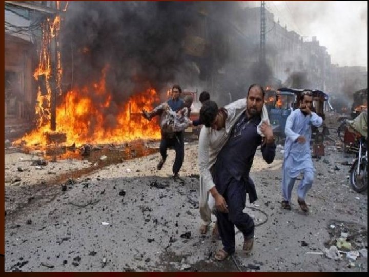
What is Trauma? l Physical injury caused by an external source. S-IV, JHL

GALLERY PAGE INFORMATION GOES HERE SNAP IFNO SNAP IFNO SNAP IFNO SNAP IFNO PAGE 1 OF 1 Best View at (1024 x 768) on Microsoft Internet Explorer Copyright © 2010 Punjab Emergengy Services(Res cue 1122), De ra Ghazi Khan Developed by Digerians Soft.

The biggest killer in Pakistan? Trauma is the leading cause of death in the first four decades of life and third overall in every country of the world For every person dying, three more get disabled
 On road")
Death following Injuries Trimodal distribution : l l Immediate within minutes (50%) On road , due to widespread damage to brain, ruptured heart or great vessels (only preventive measures). Within Hours -Golden hours – 30% Life threatening injuries like facial injuries leading to airways obstruction, altered breathing mechanism , massive blood loss. Within Days (20%) Sepsis and multi organ failure l

Deaths following Injuries l Trimodal distribution minutes golden hour days weeks lethal injuries life threatening injuries complications (sepsis, MOF)

Dangerous Injuries l Crash greater than 20 mile/h l Fall from height of 20 ft or more l Death of another person l Ejection of patient

The History of the ATLS In February 1976, a tragedy occurred that would change the first hour of trauma care for patients. Dr. Jim Styner, an orthopedic surgeon, crashed his small plane into a cornfield in rural Nebraska. Dr. Styner sustained serious injuries, three of his children sustained critical injuries, and one child sustained minor injuries. His wife was killed instantly.

Continue The care that he and his family received was less than adequate. There was an obvious lack of training for proper triage and injury treatment. The surgeon, recognizing how inadequate his treatment was stated, ” when I can provide better care in the field with limited resources than what my children and I received at the primary care facility, there is something wrong with the system and the system has to be changed. ”

Concept of ATLS He started the course in Nebraska in 1978 which was taken up by American College of surgeons and given nationally 1979.
 l Is a training program for doctors l Its")
Advanced Trauma Life Support (ATLS) l Is a training program for doctors l Its goal is to teach a simplified and standardized approach for trauma patients l The philosophy of the ATLS program is to treat the greatest threat to life first and then reassess and treat again

The ATLS -- Basic Concepts l l Treat the greatest threat to life first using the ABCDE approach, (don’t start with history) Full diagnosis is not needed to treat a life threatening emergency. Lack of a definitive diagnosis should not impede the application of indicated treatment.
 § Resuscitation § Secondary survey")
Initial Assessment § Rapid primary survey (60 sec exam) § Resuscitation § Secondary survey § Initiate a definitive care It is a continuous process

Primary Survey l QUICK HEAD TO TOE EXAMINATION l The first and key part of the assessment of patients presenting with trauma is called the primary survey. During this time, lifethreatening injuries are identified and simultaneously resuscitation is begun.

Primary Survey The ABCs v A Airway with cervical spine control v B Breathing and ventilation v C Circulation with hemorrhage control v D Disability: Neurologic status v E Exposure, prevent hypothermia

AIRWAY WITH CERVICAL SPINE CONTROL Ø Establish patent airway Ø Cervical spine control

Assume Cervical spine injury Ø Blunt injury above clavicle Ø Multi system trauma Ø Altered level of consciousness

Airway Problems are a common cause of Preventable Deaths l Failure to recognize airway need l Inability to establish airway l Failure to correct an incorrectly placed airway l Dislodgement of a correctly placed airway l Delay in establishing adequate ventilation l Aspiration of Gastric Contents

Objective Signs of Airway Obstruction l Look if agitated Agitation hypoxia, l Using accessory muscles or not? l Listen for abnormal sounds Noisy breathing is obstructed breathing l Feel the trachea

Management l A rigid suction device is essential, all trauma victims need supplemental O 2 l Airway maintenance l Chin lift l Modified Jaw thrust l Oral/nasopharyngeal airway l Surgical/needle cricothyriodotomy

Breathing & VENTILATION l The chest must be examined by inspection, palpation, percussion and auscultation. l Subcutaneous emphysema and tracheal deviation must be identified if present.

Indication for ventilation in chest injury l Tachypnea above 40 l Extensive pulmonary contusion or diffuse Infiltrative change on X-ray

S-IV, JHL

Life threatening Chest Injuries which may impair ventilation • • • Airway obstruction Open pneumothorax Tension pneumothorax Massive hemothorax >1500 ml Flail chest with pulmonary contusion Cardiac tamponade

C-Circulation with Hemorrhage Control l Hemorrhage is the predominant cause of preventable post-injury deaths. Hypovolemic shock is caused by significant blood loss. Two large-bore intravenous lines are established and crystalloid solution given. If the patient does not respond to this, type-specific blood, or Onegative should be given.

C-Circulation • Radial pulse is palpable with BP of 80 mm of Hg • Femoral pulse is palpable with BP of 70 mm of Hg Carotid Pulse is palpable with BP of 60 mm of Hg •

Regions from where patient can lose large volume of blood l Externally l The chest l The abdomen l The retroperitoneum l Into muscle compartment

Circulation Assess l blood volume loss and cardiac output l Level of consciousness l Skin color and temperature l Pulse Don’t wait for BP drop to make a diagnosis of hypovolemic shock. At least 30 % volume loss is needed for BP to drop

Causes of Shock in trauma l Hemorrhage – the commonest cause of hypovolumic shock in trauma patients l Non hemorrhagic l Cardiac Pump problems l Cardiac tamponade, l Tension pneumothorax, l Myocardial contusion l Neurogenic shock

Hypovolemia resulting from Internal Loss Ø Intra-abdominal or intra thoracic injury Ø Fractures of the pelvis and /or femur Ø Penetrating injuries with arterial or venous involvement

Disability Rapid Neurological Evaluation Ø Level of consciousness A Alert V Responds to voice P Responds to pain U Unresponsive Ø Pupils

Glasgow Coma Scale or GCS is a neurological scale that aims to give a reliable objective way of recording the conscious state of a person for initial as well as subsequent assessment.

CONTINUED l The scale comprises three tests: eye, verbal and motor responses. The three values separately as well as their sum are considered. l The lowest possible GCS (the sum) is 3 (deep coma or death), while the highest is 15 (fully awake person).

S-IV, JHL

Exposure / Environment Ø Undress patient completely Ø Do not forget the back of the patient Ø Protect from hypothermia: Intravenous fluids should be warmed and a warm environment maintained. Ø Patient privacy should be maintained.

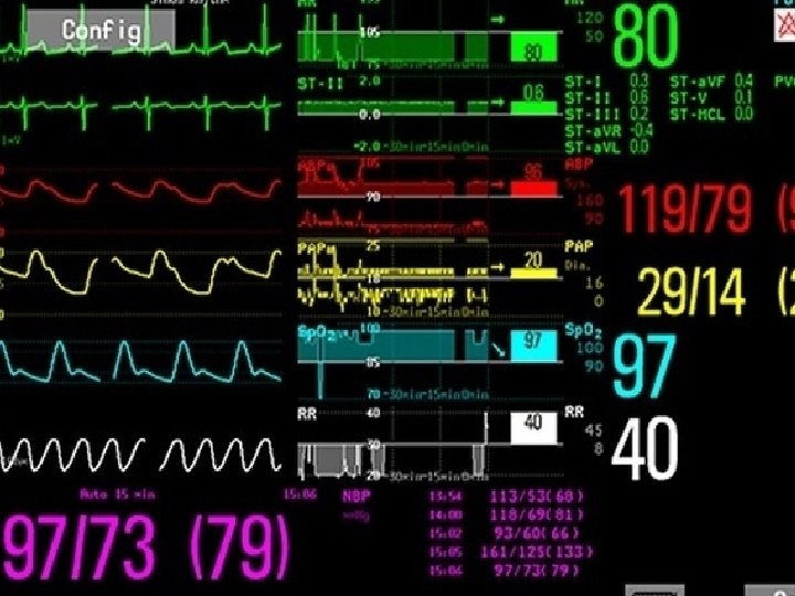
Resucitation Phase l Secure large bore IV access l Shock therapy l Continuous ECG monitoring l Blood samples – CBC , ABGs, Cross match, Glucose, coagulation studies l NG & Folley’s catheter (if not contraindicated)
 Ø Urinary output Ø ECG")
Monitoring Ø Vital signs (Temp, Pulse, Respiration & BP) Ø Urinary output Ø ECG Ø Pulse oximetery / ABGs
 l Pelvis (AP) l")
Priority X Rays Chest (AP) l Pelvis (AP) l
")
What is an adequate Cervical spine X ray? (Lateral view)

Secondary Survey Initiate resuscitation and reassess ABCs before secondary survey Ø "Head to toe" evaluation Ø Complete neurological exam Ø X- rays as indicated Ø Special procedures e. g DPL etc Ø " Tubes and fingers " in every orifice

Re-evaluation Continue Monitoring Ø New findings / deterioration / improvement Ø High index of suspicion for unapparent injuries Ø Pain relief after surgical consultation

Secondary survey Mechanism of injury Ø Blunt Direction of impact determines injury pattern History and description of events is important Ø Penetrating Energy transfer factors like velocity and calibre of bullet, trajectory and distance

Head and face Ø Head Pupils Visual acuity Injury Ø Maxillofacial Assume c-spine injury

Spine And Neck Ø Maintain immobilization of neck until injury excluded Ø Absence of neurological deficit does not exclude neck injury Ø Penetrating neck injury if going through platysma requires evaluation

Chest & Abdomen l Sequentially assessed for potential threats to life

Extremities & Back l Long bone # l Neurovascular injuries l Compound #

Adjuncts to Secondary Survey l Radiology – Plain & Contrast l USG l CT l Endoscopies (Broncho – Esophago l Angiography

Record Keeping v Meticulous record keeping is essential to evaluate patient's needs and clinical status especially since more than one doctor often caring for patient v Accurate record of fluids given flow sheet v Medicolegal problems arise frequently and precise records are helpful for all concerned

Definitive Care l Treatment by appropriate consultant

Prevention of Trauma l Primary prevention-Antidrinking driving, speed limit l Secondary-Active –Helmet-Seat belts. Passive -ABS, Air bags l Tertiary -minimize the effects of injury by improving health care delivery


- Slides: 54