Athletic Injuries ATC 222 Head Face Eyes Ears


- Slides: 70

Athletic Injuries ATC 222 Head, Face, Eyes, Ears, Nose, and Throat Chapter 27

Facial Injuries • Mandible Fracture – – – deformity malocclusion malalignment bleeding around teeth/gums lower lip anesthesia pain with biting • Treatment

Facial Injuries • Mandible Dislocation/Subluxation – commonly from lateral force – malalignment – malocclusion – open, locked jaw

Dental Injuries • Types – fracture – dislocation/subluxation • Treatment – realign subluxation – replace/preserve dislocation or fracture – 30 minute survival rate

Nasal Injuries • Fracture or Cartilage Separation – S/S • • deformity profuse bleeding immediate swelling crepitus – treatment • control hemorrhaging • referral • most return to activity in 3 -4 days

Nasal Injuries • Epistaxis (nosebleed) – – – sit upright ice (nose and ipsilateral carotid) direct pressure on nostril cotton/gauze plug refrain from nose blowing for 2 hours

Eye Injuries • Causes and Prevention • S/S or Serious Eye Injury – – – prolonged blurred vision loss of part/all of visual field sharp, stabbing, throbbing pain double vision embedded object blood in anterior chamber (hyphema)

Treatment of Serious Eye Injury • immediate referral • cover both eyes with embedded object • ice only to surrounding tissue • no pressure applied to eyes

• • Orbital Blowout Fx Blunt trauma Inability to look upward Diplopia Sunken eye

What is wrong with this picture?

Orbital Hematoma • “Black Eye” • Bleeding in orbit area and poss. Sclera • Rule out serious eye injury

Foreign Body in Eye • Embedded? • Removal – Close eye – eye rinse – removal with gauze pad

The ole finger in the eye play!!!!

Ear Injuries • Hematoma Auris (cauliflower ear) – causes – S/S • swelling • redness, warmth • pain – treatment • ice • protection • aspirate

Otitis Media and Externa • Etiology • Signs and Symptoms • Treatment

Neurological System and Evaluation

Neuron (Nerve Cell) • • • dendrites cell body axon Schwann cells motor end plate

Functional Classification of Neurons • Sensory • Associational – Inter-neurons • Motor – Upper motor neuron – Lower motor neuron

Synapse • Functional connection between 2 neurons – chemical or electrical • Neurotransmitters – acetylcholine – norepinephrine • Motor Unit

Nervous System Divisions • Central Nervous System (CNS) – Brain – Spinal Cord • Peripheral Nervous System (PNS) – Cranial Nerves – Spinal Nerves • R. T. D. C. B.

PNS – Somatic NS – Autonomic NS • sympathetic • parasympathetic • enteric NS

Somatic Nervous System • Functions – voluntary control of skeletal muscle – convey conscious/unconscious sensory (afferent) information • vision, pain, touch, unconscious muscle sense

Autonomic Nervous System • Functions • convey sensory input from visceral organs, glands and cardiovascular system • involuntary control of smooth and cardiac muscle • maintain homeostasis • Divisions of ANS – Sympathetic Nervous System • thoracolumbar – Parasympathetic Nervous System • craniosacral – Enteric Nervous System

Sympathetic Nervous System • Dominates in stress conditions – physical and psychological – very rapid effects • “Fight or Flight” theory – increased sweating, HR, RR – blood diverted to skeletal muscles – pupil dilation – conversion of glycogen to glucose

Parasympathetic Nervous System • Opposite actions of sympathetic nervous system • dominates in relaxed states – decreased HR and RR – increased peristalsis – increased saliva and intestinal secretions – pupil constriction

Enteric Nervous System • innervates GI tract, pancreas, gall bladder

CNS • Gray matter = nerve cell bodies • White matter = axons • Efferent neurons – motor neurons • Afferent neurons – sensory neurons

Meninges • Dura Mater – tough, inelastic membrane – adheres to inner part of cranium • Arachnoid Mater – delicate, web-like tissue – avascular • Pia Mater – thin, delicate tissue hugging brain – no space between pia mater and brain – capillary rich to supply brain with blood

Meninges Cont. • Epidural Space – “potential space” – between cranium and dura mater – space created due to epidural hematoma – middle meningeal artery • Subdural Space – filled with a serous lubricant – prevents dura mater and arachnoid from adhering to each other • Subarachnoid Space – relatively large – filled with cerebrospinal fluid – ventricles

Cerebrum • Basal Ganglia • Limbic system

Cerebrum • general appearance and behavior • level of consciousness (loc) • intellectual performance – short term memory (STM) – long term memory (LTM) • amnesia? – calculation – reasoning • emotional control • language skills • voluntary movement (cerebral cortex)

Basal Ganglia and Limbic System • Basal Ganglia – part of extra-pyramidal system – inter-connects several part of CNS – fine tune motor control • Limbic System – emotion, hunger, biological rhythms, smell

Diencephalon • • epithalamus hypothalamus subthalamus

Thalamus/Hypothalamus • Thalamus – receives input from every sensory system – sensory and motor integration • Hypothalamus – homeostasis (temp), hunger, thirst, emotions

Cerebellum • Coordination – control of timing, speed, and direction of movement • Equilibrium – balance, posture

Brain Stem • midbrain – eye tracking; voluntary movement • medulla – decussation of UMN • pons – relay info. from cortex to cerebellum; respiration • medulla oblongata – reflexes for vomiting, swallowing, coughing, salivation, pupils • cranial nerves III, IV, V, VII, VIII, IX, X, XII

Movie Time Rated PG

Reticular Formation • Extends throughout the length of the brain stem • Reticular activating system – wakefulness – modification of sensory input – controls motor function via reticulospinal tract – receives input from hypothalamus and limbic system (emotion)

Vestibular Nuclei • located in brain stem • receive input from labyrinthine system, reticular formation, and cerebellum • controls/interprets balance, head control, and eye tracking

Spinal Cord • Function – pathway for efferent and afferent nerve fibers • ascending and descending spinal tracts – connects peripheral and spinal nerves to brain – center for spinal (monosynaptic) reflexes • Location – foramen magnum to app. L 2 • Gives rise to 31 pair of spinal root nerves • Cauda equina – lumbrosacral plexus from L 2 on down

Spinal Nerves • 31 pair – dorsal spinal root = afferent = sensory – ventral spinal root = efferent = motor • Doral and ventral root join to form the peripheral nerve • Spinal nerves exit below respective vertebral level except for cervical • Myotome – voluntary muscle group receiving motor innervation from a specific spinal nerve • Dermatome – section of skin that receives sensory innervation from a specific spinal nerve – adjacent dermatomes overlap – partial loss = peripheral complete loss = cord


Descending Tracts “Motor” • corticospinal (Pyramidal Tract) – voluntary skilled movement in extremities • reticulospinal – facilitate or inhibit motor neurons; – posture • tectospinal – postural reflexes of head for vision • rubrospinal – facilitate/inhibit motor neurons – posture • vestibulospinal – facilitate/inhibit postural muscles of abdomen, back, neck

Ascending Tracts • Exteroceptive, Proprioceptive, and Interoceptive • ventral and lateral spinothalamic – pain and temperature • spinocerebellar – proprioceptive and exteroceptive – vestibular nuclei and joint receptors • spinoreticular – muscle, joints, and skin • gracile and cuneate – touch, pressure, conscious joint sense

Cranial Nerves • Sensory and/or Motor Function – 12 pairs • On Old Olympus’ Towering Top A Fin And German Viewed Some Hops • Oh Oh Oh To Touch and Feel a Girl/Guy Very Sexy and Hot • Motor and/or Sensory Function – Some say marry money but my brother says bad boys marry money.

Cranial Nerves • I. Olfactory • function: smell • testing: identify common odors • II. Optic • function: vision • testing: check visual fields, check vision • III. Oculomotor • function: eye movement, pupil reflex • testing: tracking, direct/consensual pupil reflex, accommodation, nystagmus, drooping eyelid • IV. Trochlear • function: eye movement • testing: tracking, nystagmus

Cranial Nerves Cont. • V. Trigeminal • function: muscles of mastication, facial sensation, corneal reflex • testing: check facial sensation, muscles of mastication • VI. Abducens • function: eye movement • testing: tracking, nystagmus • VII. Facial • function: muscles of facial expression, taste to anterior tongue • testing: facial expressions, taste • VIII. Vestibulocochlear • function: hearing, equilibrium • testing: hearing, check for tinnitus, check balance

Cranial Nerves Cont. • IX. Glossopharyngeal • function: taste to posterior tongue, muscles of larynx/pharynx • testing: taste, gag reflex, speak/swallowing, coughing • X. Vagus • function: swallowing, phonation, taste • testing: speak/swallowing, gag reflex, taste, cough • XI. Spinal Accessory • function: motor control of upper trap and sternocleidomastoid • testing: SMT/DMT of trap and SCM • XII. Hypoglossal • function: tongue movement • testing: tongue protrusion (deviation? )

Cranial Nerve Quick Test • • • Vision Visual Fields Eye Tracking Facial Sensation Muscles of Facial Expression Muscles of Mastication Hearing/Balance Swallowing Upper Trap/SCM Strength Tongue Protrusion Pupil Reflexes

Movie Time Rated PG

Proprioception • The awareness of posture, movement, muscle length/tension, changes in equilibrium, weight, resistance of objects, and speed/range, angle of movement • Proprioceptors – muscle spindle – Golgi tendon organ (GTO) – mechanoreceptors

Muscle Spindle • Detects length and rate of length • Extrafusal vs. Intrafusal fibers • extrafusal = skeletal muscle fibers – innervated by alpha motor neurons • intrafusal = muscle spindle fibers – innervated by gamma motor neurons

Muscle Spindles • Intrafusal fibers – located within muscle belly – stretching a muscle also stretches the muscle spindle – most sensitive to rapid stretching • Types – Nuclear Bag 1 (Dynamic) • rate of change in length • Ia afferent; fires rapidly but adapts quickly – Nuclear Chain (Static) • overall length • II afferent; slow firing and non-adapting

Golgi Tendon Organ – located within tendons – Ib afferent • slow firing and non-adapting – most sensitive to excessive stretch – sensitive to excessive tension due to muscle contraction – excessive tension will cause a reflexive inhibition of alpha mn

Myotatic or Stretch Reflexes • Deep Tendon Reflex – biceps brachii: C 5 -C 6 – brachioradialis: C 5 -C 6 – triceps brachii: C 7 – infrapatellar: L 3 -L 4 – posterior tibialis: L 5 – achilles: S 1 • Jendrassik Maneuver – increases facilitative activity of spinal cord

Superficial Reflexes • Abdominal – upper: T 6 -T 9 – lower: T 9 -T 12 • • Cremasteric: L 1 -L 2 Plantar: S 1 -S 2 Gag Corneal

Visceral Reflexes • Pupillary reflex – direct – consensual – accommodation • Blink reflex

Pathological Reflexes • • Babinski sign Oppenheim sign Decorticate rigidity ** Decerebrate rigidity ** • ** due to lack of cortical/cerebral control

Lower Motor Neuron Lesions • weakness/paralysis/paresis of a voluntary motor group • decreased tone (flaccidity) of involved motor group • decreased/absent deep tendon reflex (hyporeflexia or areflexia) • atrophy of muscle/muscle group • radicular pain specific to a spinal nerve path • decreases/absent sensation of specific dermatomes (hypoesthesia or anesthesia)

Upper Motor Neuron Lesion • pathological reflex present (eg: Babinski sign) • weakness distal to lesion • hemiplegia/paraplegia • increased deep tendon reflex (hypereflexia) ** • hypertonicity – spasticity – rigidity • decreased/absent superficial reflexes • ** due to lack of cortical control

Reflex Grading Scale • • • 0 = 1+ = 2+ = 3+ = 4+ = Absent Decreased (elicited with reinforcement) Normal Increased Clonus

Movie Time Rated R


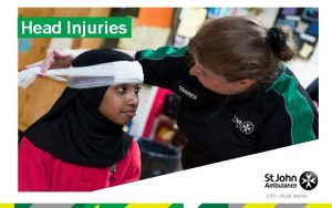
Head Injuries • Incidence of serious injury has decreased – neck injuries? – protective gear • Appr. 250, 000 concussions/year • Focal vs. Diffuse Injuries

Concussion • Definition – clinical syndrome characterized by immediate and transient impairment of normal neurological function • Causes – coup Vs. contrecoup • • Grades Return to Play Criteria Post-concussion Syndrome Second Impact Syndrome

Intracranial Hemorrhaging • Epidural hematoma – arterial bleeding – rapid onset (poss. 10 -20 min. ) • Subdural hematoma – venous/capillary bleeding – slow onset • Intracerebral hematoma – compressive mechanism/aneurysm – rapid onset

Evaluation Process • Primary Assessment? – ABC’s • Secondary Assessment – Mental Status – Cranial Nerve Exam – Motor System Exam – Proprioception, balance, and coordination – Sensory Exam – Reflex Examination

Cranial Nerve Exam • Test for: – Vision – Tracking – Visual Fields – Pupil Reflex – Hearing – Swallowing – Shoulder Shrug – Facial Sensation – Facial Expression – Tongue Protrusion – Mastication

Other Signs and Symptoms • • • headache nausea, vomiting seizures unequal pupils tinnitus unusual drowsiness

Treatment • Recheck athlete on regular basis • Refer if in doubt or in more severe cases • Monitor throughout the night • No alcohol, aspirin, ibuprofen • No activity until asymptomatic • No new s/s or no worsening of current s/s
 Dns 208 67 222 222
 Slanted ears
 Impaired eyes and ears
 Hear us from heaven jared anderson
 Special senses the eyes and ears
 Eyes that see and ears that hear
 Eyes are to spectacles as ears are to
 Whoever has ears let them hear
 Medical terminology chapter 11 learning exercises answers
 A short backboard or vest-style immobilization
 Chapter 21 caring for head and spine injuries
 Animal whose ears cannot be seen
 Pf
Pf What colour is hazel eyes
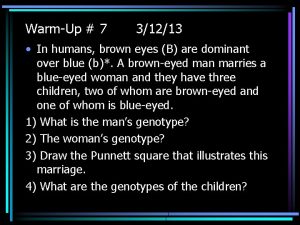
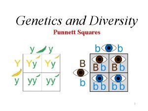
 Brown eyes are dominant to blue eyes
 Brown eyes phenotype
 Can two blue eyed parents have a brown eyed child
 Face to face class
 Face-to-face narrative examples
 Whats this shape
 Ecdl.com
 Lesson 2 elements of communication
 Onomatopoeia in romeo and juliet act 2
 Politeness and interaction
 Pros and cons of telephone interviews
 Phospholipid bilayer
 Brown and levinson
 Kiran sanghera
 29 cfr 1910 regarding face head foot and hand protection
 Regional write up head face and neck
 Mism 222
 222$ in euro
 Cs-222
 Round to it
 Blk 222
 Google tradutor
 Infosys 222
 Item 222
 Flogel fg 1000
 Ece 222 github
 Xxaxis matlab 222
 Unit 222 communication in a business environment
 Cs 222
 Psych 222
 Mide-222
 Infosys 222
 Cpcs 222
 Tio mempunyai kelereng sebanyak 24 butir
 Navadmin 222/15
 Pineer species
 222 telephone
 Cs-222
 Tamu csce 222
 Psalm 222
 Eco 222
 Funk radius
 Nerologopeda
 Bio 222
 Infosys 222
 Restriction codes in texas
 10000/222
 Infosys 222
 Mekila 222
 Csce 222
 Operation of moving head disk storage
 Condyles of femur
 Indexing head formula
 Parts of the chest
 The tone unit
 The attacking firm goes head-to-head with its competitor.
 Pre-head head tonic syllable tail


