ASSOCIATE PROFESSOR IOLANDA BLIDARU MD Ph D Epidemiology

ASSOCIATE PROFESSOR IOLANDA BLIDARU MD, Ph. D
. ® 20% of female >")
Epidemiology ® The commonest of all pelvic T. (1/3). ® 20% of female > 35 years have fibroid. ® Childbearing life. ® Often enlarge during pregnancy or during oral contraceptive use, and regress after menopause

®Uterus deprived from a baby consoles itself with a fibroid.

Causes ®Unknown ®Hyperestrogenemia – E 2 / ER, P / PR, Gn. RH, growth factors (IGF-1, EGF< PDGF< FGF) ®Race ® Obesity ® Chromosomal 14) abnormalities (7, 12,

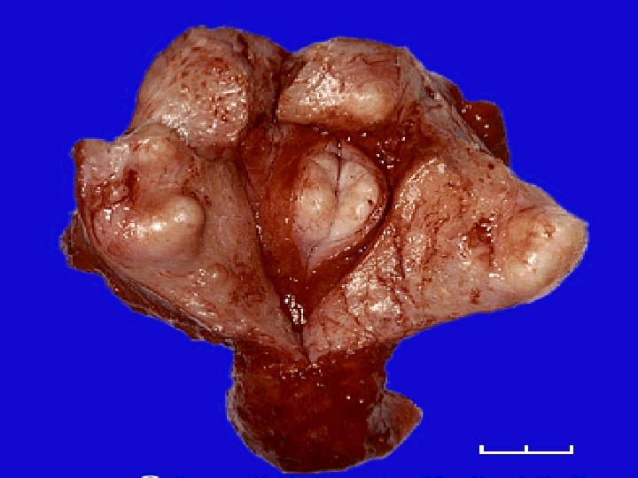
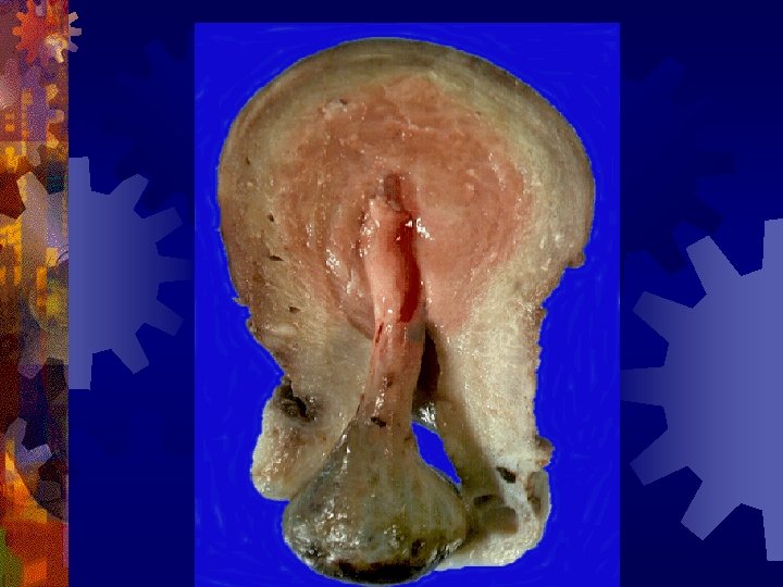
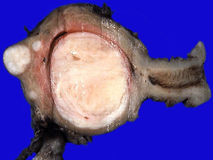
Pathology ®MACROSCOPY q site q shape q size q consistency q cut section q capsule q number q varieties

Uterine leiomyoma Cervical • 1 -2% • solitary Corporeal • 98% • multiple

Corporeal leiomyoma Interstitial • 58% Subserous • 18% submucus • 24% • not capsulated
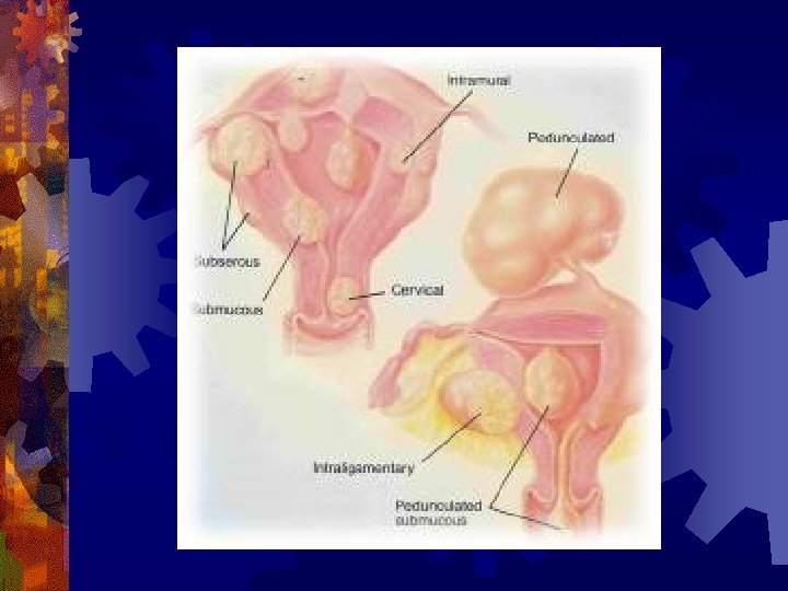

Cervical leiomyoma Exocervix • small • sessile • polypoid Supravaginal cervix • sessile • pedunculated
. ®Soft (pregnancy-cystic degeneration). ®Stony hard (Calcification)")
CONSISTENCY ®Firm ®Harder (hyaline degeneration). ®Soft (pregnancy-cystic degeneration). ®Stony hard (Calcification)

Leiomyomata Uterus

CUT SECTION ® well demarcated surrounding muscle. ® whorly (intermingling muscle fibers and fibrous tissue). ® paler than surrounding (ischaemia).
. ®Smooth muscle cells and fibrous tissue")
Microscopic Examination ®Few formed blood vessels (blood lakes). ®Smooth muscle cells and fibrous tissue cells.

Leiomyoma:

Changes occuring with fibroid General Genital tract Tumor itself
 ®Endometriosis (30 -40%)")
Genital tract ®Endometrium - hyperplasia ®Tubes - inflammation (salpingitis) ®Endometriosis (30 -40%)

Tumour itself ®Benign Ø Ø Ø Ø degeneration atrophic hyaline red cystic fatty calcification necrosis with or without infection vascular (edema, lymphangiectasia) ®Malignant degeneration (0. 1 -0. 5 % - growth after menopause, rapid enlargement, recurrent fibroid polyp).

DIAGNOSIS ®History ®Examination. ®Investigation. ®D. D.
. ® Pain - uncomplicated →")
SYMPTOMS ® No symptom ® Bleeding (menorrhagia - metrorrhagia). ® Pain - uncomplicated → congestion → dysmenorrhea; complicated → degeneration (malignant, infection, torsion) ® Infertility ® Mass ® Discharge ® Pressure symptoms (urinary, lower limb edema, constipation)
 • Asymmetrically enlarged uterus (subserous")
Signs • Symmetrically enlarged uterus (submucosal fibroid) • Asymmetrically enlarged uterus (subserous
 ® Laboratory (Hb, Ht, urinary tests, pregnacy test, Pap test")
Investigations ® Clinical (examination) ® Laboratory (Hb, Ht, urinary tests, pregnacy test, Pap test etc) ® Imaging & instrumental techniques (US, hysteroscopy, hysterography, colposcopy, fractional curettage, Ct scan) ® Miscellaneous (intravenous urography, etc)
 ® Ademomyosis. Leiomyomas - myomectomy, adenomyosis -")
DIFFERENTIAL DIAGNOSIS ® Pregnancy (normal / abnormal) ® Ademomyosis. Leiomyomas - myomectomy, adenomyosis - hysterectomy ® Solid Adnexal Mass (fibromas, Brenner tumors, inflammatory mass) ® Uterine Leiomyosarcoma ( histologically - the presence of infiltrative margins, nuclear atypia, and increased mitotic figures )

Uterus Adenomyosis:

DIFFERENTIAL DIAGNOSIS

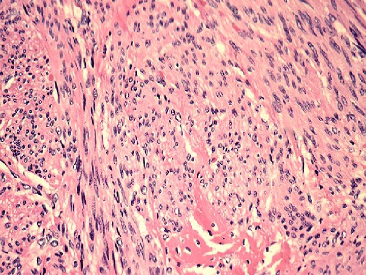
Uterine Leiomyosarcom
. ® Conservative ®")
Treatment of Leiomyoma ® No treatment ü Patient (age, parity, symptoms). ® Conservative ® Radiological ü Tumor (number, size, type) ® Surgical ® Gn. RH agonistsü Complications. ® Uterine artery embolization.
")
Treatment of Leiomyoma MEDICAL ®Progesterone / Progestins ®Selective PR modulator / antagonist (Mifepristone, Ulipristal) ®Gn. RH agonists (Buserelin, Triptorelin, Leuprolid, Histerelin, Goserelin)
 ®Hysterectomy (Hysteroscopy,")
SURGICAL ®Myomectomy laparoscopy, laparotomy) ®Hysterectomy (Hysteroscopy,
- Slides: 32