
Assignment Topic Enlist five developmental disturbances of teeth
Assignment Topic: Enlist five developmental disturbances of teeth affecting the structure By Muhammed. Ramshad. P BDS 1 st year Yenepoya Dental Collage
Developmental disturbances of teeth affecting the structure • • Amelogenesis imperfecta Dentin dysplasia Regional odontodysplasia
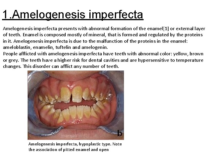
1. Amelogenesis imperfecta presents with abnormal formation of the enamel[1] or external layer of teeth. Enamel is composed mostly of mineral, that is formed and regulated by the proteins in it. Amelogenesis imperfecta is due to the malfunction of the proteins in the enamel: ameloblastin, enamelin, tuftelin and amelogenin. People afflicted with amelogenesis imperfecta have teeth with abnormal color: yellow, brown or grey. The teeth have a higher risk for dental cavities and are hypersensitive to temperature changes. This disorder can afflict any number of teeth. Amelogenesis imperfecta, hypoplastic type. Note the association of pitted enamel and open
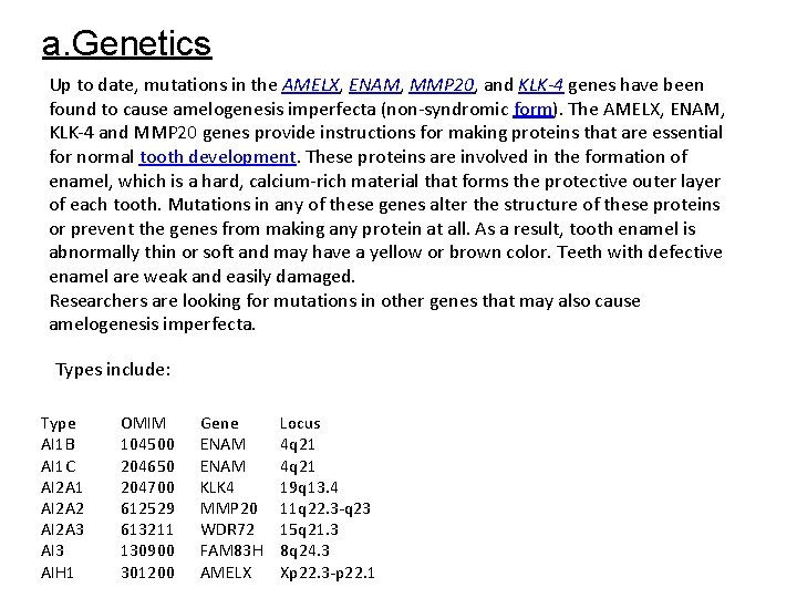
a. Genetics Up to date, mutations in the AMELX, ENAM, MMP 20, and KLK-4 genes have been found to cause amelogenesis imperfecta (non-syndromic form). The AMELX, ENAM, KLK-4 and MMP 20 genes provide instructions for making proteins that are essential for normal tooth development. These proteins are involved in the formation of enamel, which is a hard, calcium-rich material that forms the protective outer layer of each tooth. Mutations in any of these genes alter the structure of these proteins or prevent the genes from making any protein at all. As a result, tooth enamel is abnormally thin or soft and may have a yellow or brown color. Teeth with defective enamel are weak and easily damaged. Researchers are looking for mutations in other genes that may also cause amelogenesis imperfecta. Types include: Type AI 1 B AI 1 C AI 2 A 1 AI 2 A 2 AI 2 A 3 AIH 1 OMIM 104500 204650 204700 612529 613211 130900 301200 Gene ENAM KLK 4 MMP 20 WDR 72 FAM 83 H AMELX Locus 4 q 21 19 q 13. 4 11 q 22. 3 -q 23 15 q 21. 3 8 q 24. 3 Xp 22. 3 -p 22. 1
Amelogenesis imperfecta can have different inheritance patterns depending on the gene that is altered. Most cases are caused by mutations in the ENAM gene and are inherited in an autosomal dominant pattern. This type of inheritance means one copy of the altered gene in each cell is sufficient to cause the disorder. Amelogenesis imperfecta is also inherited in an autosomal recessive pattern; this form of the disorder can result from mutations in the ENAM or MMP 20 gene. Autosomal recessive inheritance means two copies of the gene in each cell are altered. About 5% of amelogenesis imperfecta cases are caused by mutations in the AMELX gene and are inherited in an X-linked pattern. A condition is considered X-linked if the mutated gene that causes the disorder is located on the X chromosome, one of the two sex chromosomes. In most cases, males with an X-linked form of this condition experience more severe dental abnormalities than affected females. Other cases of this condition result from new gene mutations and occur in people with no history of the disorder in their family. b. Treatment Crowns are sometimes being used to compensate for the soft enamel. Usually stainless steel crowns are used in children which may be replaced by porcelain once they reach adulthood. In the worst case scenario, the teeth may have to be extracted and implants or dentures are required.
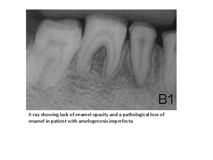
X-ray showing lack of enamel opacity and a pathological loss of enamel in patient with amelogenesis imperfecta

2. Dentinogenesis imperfecta (hereditary Opalescent Dentin) is a genetic disorder of tooth development. This condition causes teeth to be discolored (most often a blue-gray or yellow-brown color) and translucent. Teeth are also weaker than normal, making them prone to rapid wear, breakage, and loss. These problems can affect both primary (baby) teeth and permanent teeth. This condition is inherited in an autosomal dominant pattern, which means one copy of the altered gene in each cell is sufficient to cause the disorder. Dentinogenesis imperfecta affects an estimated 1 in 6, 000 to 8, 000 people.
a. Types of dentinogenesis imperfecta with similar dental formalities usually an autosomal dominant trait with variable expressivity but can be recessive if the associated osteogenesis imperfecta is of recessive type. This is type l. Type II : Occurs in people without other inherited disorders (i. e. Osteogenesis imperfecta). It is an autosomal dominant trait. A few families with type II have progressive hearing loss in addition to dental abnormalities. Mutations in the DSPP gene have been identified in people with type II and type III dentinogenesis imperfecta. Type I occurs as part of osteogenesis imperfecta. b. Clinical features Clinical appearance is variable. However, the teeth usually involved and more severely affected are deciduous teeth in type 1; whereas in type 2 both the dentitions are equally affected. The teeth may be gray to yellowish brown. They exhibit translucent or opalescent hue. Enamel is usually lost early due to loss of scalloping at the DEJ. However, the teeth are not more susceptible to dental caries than normal ones. c. Radiographic features Type I and II show total obliteration of the pulp chamber. Type III shows thin dentin and extremely enormous pulp chamber. These teeth are usually known as Shell Teeth.
d. Histology Dentinal tubules are irregular and are bigger in diameter. Areas of uncalcified matrix are seen. Sometimes odontoblasts are seen in dentin. e. Treatment One treatment option is bonding, putting lighter enamel on the weakened enamel of the teeth and with lots of treatments of this bonding, the teeth appear whiter to the eye, but the teeth on the inside and under that cover are still the same. Due to the weakened condition of the teeth, many common cosmetic procedures such as braces and bridges are inappropriate for patients with Dentinogenesis imperfecta and are likely to cause even more damage than the situation they were intended to correct.
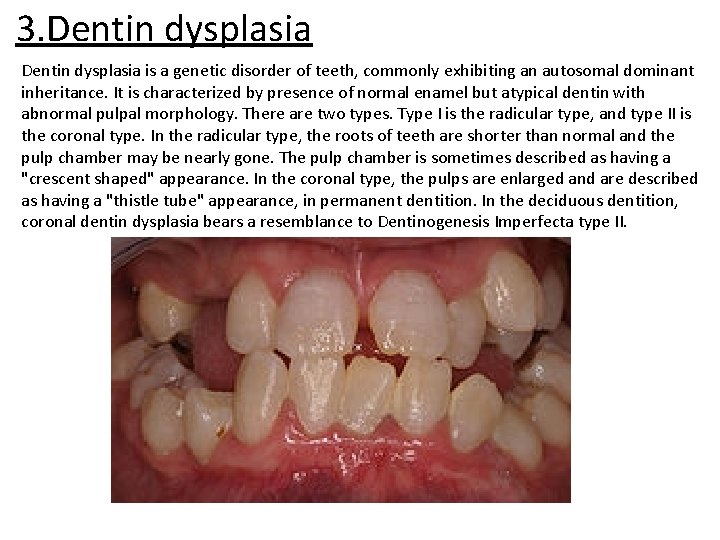
3. Dentin dysplasia is a genetic disorder of teeth, commonly exhibiting an autosomal dominant inheritance. It is characterized by presence of normal enamel but atypical dentin with abnormal pulpal morphology. There are two types. Type I is the radicular type, and type II is the coronal type. In the radicular type, the roots of teeth are shorter than normal and the pulp chamber may be nearly gone. The pulp chamber is sometimes described as having a "crescent shaped" appearance. In the coronal type, the pulps are enlarged and are described as having a "thistle tube" appearance, in permanent dentition. In the deciduous dentition, coronal dentin dysplasia bears a resemblance to Dentinogenesis Imperfecta type II.
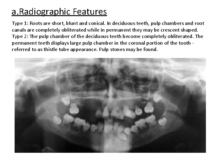
a. Radiographic Features Type 1: Roots are short, blunt and conical. In deciduous teeth, pulp chambers and root canals are completely obliterated while in permanent they may be crescent shaped. Type 2: The pulp chamber of the deciduous teeth become completely obliterated. The permanent teeth displays large pulp chamber in the coronal portion of the tooth - referred to as thistle tube appearance. Pulp stones may be found.
b. Histologic Features Type 1: Normal dentinal tubule formation is blocked and new dentin forms around obstacles. . . known as lava flowing around boulders.
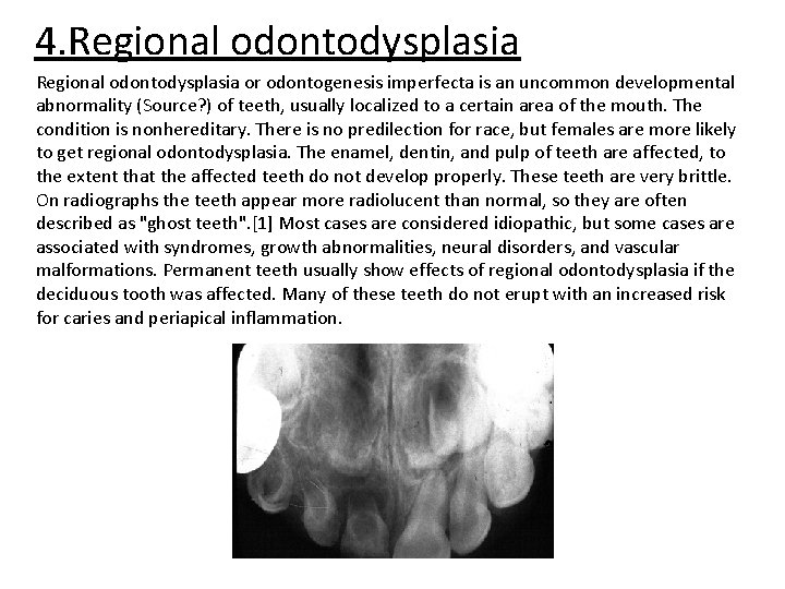
4. Regional odontodysplasia or odontogenesis imperfecta is an uncommon developmental abnormality (Source? ) of teeth, usually localized to a certain area of the mouth. The condition is nonhereditary. There is no predilection for race, but females are more likely to get regional odontodysplasia. The enamel, dentin, and pulp of teeth are affected, to the extent that the affected teeth do not develop properly. These teeth are very brittle. On radiographs the teeth appear more radiolucent than normal, so they are often described as "ghost teeth". [1] Most cases are considered idiopathic, but some cases are associated with syndromes, growth abnormalities, neural disorders, and vascular malformations. Permanent teeth usually show effects of regional odontodysplasia if the deciduous tooth was affected. Many of these teeth do not erupt with an increased risk for caries and periapical inflammation.
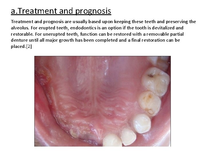
a. Treatment and prognosis are usually based upon keeping these teeth and preserving the alveolus. For erupted teeth, endodontics is an option if the tooth is devitalized and restorable. For unerupted teeth, function can be restored with a removable partial denture until all major growth has been completed and a final restoration can be placed. [2]

- Slides: 15