Assessment of the Breast Dr Freida FullerJonap fulerjonapfmercer

Assessment of the Breast Dr. Freida Fuller-Jonap fulerjonap_f@mercer. edu

Risk Factors for Breast Cancer • • • gender age genetics family history personal history early menarch and late menopause • no natural children • first child born to mother older than age 30 • oral contraceptive use • regular alcohol intake • higher education and socioeconomic status • previous breast irradiation

Subjective Data Collection • • • History Surgeries involving the breast Medication history History of fibrocystic breast disease Any changes in the breasts


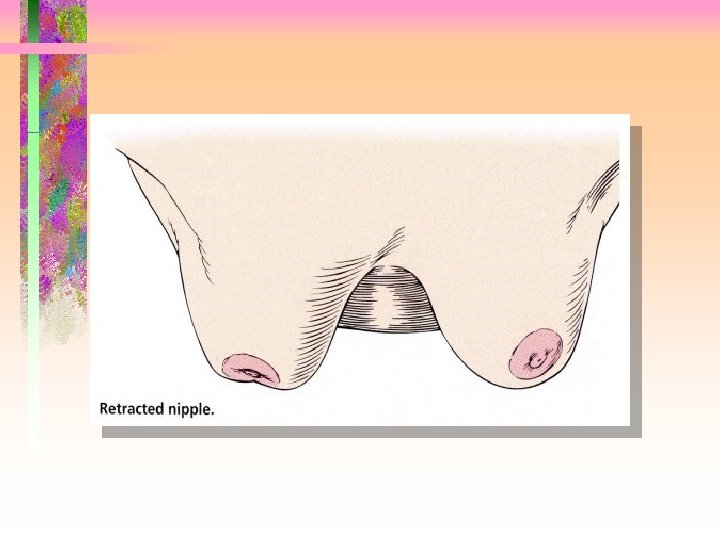
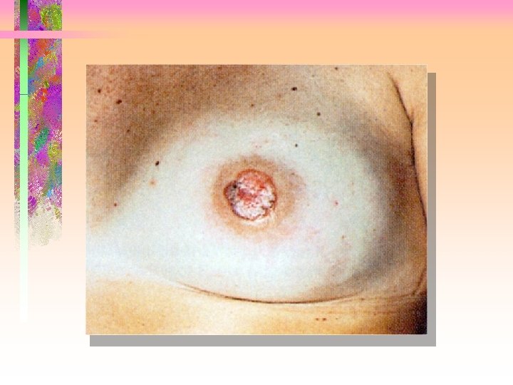
Observe for • masses • discharge • skin texture, rashes, • Paget’s disease pigmentation • Peau d’orange • Retraction or dimpling • venous patterns • areolar and nipple characteristics


Palpation • Palpate axilla in sitting position preferrably


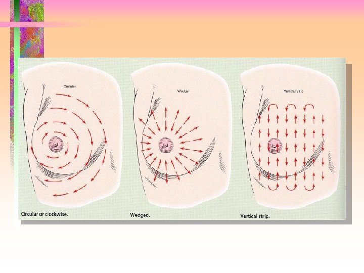
Breast Palpation • Breast palpation position • Use the flat pads of three fingers • Vary the levels of pressure: light, medium, firm • Use systematic pattern of examination


If a mass is detected the following should be noted • Size in centimeters and its position • Shape • Delimitation or discreteness • Consistency • Mobility • Tenderness • Erythema • Dimpling • Depth


Examination of the Male Breast • Essentially the same as for women, but can be done sitting up since not a large amount of tissue

Developmental Considerations • Before 10: small nipples, small and slightly elevated • Between 10 and 14: areola enlarges • 14 and above: areola recedes into breast contour, adult female breast forms • During reproductive years: cycle of size change, nodularity and tenderness • Post-menopausal: more flabby

Differential Diagnoses • Cancerous tumors • Fibroadenomas • Benign breast disease: fibrocystic breast disease

Diagnostic Testing • Mammography • Ultrasound
- Slides: 20