Assessment of fetal wellbeing in pregnancy and labour

Assessment of fetal wellbeing in pregnancy and labour Dr Ban Hadi

Green-top Guideline 57 February 2011 Most women are aware of fetal movements by 20 weeks of gestation. Clinicians should be aware (and should advise women) that although fetal movements tend to plateau at 32 weeks of gestation, there is no reduction in the frequency of fetal movements in the late third trimester.

Green-top Guideline 57 February 2011 Although some multiparous women may perceive fetal movements as early as 16 weeks of gestation , some primiparous women may perceive movement much later than 20 weeks of gestation

Factors which influence a woman’s perception of this activity 1. Fetal ‘sleep’cycles 2. Maternal position 3. Drugs, smoking 4. Maternal blood glucose 5. Administration of corticosteroids 6. Fetal malformations, position

Fetal movements are usually absent during fetal ‘sleep’cycles, which occur regularly throughout the day and night and usually last for 20– 40 minutes. These sleep cycles rarely exceed 90 minutes in the normal, healthy fetus

women perceive most fetal movements when lying down, fewer when sitting and fewest while standing. It is therefore not surprising that pregnant women who are busy and not concentrating on fetal activity often report a misperception of a reduction of fetal movements. an anteriorly positioned placenta may decrease a woman’s perception of fetal movements

cigarette smoking is associated with a decrease in fetal activity Raised maternal blood glucose may increase fetal movements

The administration of corticosteroids to enhance fetal lung maturation has been reported by some authors to decrease fetal movements and fetal heart rate variability detected by cardiotocography (CTG) over the 2 days following administration

Fetuses with major malformations are generally more likely to demonstrate reduced fetal activity (anencephaly has more activity) Fetal presentation has no effect on perception of movement

Fetal position might influence maternal perception: 80% of fetal spines lie anteriorly in women who were unable to perceive fetal movements despite being able to visualise them when an ultrasound scan was performed

How can fetal movements be assessed? 1. Subjective maternal perception of fetal movements 2. Objective assessments of fetal movements use Doppler or real-time ultrasound

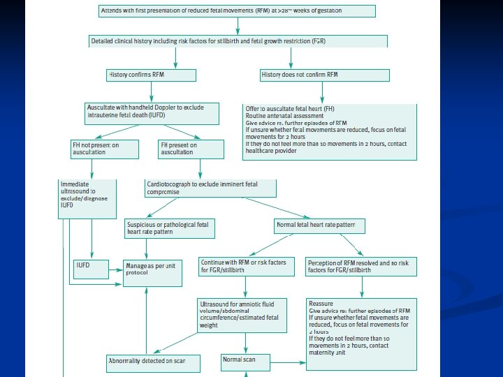
If women are unsure whether movements are reduced after 28+0 weeks of gestation, they should be advised to lie on their left side and focus on fetal movements for 2 hours. If they do not feel 10 or more discrete movements in 2 hours, they should contact their midwife or maternity unit immediately.

There is lack of clinical data of precise normal movement count Maternal anxiety is a draw back of maternal fetal movement counting

Management of women with RFM 1. History : a. Risk factors for FGR and IUD, duration of RFM b. Kick count: reassuring if 4 or more movements in 1 hr. 3 or less indicate further assessment A second approach is to have the mother begin counting fetal movements when she wakes up in the morning and record the number of hours required to feel 10 movements. On average, this takes 2 to 3 hours. Again, maternal reports of decreased movement should prompt further testing

2. Examination: The key priority when a woman presents with RFM is to confirm fetal viability. In most cases, a handheld Doppler device will confirm the presence of the fetal heart beat General: BMI, general health Vital signs: blood pressure Abdominal exam. measurement of symphysis– fundal height to detect SGA fetuses

3. CTG : Non stress test After fetal viability has been confirmed and history confirms a decrease in fetal movements, arrangements should be made for the woman to have a CTG to exclude fetal compromise if the pregnancy is over 28+0 weeks of gestation

At least two or more accelerations with fetal movement in 20 min. by at least 15 bpm for 15 sec. is considered as reactive NST Before 32 weeks, accelerations are defined as having an acceleration that is 10 bpm or more above baseline for 10 seconds or longer.

A reactive NST is highly predictive of low risk for fetal mortality in the subsequent 72 to 96 hours and is still predictive at 1 week. Fetuses do not routinely demonstrate reactivity before 28 weeks, and it may be normal to have a nonreactive tracing as late as 32 weeks' gestation. After 32 weeks, a nonreactive tracing should prompt further evaluation of fetal wellbeing, such as measuring a biophysical

4. Ultrasound: Ultrasound scan assessment should be undertaken as part of the preliminary investigations of a woman presenting with RFM after 28+0 weeks of gestation if: 1. The perception of RFM persists despite a normal CTG or 2. If there any additional risk factors for FGR/stillbirth.

Ultrasound scan assessment should include fetal biometry: the assessment of abdominal circumference and/or estimated fetal weight to detect the SGA fetus, and the assessment of amniotic fluid volume. Ultrasound should include assessment of fetal morphology if this has not previously been performed

4. Ultrasound Biophysical profile: There may be a role for the selective use of BPP in the management or investigation of RFM There is evidence from uncontrolled observational studies that BPP in high-risk women has good negative predictive value; that is, fetal death is rare in women in the presence of a normal BPP


A score of 6 raises concern, and the BPP should be repeated in 6 to 24 hours, especially in fetuses over 32 weeks' gestation. If the score does not improve, delivery should be considered, depending on gestational age and individual circumstances. Scores of 4 or below are worrisome, and delivery should be considered, again depending on gestational age and clinical context
. This test is also known as")
Non stress Test and Amniotic Fluid Index (AFI). This test is also known as the modified biophysical profile. In the third trimester, an AFI and NST are often used together to assess fetal well-being. The AFI is the sum of the maximum vertical pockets of umbilical-cord-free amniotic fluid in each of the four quadrants of the uterus. In general, the AFI reflects fetal perfusion, and, if decreased, raises suspicion for placental insufficiency. A normal test has a reactive NST and an AFI greater than 5 (and less than 25). An abnormal test lacks one or both of these findings.

Doppler study: of the umbilical and middle cerebral art.
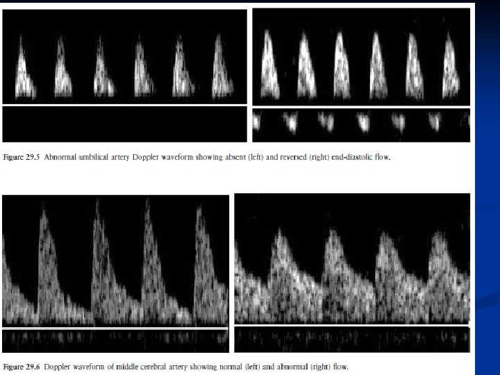
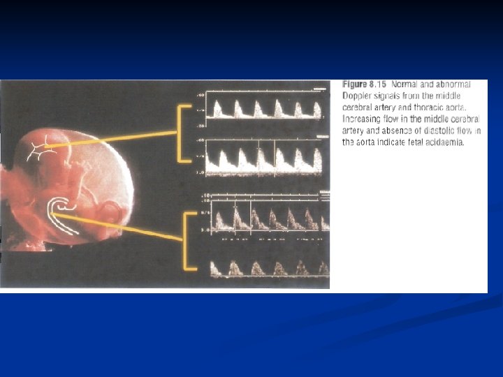

A measure of the amount of diastolic flow relative to systolic is provided by several indices, such as the pulsatility index or resistance index, which essentially compare the amount of diastolic flow to systolic flow. When these indices are high, this indicates high resistance to flow; when the indices are low, resistance to flow is low

while a rising resistance in the fetal aorta reflects compensatory vasoconstriction in the fetal body. Absent diastolic flow in the fetal aorta implies fetal acidaemia. Perhaps the most sensitive index of fetal acidaemia and incipient heart failure is demonstrated by increasing pulsatility in the central veins supplying the heart, such as the ductus venosus and inferior vena cava.

Doppler ultrasound studies of the uterine arteries may demonstrate markers of increased resistance to flow including the diastolic ‘notch’ in the waveform

Uterine artery waveform with diastolic notch

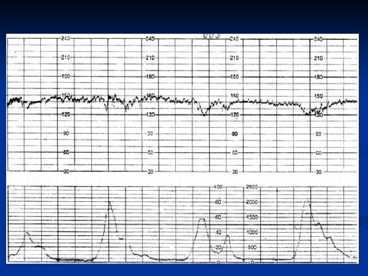
5. Contraction stress test: A positive CST is one in which late decelerations occur with more than 50% of contractions. Late decelerations are decelerations that reach their nadir after the peak of the contraction. A negative CST is one in which no late decelerations occur. A CST with non repetitive late decelerations is considered equivocal, and further evaluation of the pregnancy is performed. An inadequate or unsatisfactory CST is one in which adequate contractions are not achieved. Relative contraindications to CST include preterm labor, preterm premature rupture of membranes, placenta previa, and high risk for uterine rupture. Previous low transverse cesarean section is not a contraindication

Criteria for Interpretation of the Contraction Stress Test Negative: no late or significant variable decelerations Positive: late decelerations following 50% or more of contractions (even if the contraction frequency is fewer than three in 10 minutes) Equivocal-suspicious: intermittent late decelerations or significant variable decelerations Equivocal-hyperstimulatory: fetal heart rate decelerations that occur in the presence of contractions more frequent than every 2 minutes or lasting longer than 90 seconds Unsatisfactory: fewer than three contractions in 10 minutes or an uninterpretable tracing

Assessment of fetal wellbeing during labour 1. History and examination

2. Amount and color of amniotic fluid Clear liquore of normal amount is reassuring Absent liquore, blood stained and meconium stained liquore raise concern

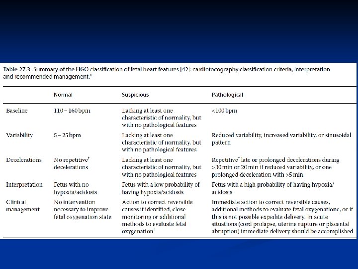
3. CTG: To interpret a CTG you need a structured method of assessing it’s various characteristics. The most popular structure can be remembered using the acronym DR C BRAVADO DR – Define Risk C – Contractions BRa – Baseline Rate V – Variability A – Accelerations D – Decelerations

Define risk You first need to assess if this pregnancy is high or low risk This is important as it gives more context to the CTG reading

In the low-risk situation, intermittent auscultation, either by Pinard stethoscope or by handheld Doppler, is often advocated. Current guidelines suggest auscultating the FHR every 15 minutes in the active phase of the first stage of labour. This should be for 60 seconds following a contraction, in order to detect significant decelerations. Maternal pulse should also be recorded by palpation to avoid confusion, particularly when an FHR abnormality is suspected.

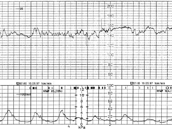
Contractions Record the number of contractions present in a 10 minute period – e. g. 3 in 10 Each big square is equal to 1 minute, so you look how many contractions occurred in 10 squares Individual contractions are seen as peaks on the part of the CTG monitoring uterine activity

Duration – how long do the contractions last? Intensity – how strong are the contractions?
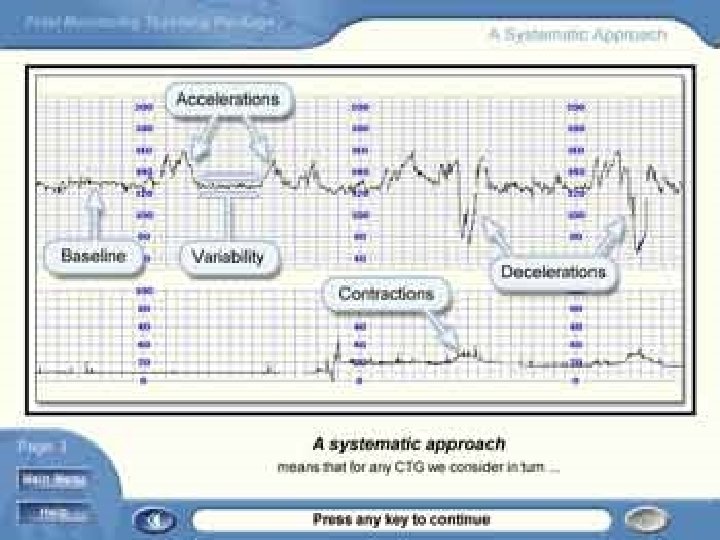

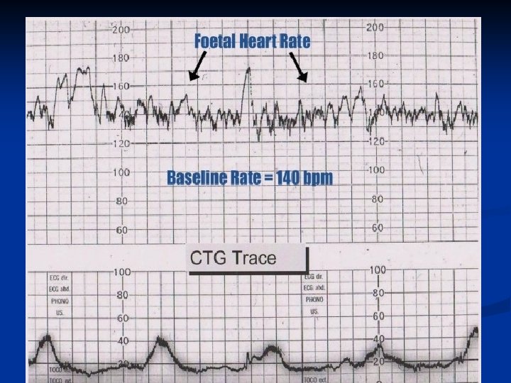

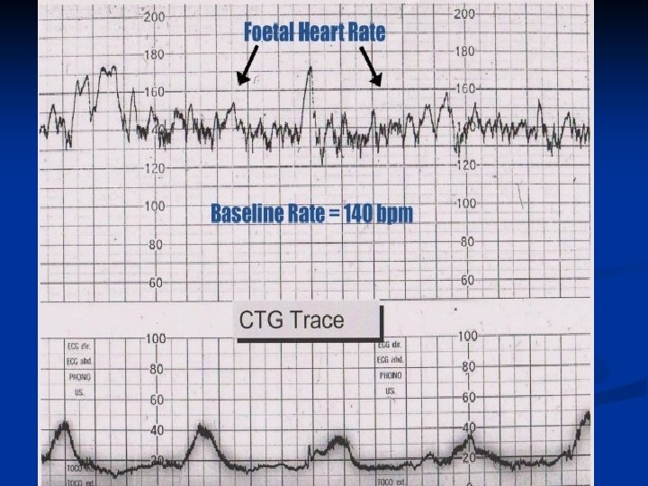
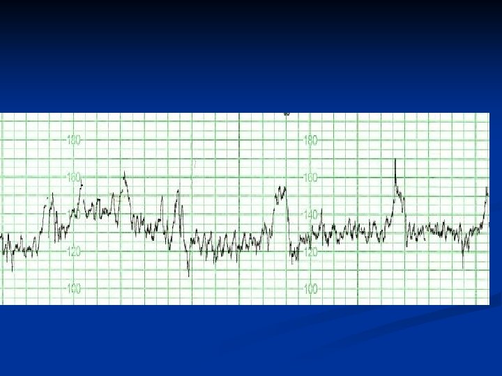
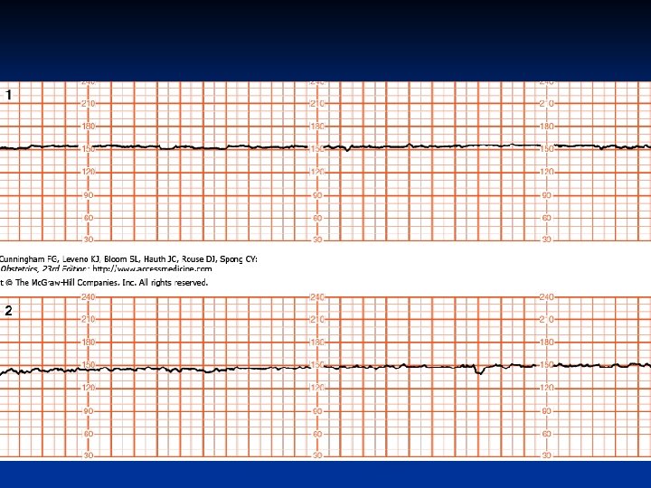
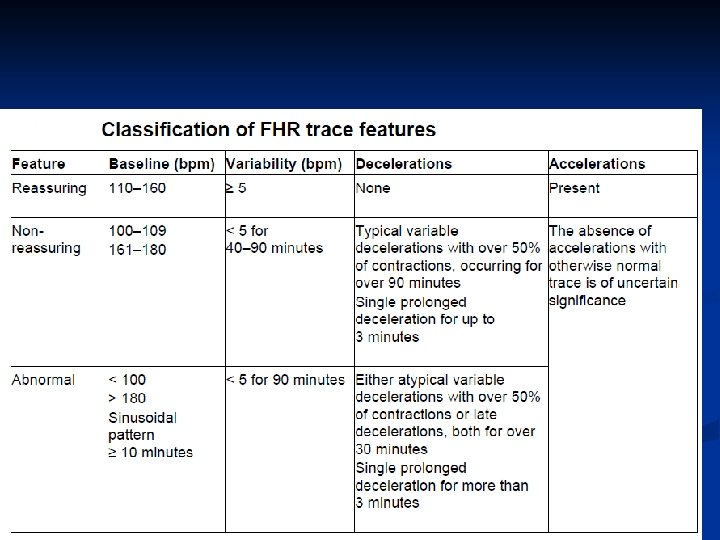
The baseline rate: is the average heart rate of the fetus in a 10 minute window Look at the CTG & assess what the average heart rate has been over the last 10 minutes Ignore any Accelerations or Decelerations A normal fetal heart rate is between 110160 bpm

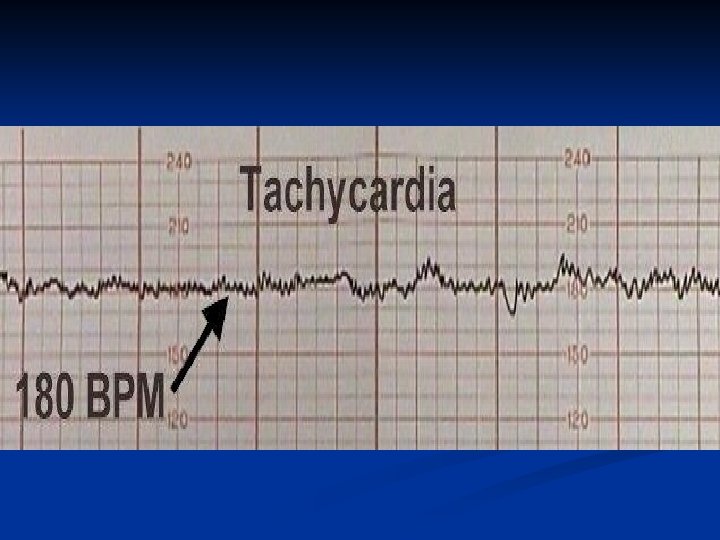
Foetal Tachycardia Foetal tachycardia is defined as a baseline heart rate greater than 160 bpm It can be caused by: Foetal hypoxia Chorioamnionitis – if maternal fever also present Hyperthyroidism Foetal or Maternal Anaemia Foetal tachyarrhythmia

Foetal Bradycardia Foetal bradycardia is defined as a baseline heart rate less than 110 bpm. Mild bradycardia of between 100 -110 bpm is common in the following situations: Post-date gestation Occiput posterior or transverse positions Severe prolonged bradycardia (< 80 bpm for > 3 minutes) indicates severe hypoxia

Causes of prolonged severe bradycardia are: Prolonged cord compression Cord prolapse Epidural & Spinal Anaesthesia Maternal seizures Rapid fetal descent If the cause cannot be identified and corrected, immediate delivery is recommended

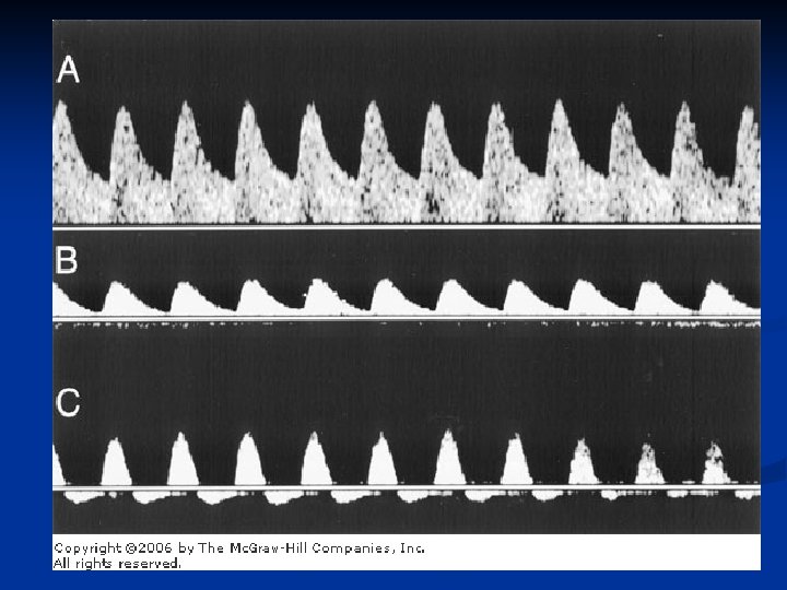
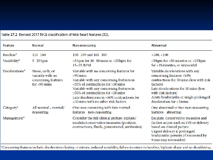
Variability can be categorised as: Reassuring – ≥ 5 bpm Non-reassuring – < 5 bpm for between 4090 minutes Abnormal – < 5 bpm for >90 minutes


Reduced variability can be caused by: Foetus sleeping – this should last no longer than 40 minutes – most common cause Foetal acidosis (due to hypoxia) – more likely if late decelerations also present Foetal tachycardia Drugs – opiates, benzodiazipine’s, methyldopa, magnesium sulphate Prematurity – variability is reduced at earlier gestation (<28 weeks) Congenital heart abnormalities

Accelerations are an abrupt increase in baseline heart rate of >15 bpm for >15 seconds The presence of accelerations is reassuring Antenatally there should be at least 2 accelerations every 15 minutes Accelerations occurring alongside uterine contractions is a sign of a healthy fetus However the absence of accelerations with an otherwise normal CTG is of uncertain significance


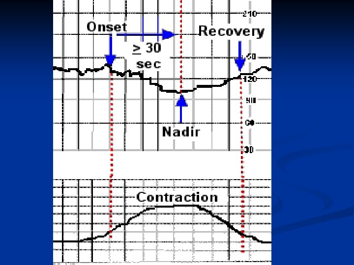
Decelerations are an abrupt decrease in baseline heart rate of >15 bpm for >15 seconds but < 2 min There a number of different types of decelerations, each with varying significance

Early decelerations start when uterine contraction begins & recover when uterine contraction stops. Nadir of the deceleration occurs at the same time as the peak of the contraction This is due to increased foetal intracranial pressure causing increased vagal tone It therefore quickly resolves once the uterine contraction ends & intracranial pressure reduces This type of deceleration is therefore considered to be physiological & not pathological

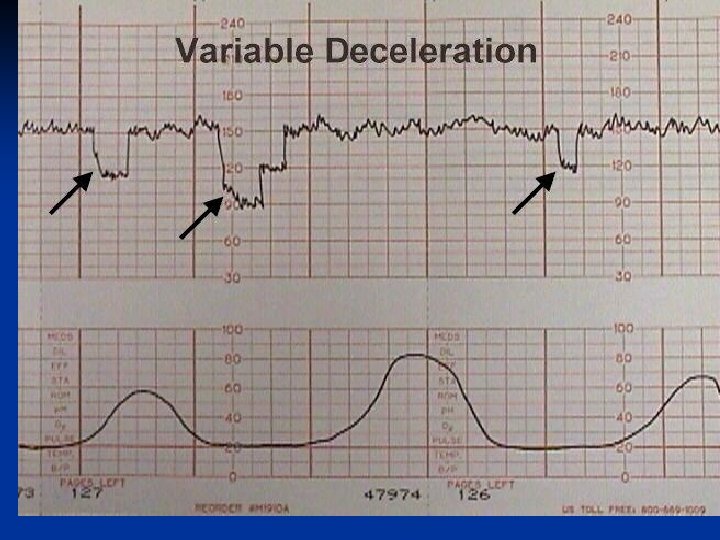
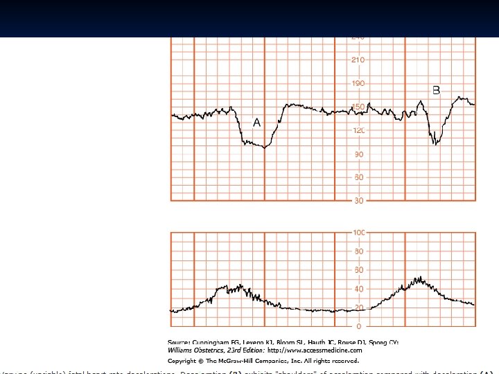
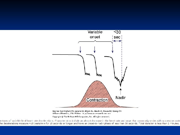
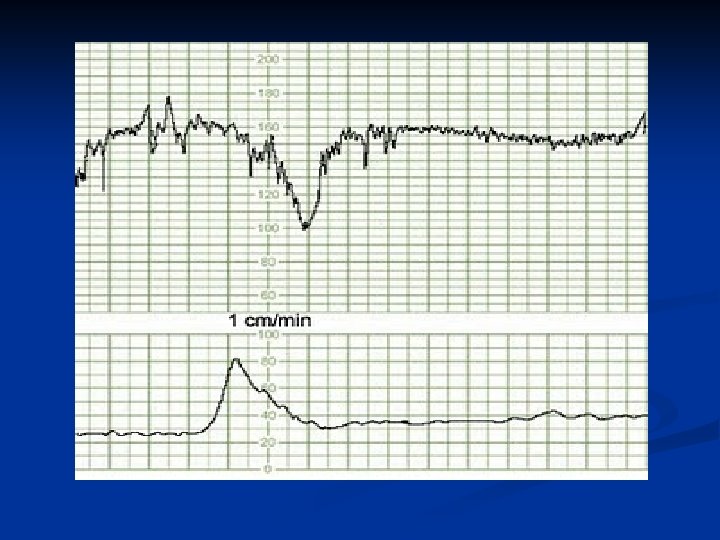
Variable decelerations are seen as a rapid fall in baseline rate with a variable recovery phase They are variable in their duration & may not have any relationship to uterine contractions They are most often seen during labour & in patients with reduced amniotic fluid volume

Variable decelerations are usually caused by umbilical cord compression The umbilical vein is often occluded first causing an acceleration in response Then the umbilical artery is occluded causing a subsequent rapid deceleration When pressure on the cord is reduced another acceleration occurs & then the baseline rate returns Accelerations before & after a variable deceleration are known as the “shoulders of deceleration” There presence indicates the foetus is not yet hypoxic & is adapting to the reduced blood flow.

Variable decelerations can sometimes resolve if the mother changes position The presence of persistent variable decelerations indicates the need for close monitoring Variable decelerations without the shoulders is more worrying as it suggests the fetus is hypoxic

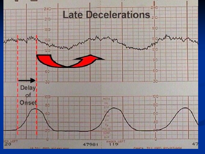
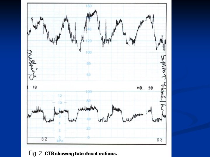
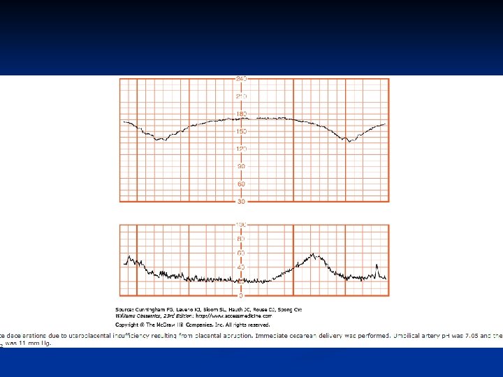
Late decelerations begin at the peak of uterine contraction & recover after the contraction ends. Onset, nadir, and recovery of the deceleration occur after the beginning, peak, and end of the contraction This type of deceleration indicates there is insufficient blood flow through the uterus & placenta As a result blood flow to the fetus is significantly reduced causing fetal hypoxia & acidosis. Reduced utero-placental blood flow can be caused by: Maternal hypotension Pre-eclampsia Uterine hyper-stimulation

Onset, nadir, and recovery of the deceleration occur after the beginning, peak, and end of the contraction

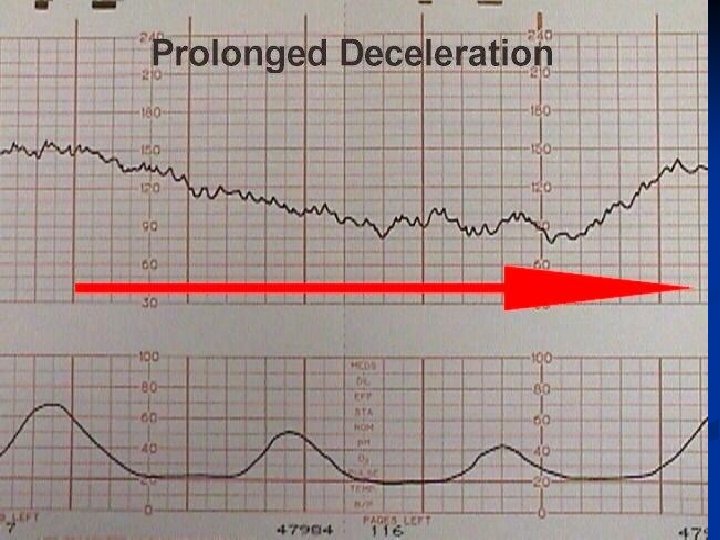
Prolonged deceleration A deceleration that last more than 2 minutes If it lasts between 2 -3 minutes it is classed as Non-Reassurring If it lasts longer than 3 minutes it is immediately classed as Abnormal Action must be taken quickly – e. g. Fetal blood sampling / emergency C-section n

A sinusoidal pattern indicates: Severe foetal hypoxia Severe foetal anaemia Foetal/Maternal Haemorrhage. Immediate C-section is indicated for this kind of pattern. Outcome is usually poor


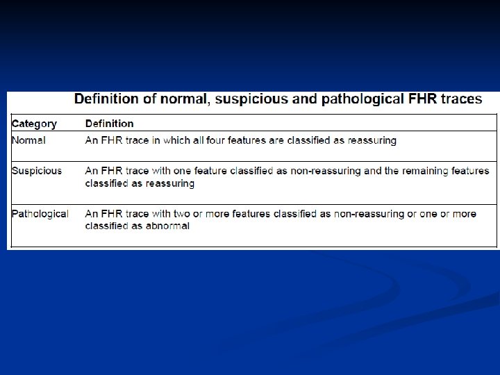
The overall impression can be described as either: Reassuring Suspicious Pathological
 A. NONSURGICAL n Lateral positioning avoids compression")
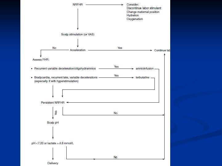
Management of non-reassuring fetal status (Fetal Distress) A. NONSURGICAL n Lateral positioning avoids compression of vena cava and aorta by the gravid uterus. This increases cardiac output and uteroplacental perfusion. n Oxygen is administered (6 -8 L/min) to the mother with mask to improve fetal Sa. O 2. n Correction of dehydration by IV fluids (crystalloids) improves intravascular volume and uterine perfusion. n Correction of maternal hypotension (following epidural analgesia) with immediate infusion of 1 L of crystalloid (Ringer’s solution).

Stoppage of oxytocin to improve fetal oxygenation. Fetal hypoxia may be due to strong and sustained uterine contractions. With reassuring FHR and in absence of fetal acidemia, oxytocin may be restarted. Tocolytic (Inj terbutaline 0. 25 mg SC) is given when uterus is hypertonus and there is nonreassuring FHR. Amnioinfusion is the process to increase the intrauterine fluid volume with transcervical infusion of warm normal saline (500 m. L). Indications are: (a) Oligohydramnios and cord compression (b) To dilute or to wash out meconium (c) To improve variable or prolonged decelerations. Advantages: Reduces cord compression, meconium aspiration, and improves Apgar score. It also reduces

If the fetal heart rate pattern remains nonreassuring, further tests are performed to rule out metabolic acidosis. Tests are: 1. Fetal heart acceleration 2. Scalp blood p. H 3. Fetal pulse oximetry (not supported by ACOG) 4. Fetal ECG/ST segment analysis (STAN). If acidosis is excluded → labor is monitored with repeat testing (every 30 min) to exclude acidosis. If the fetus is acidaemic → urgent delivery by safest method (vaginal or abdominal) depending on the
 of the fetus is done using an electronic larynx placed on")
Vibroaccoustic stimulation (VAS) of the fetus is done using an electronic larynx placed on the maternal abdomen. Presence of FHR accelerations indicates normal blood p. H. Fetal scalp stimulation by pinching with an Allis forceps or by gentle digital stroke is done before scalp blood p. H test. Presence of FHR accelerations is associated with normal scalp blood p. H.

Fetal blood sampling:

Procedure: Mother is in left lateral position. An illuminated plastic cone is inserted through the dilated cervix against the fetal head. An incision of 2 mm depth is made with a lancet. Blood is collected (100 μL) with a long capillary tube for p. H and PCO 2 estimation
 When delivery is urgently indicated or spontaneous delivery is imminent (ii) Maternal")
Contraindications: (i) When delivery is urgently indicated or spontaneous delivery is imminent (ii) Maternal infection (HIV, hepatitis or herpes simplex virus) (iii) Fetal coagulation disorders (iv) Prematurity (< 34 weeks). Risks involved are: Fetal bleeding from the incision site and maternal injury.

indicates delivery

p. H definitions Acidemia: increased concentration of hydrogen ions in blood. • Acidosis: a pathologic condition marked by increased concentration of hydrogen ions in tissue. • Hypoxemia: decreased oxygen content in blood. • Hypoxia: a pathologic condition marked by decreased level of oxygen in tissue. • Asphyxia: usually acidemia, hypoxia, and metabolic acidosis. All of these criteria must be present to entertain a diagnosis of possible intrauterine asphyxia: (1) p. H< 7. 00; (2) Apgar ≤ 3 at > 5 minutes, and (3) neonatal neurologic sequelae (e. g. seizures, coma,
 for a")
Fetal Pulse Oximetry determines fetal oxyhemoglobin saturation. Oxygen saturation (Sa. O 2) for a normal fetus in labor ranges between 40 and 70 percent. A saturation (Sa. O 2) of 30% (cut off value) correlates with the scalp p. H of 7. 20. Reassuring oxygen saturation (> 30%) even in the presence of nonreassuring FHR tracing indicates normal fetal oxygenation. Procedure : The sensor is placed against the fetal cheek trans-cervically when the membranes are ruptured

Fetal pulse oximeter sensor placement may allow assessment of fetal oxyhemoglobin saturation once membranes are ruptured. Saturation values below 30 percent, however, when persistent for 2 minutes or longer, were associated with an increased risk of potential fetal compromise.

B. SURGICAL: Cesarean delivery should be done with a 15° lateral tilt till the baby is delivered. Pediatrician should be made available.

Thank you
- Slides: 100