Assessment of Fetal Well Being Heather M Said

Assessment of Fetal Well Being Heather M. Said, DO Assistant Professor, Maternal Fetal Medicine

Learning Objectives Discuss advantages and disadvantages of external versus internal fetal monitoring Be able to interpret fetal heart tracing Baseline, variability, accelerations, decelerations Be able to interpret contraction patterns Be able to classify fetal heart rate tracings into one of three categories Discuss common modes of antenatal surveillance NST, BPP, Modified BPP
 Benefits Non-invasive Can be")
Ways to Monitor: External Uterine contractions Fetal heart rate (FHR) Benefits Non-invasive Can be used with intact membranes Disadvantages Sometimes not able to adequately trace heart rate or contractions Detects frequency of contractions but says nothing about their strength
 Benefits Ability to trace")
Ways to Monitor: Internal Uterine contractions Fetal heart rate (FHR) Benefits Ability to trace regardless of maternal habitus or position Able to quantify strength of contractions Disadvantages More invasive Only used with ruptured membranes Caution with patients who have HIV, hepatitis B/C Rare complications with placement

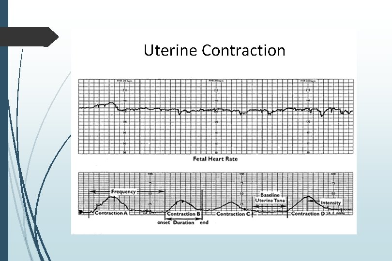
The Basics Each dark line represents one minute intervals BPM Fetal heart rate MVUs Contractions

Features to Describe FHTs Baseline Variability Accelerations Decelerations Interpret into 1 of 3 categories Contractions Frequency Amplitude (if IUPC)

Baseline Mean fetal heart rate Rounded to increments of 5 During a 10 minute period Excluding accelerations and decelerations Normal baseline 110 -160 BPM

Baseline

Baseline Bradycardia <110 BPM Tachycardia >160 BPM Indeterminate less than 2 minutes of baseline is present
")
Fetal Tachycardia Normal variant prematurity Intra-amniotic infection Response to maternal condition (fever, dehydration, etc) Fetal anemia Fetal cardiac arrhythmia (SVT) Fetal hypoxia Thyrotoxicosis

Fetal Tachycardia

Fetal Bradycardia

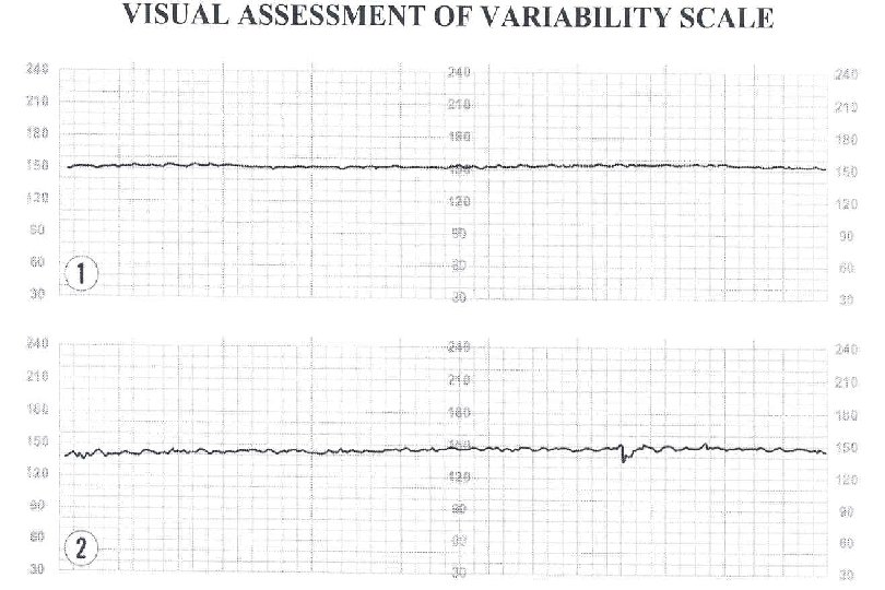
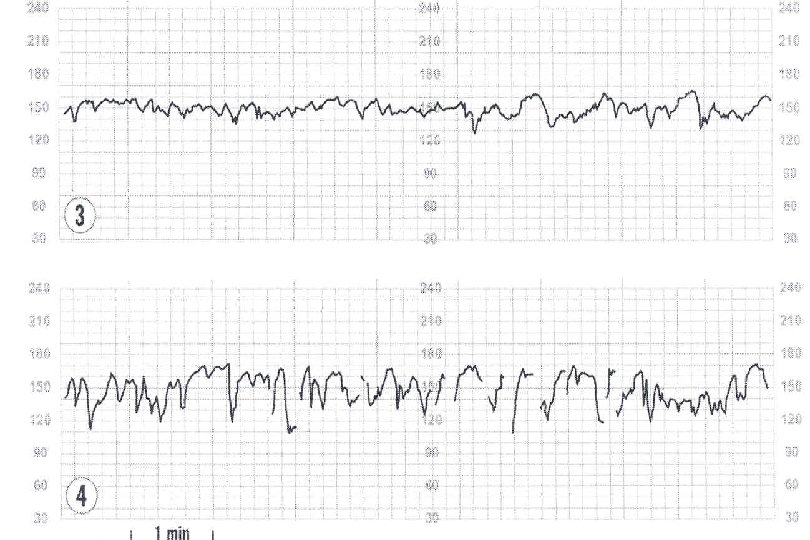
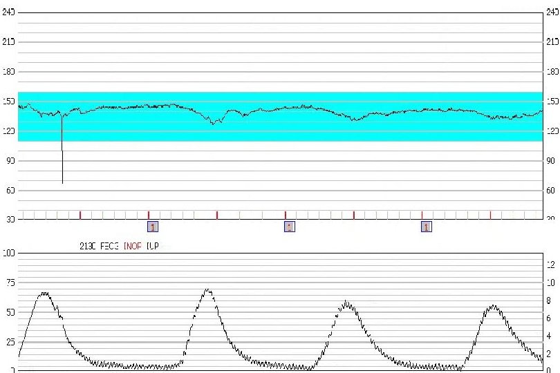
Variability Fluctuations in FHR Beat to beat variation Descriptors are: Absent: undetectable amplitude range Minimal: Up to 5 BPM Moderate: amplitude range 6 to 25 BPM Reassuring Marked: amplitude range greater than 25 BPM
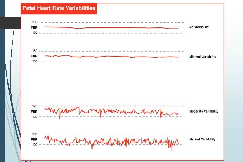

Marked Variability

Accelerations Abrupt increase in FHR Gestational age > 32 weeks At least 15 BPM above baseline Must last at least 15 seconds (15 x 15 s) Gestational age <32 weeks 10 x 10 s unless previously demonstrated 15 x 15 s Prolonged accelerations Last at least 2 minutes but fewer than ten minutes Baseline change Last 10 minutes or longer
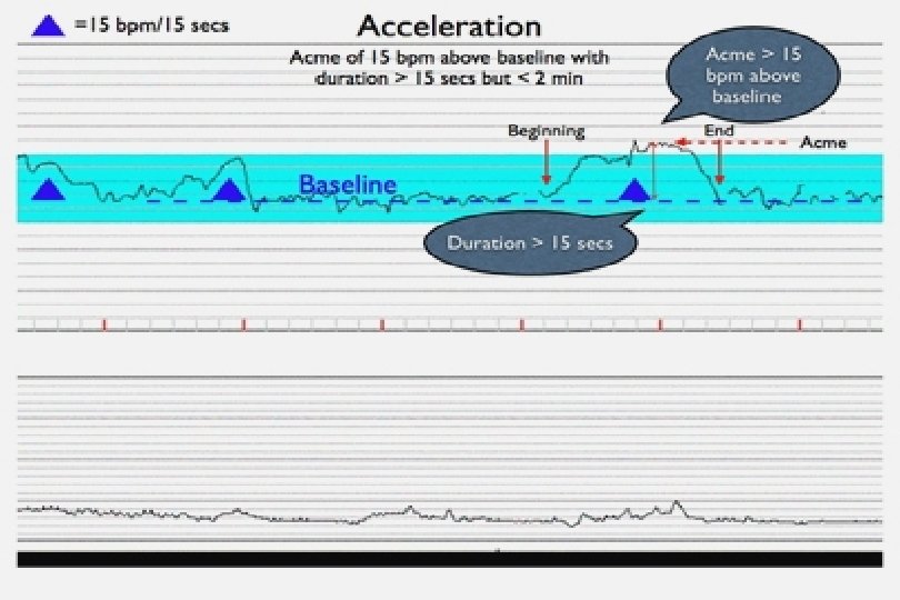

Accelerations

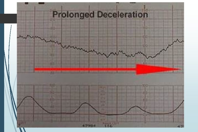
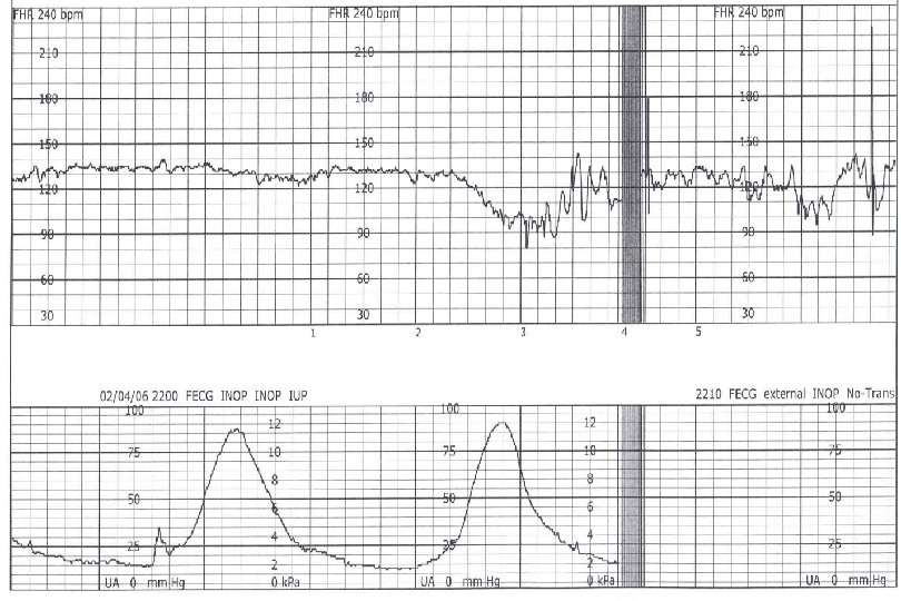
Decelerations Decrease of FHR below baseline 3 Types Early Variable Late Recurrent if occurring with >50% contractions Otherwise intermittent Prolonged if >2 minutes Baseline change if >10 minutes

Deceleration

Early Deceleration Symmetrical to contraction Mirror image of contraction Gradual decrease in FHR 30 secs or more from onset to nadir Represent vagal response to head compression Not concerning

EARLY DECELERATION v Gradual FHR decrease v Onset to nadir 30 seconds or more v Nadir of deceleration occurs with peak of contraction v Mirror contraction

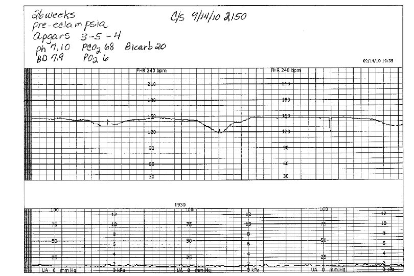
Late Decelerations Deceleration is delayed in timing Nadir following the peak of the contraction A gradual FHR decrease Onset to nadir > 30 second Represents uteroplacental insufficiency Non-reassuring

Late Decelerations

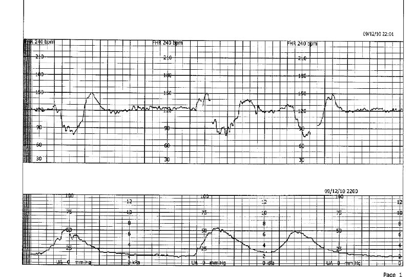
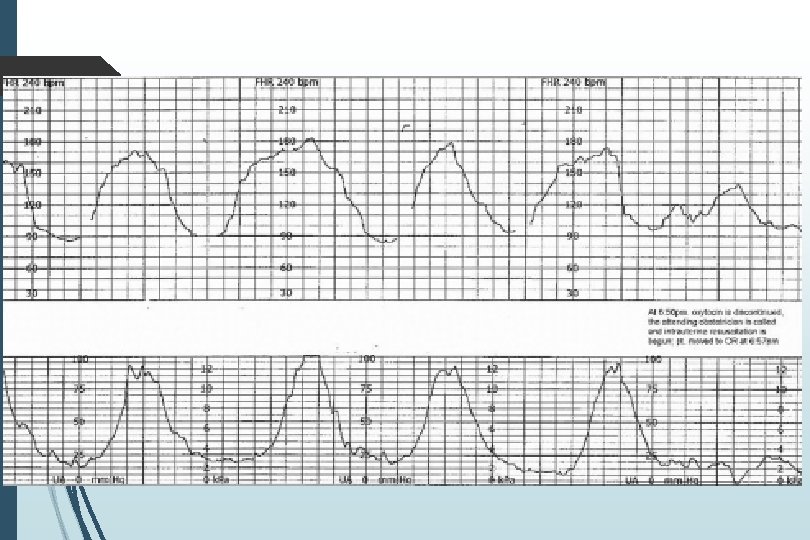
Variable Decelerations Abrupt decrease in fetal heart rate Onset to nadir less than 30 seconds Decrease in FHR May or may not be associated with contractions Represent cord compression More likely with oligohydramnios Amnioinfusion Common during labor Can be concerning if recurrent

Variable Decelerations


Sinusoidal Pattern Smooth sine-wave pattern Cycle frequency 3 -5 per minute Persists for 20 minutes or longer Non-reassuring Fetal anemia Pseudosinusoidal pattern can occur after IV narcotic administration during labor

Sinusoidal Pattern

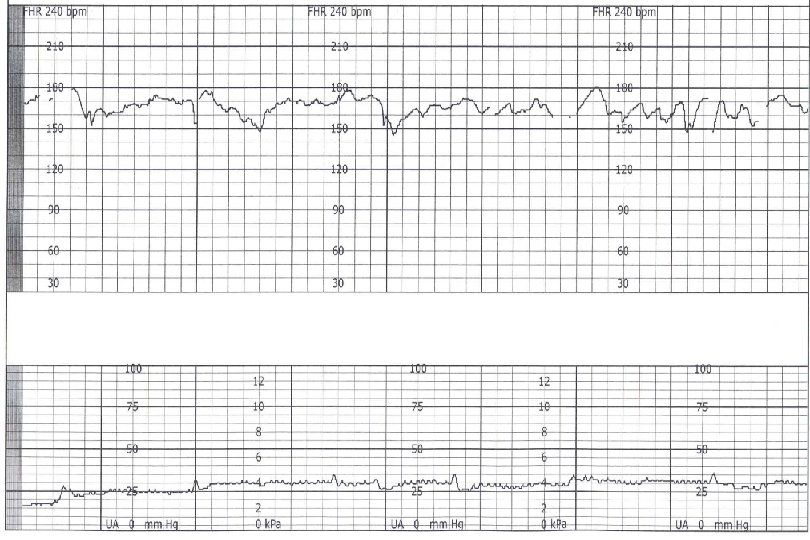
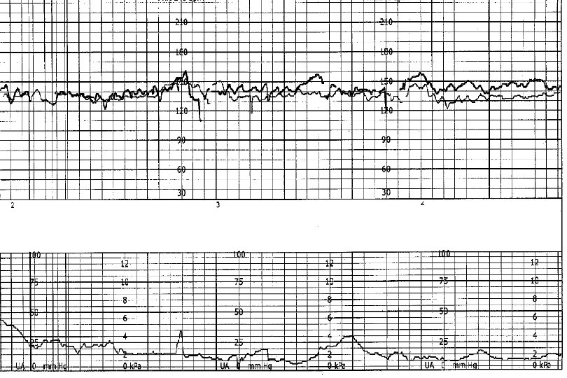
Uterine Contractions Number of contractions in 10 minutes averaged over thirty minutes Components to note Frequency Intensity Duration Relaxation time between contractions

Monitoring of Contractions

Tachysystole >5 contractions in 10 minutes Averaged over 30 minutes If sustained, could lead to fetal distress

Contraction Intensity Can only be assessed with IUPC Look at ten minute window of contractions For each contraction Subtract nadir from peak of contraction Add up these values, which represent contraction strength Measured in MVUs (Montevideo units) 200 MVUs or greater is considered an adequate contraction pattern that should in theory be enough to promote cervical change

Contraction Intensity

Categorization of FHR Tracings Recommendation of three-tiered system April 2008 More standardized interpretation Concept: Interpretation of a FHR monitor strip is a dynamic process, with determination of whether a particular strip is reassuring and what action plans should be taken… and then to evaluate at a later time

Categorization of FHR Patterns An evaluation of the fetus at a particular point in time Categories I, II, and III

3 Categories

Category I Normal baseline 110 -160 BPM FHR Variability moderate Late or Variable decelerations none

Category II Not enough evidence to place into either Category I or III Various extremes of category II Reactive tracing with one variable deceleration Minimal variability with recurrent late decelerations

Category III Abnormal tracing Predictive of abnormal fetal acid-base status Requires prompt intervention

The ABCD’s of Fetal Monitoring
 Assess maternal blood")
Intrauterine Resuscitation Oxygen IV fluids Reposition the mother (left side, etc) Assess maternal blood pressure Especially if shortly after epidural placement Check cervix Delivery imminent? Cord prolapse? Stop the Pitocin if this agent is being used Terbutaline can be given to space out contractions, especially if tachysystole is a concern
 Biophysical profile (BPP) Modified BPP (NST/AFI)")
Common Methods of Antenatal Surveillance Non-stress test (NST) Biophysical profile (BPP) Modified BPP (NST/AFI)

Non-Stress Test Monitor fetal heart rate at least 20 minutes in the absence of contractions With contractions, technically contraction stress test False negative (i. e. reassuring test with fetal demise within one week) 2/1000 Higher false positives Fetal sleep-wake cycles Maternal narcotics Outcomes Reactive 2 accelerations/20 minutes Non-reactive Less than 2 accelerations/20 minutes

Reactive NST
 NST + ultrasound markers Score linearly correlated with fetal p. H")
Biophysical Profile (BPP) NST + ultrasound markers Score linearly correlated with fetal p. H Risk of fetal death within one week of normal BPP is 0. 8/1000

Biophysical Profile • Performed during a thirty minute timeframe but can be extended • Zero or 2 points for each (all or none) • NST (sometimes not performed) • • Fetal breathing • • 3 distinct movements Fetal tone • • >30 seconds Fetal gross body movements • • 2 accelerations/20 minutes 1 episode of flexion with return to extension Amniotic fluid volume • Deepest vertical pocket at least 2 cm
 and")
Biophysical Profile In the absence of oligohydramnios Scores of 10/10, 8/8 (no NST) and 8/10 all reassuring 6/8 is NOT a score! Get NST 6/10 requires further evaluation 4/10, 2/10, 0/10 or oligohydramnios not reassuring

Modified Biophysical Profile Combination of NST/AFI NST represents acute fetal well being AFI represents chronic fetal well being 0 or 2 points NST: 0 if non-reactive, 2 if reactive AFI: 0 if oligohydramnios; 2 if not Score of 4/4 as reassuring as full BPP False negative 0. 8/1000 Score of 2/4 requires full BPP Score of 0/4 requires further evaluation

Guidelines for Reviewing FHR Monitoring Normal patient Reviewed every 30 minutes in the first stage of labor Every 15 minutes in the second stage Complicated patients Every 15 minutes in first stage Every 5 minutes in second stage

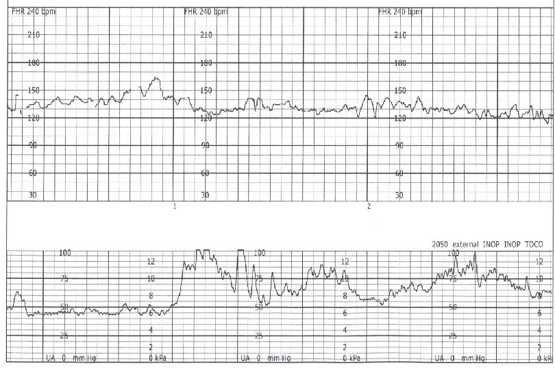
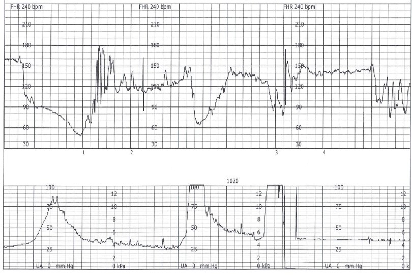
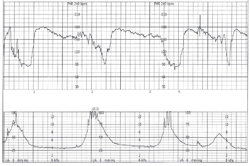
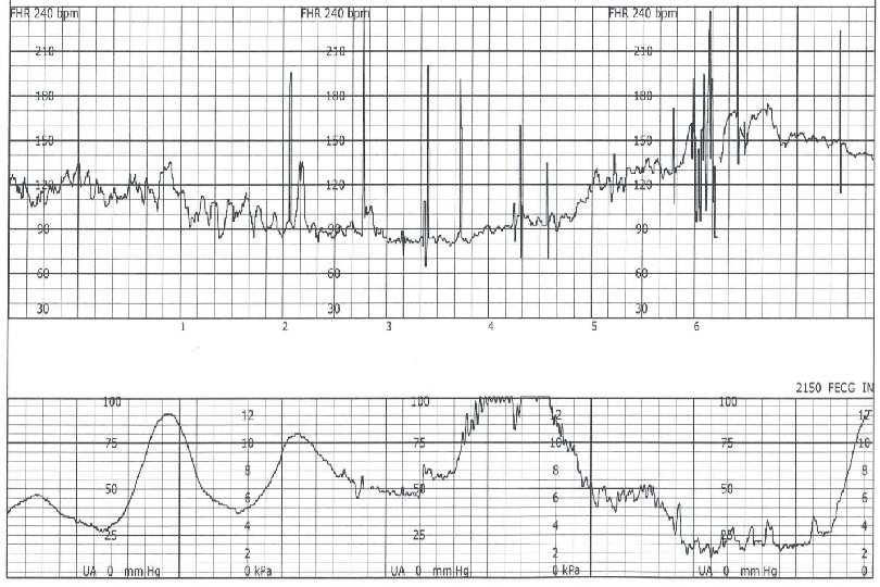
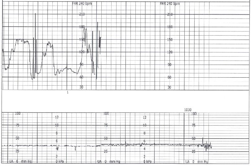
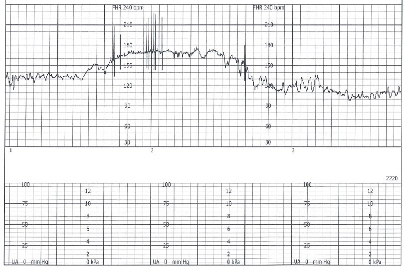
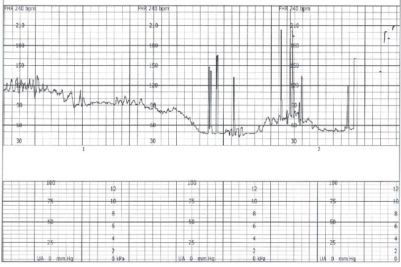
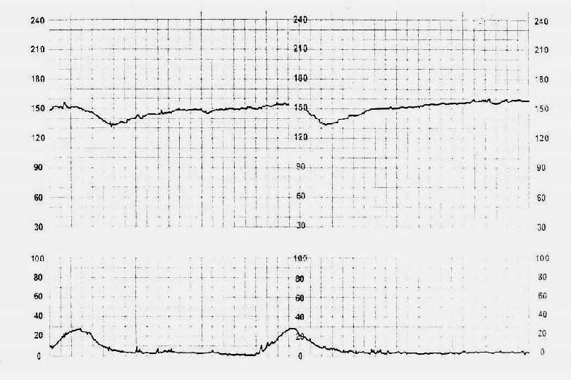
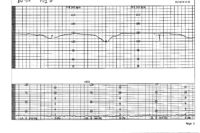
Examples of Tracings
- Slides: 71