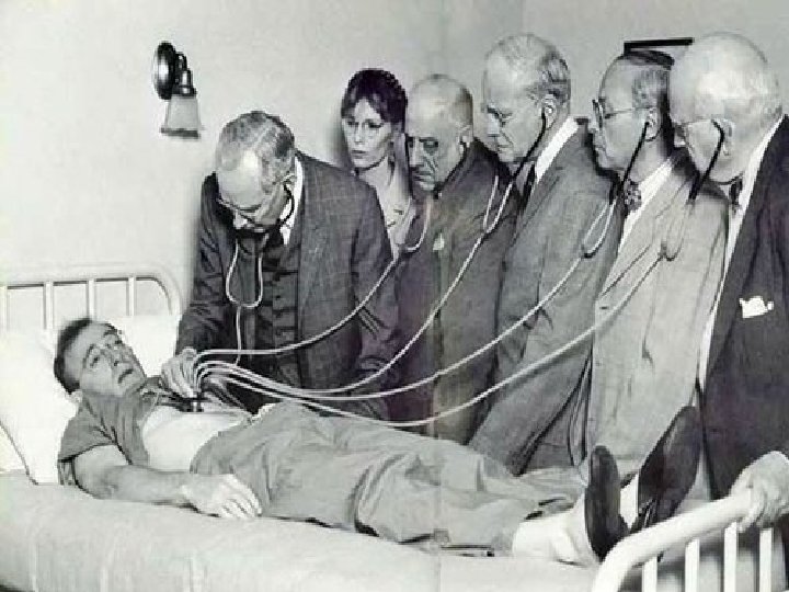
Assessment of Breast Dr Hayan Bismar MD FACS

Assessment of Breast Dr Hayan Bismar , MD , FACS

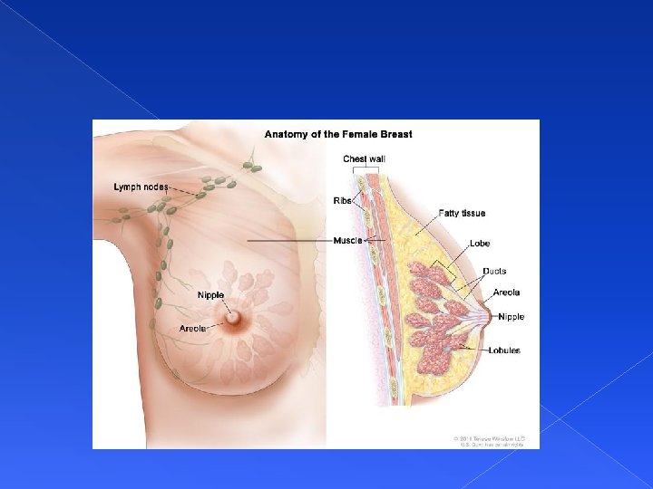
Anatomy of the breast Composed of : Muscles Ligaments Glandular tissue Fatty tissue Lymphatic tissue

Assessing: Subjective Data • • • History of Breast Disease/Surgery Lumps or thickening Discharge/Rash Swelling/Trauma Pain Does pt. perform SBE monthly? Date of last clinical breast exam CBE Date of last mammogram Axillary tenderness, lumps swelling, rash

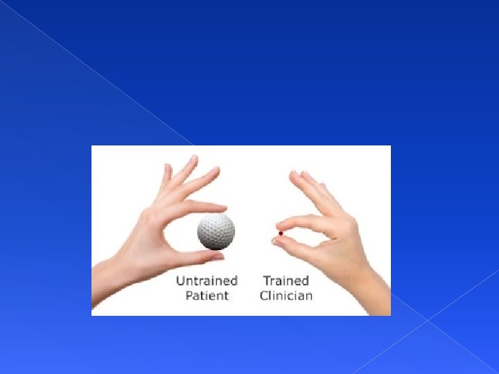
Assessing Breast Cancer Risk Examiner must ask focused questions regarding risk factors: Family history: 1 st degree relatives Estrogen exposure: Age onset of menses / Age menopause Obstetric history / Use of HRT Personal habits: Alcohol / Dietary Fat / Exercise Ever tested for gene mutation: BRCA 1/ BRCA 2 Age: Over 50 => risk

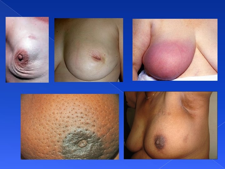
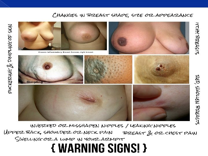
Assessing: Objective Data Inspection- patient sitting, disrobed to waist Note symmetry, size and shape Skin normally smooth &even in color. Observe the axillary and supra clavicular areas for any bulging, discoloration or edema Nipples- symmetrical? Flat? Inverted? Discharge? Bleeding?

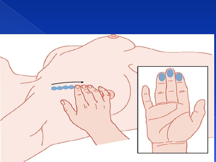
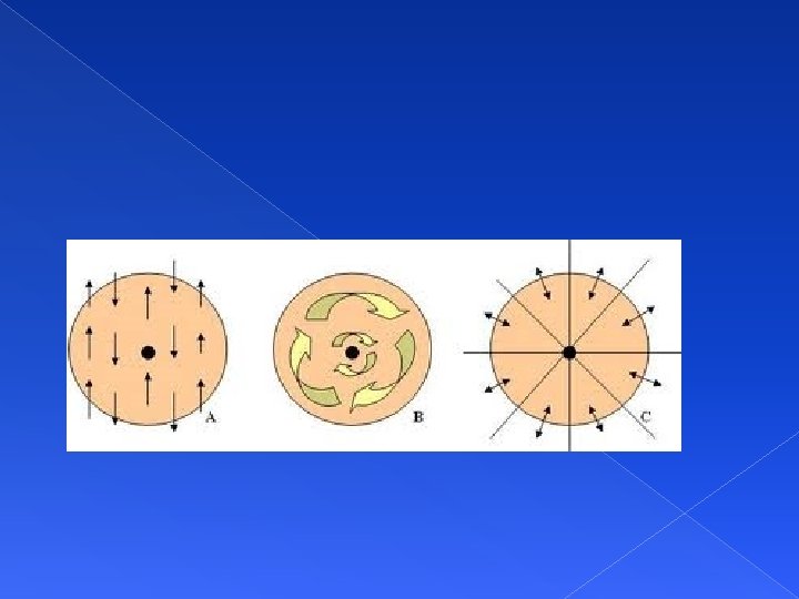
Breast Palpation Supine position with small pad/pillow under side to be palpated Arm raised over head Use pads of fingers and make gentle rotary movement on breast Use a pattern of concentric circles or laterally, like spokes of wheel. Palpate all areas of breast, clockwise fashion
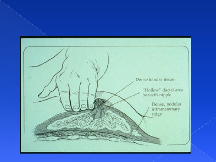

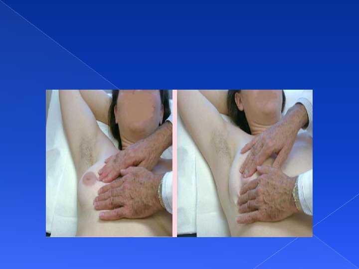
Bimanual Breast Palpation For pendulous breasts Patient sitting, leaning forward Support inferior part of breast with one hand. Use other hand to palpate breast tissue against supporting hand.

Inspect and Palpate Axillae While patient is sitting, lift and support the arm so patient’s muscles are relaxed Use the right hand to palpate left axillae Reach fingers high into axillae Move fingers firmly down in four directions: Down the chest wall, along the anterior and posterior borders of axillae and around the inner aspect of the arm Move arm through ROM to have access to areas.

Palpation of Axilla

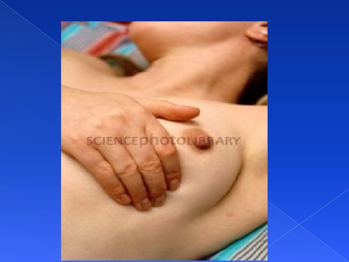
Examination of Nipples Performed after breast palpation. Palpate nipple, noting any indurations or mass. Use thumb and forefinger to apply gentle pressure to note any discharge.

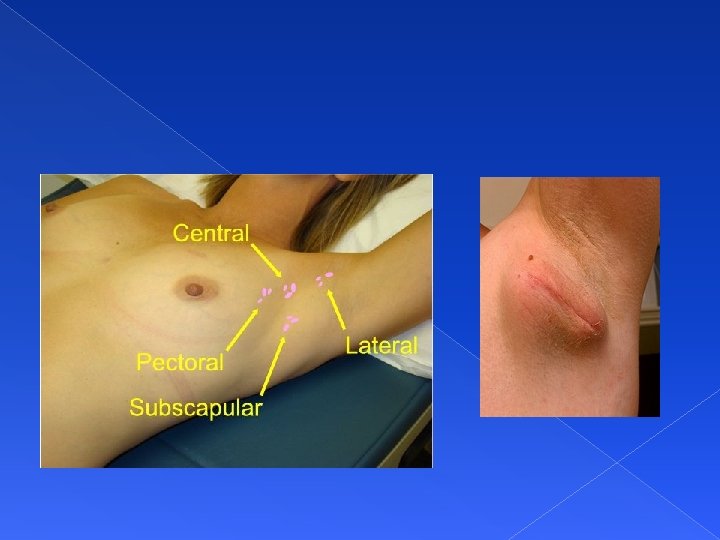
Description of breast lump Location- use breast as clock face to describe distance from nipple in centimeters (use diagram to locate). Size- in centimeters Shape Consistency Movable Tenderness

Comparison of Breast Lumps § § § § Benign Breast Disease Multiple or single Rubbery texture Mobile / slippery Regular borders Tenderness (cyclic) No retraction May increase/decrease in size rapidly § § § § Cancer Unilateral Firm texture Fixed firmly Irregular border Usually painless Usually w/retraction Grows constantly

Teaching BSE Assist patient to establish a schedule Regular monthly exams Majority of women never get breast cancer, majority of lumps are benign Early detection is important. In non-invasive cancer, survival is close to 100%

Self Breast Exam Teaching positions: 1. Standing in front of mirror 2. In the shower – soap and water assist palpation 3. Supine Keep teaching simple Demonstrate to patient and use return demonstration

The Male Breast Examination can be abbreviated but not omitted. Inspect the chest wall noting skin surface and any lumps or swelling. Palpate nipple area for lumps or enlargement. Normal male breast has a flat disc of undeveloped breast tissue beneath the nipple. Should be even with no nodules.

Summary Assessing the breast includes: Take into account developmental level Remembering to assess both females and males Inspecting & palpating breasts, nipples, lymph nodes and axillary Teaching BSE

Thank you
- Slides: 28