
Asexual Reproduction o One parent o Offspring are


- Slides: 95
Asexual Reproduction o One parent o Offspring are identical to parent/ No genetic variation o Examples: Vegetative propagation, budding, cloning, mitosis (remember ? ) o Why is this beneficial? n Fast n If it is not broke, don’t fix it!
Sexual Reproduction o Why sex? n n Energetically expensive Health risks of childbearing Genetic diversity/Genetic Recombination Overall health of the species o Meiosis makes the gametes n Spermatogenesis=4 identical sperm cells, puberty to death n Oogenesis=1 egg cell per month in humans, initial egg production before birth then freezing at Prophase I n Governed by two laws: Segregation and Independent Assortment
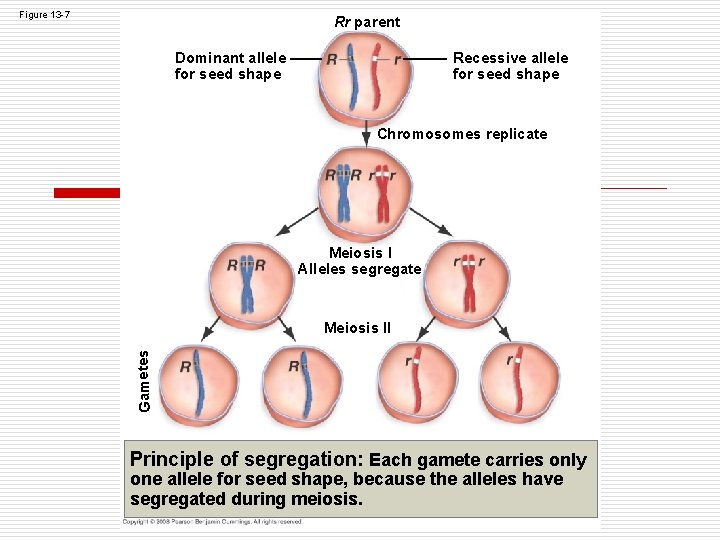
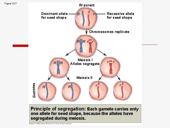
Figure 13 -7 Rr parent Dominant allele for seed shape Recessive allele for seed shape Chromosomes replicate Meiosis I Alleles segregate Gametes Meiosis II Principle of segregation: Each gamete carries only one allele for seed shape, because the alleles have segregated during meiosis.
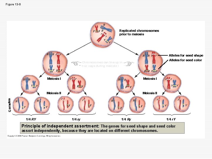
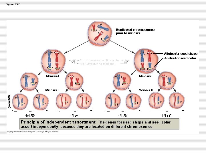
Figure 13 -8 R y y R r r Y R Rr Replicated chromosomes prior to meiosis Y r R R r Alleles for seed shape Alleles for seed color r Chromosomes can line up in two ways during meiosis I Y Y y y R Meiosis I R Y y y. Y Y r r R y y Y Meiosis I R Gametes Y Y 1/4 RY Y Meiosis II r R r Y y y Meiosis II R r r y y 1/4 ry r R R y y 1/4 Ry r Y Y 1/4 r. Y Principle of independent assortment: The genes for seed shape and seed color assort independently, because they are located on different chromosomes.
Chromosome number and fertilization o Diploid (2 n)=full set of chromosomes, is the result of fertilization with gametes n Homologs for each chromosome, 1 from mom, 1 from dad n In humans-22 autosome pairs, 1 sex-determing pair o Haploid (n)=1/2 set of chromosomes, is the result of meiosis o See examples and practice.
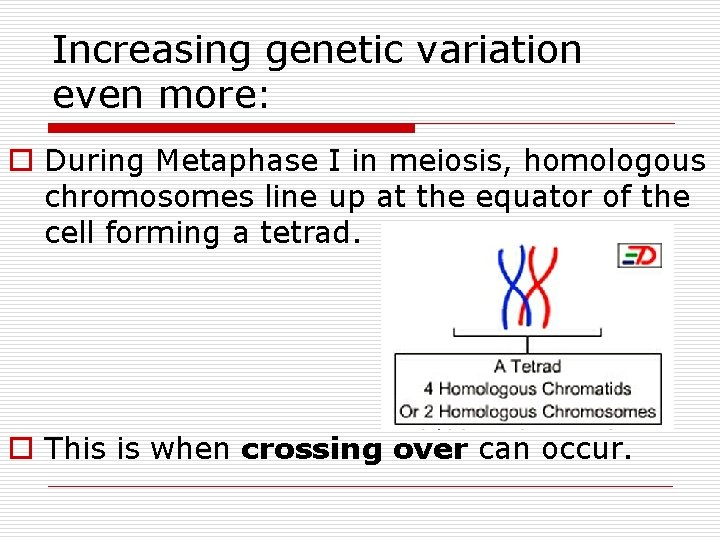
Increasing genetic variation even more: o During Metaphase I in meiosis, homologous chromosomes line up at the equator of the cell forming a tetrad. o This is when crossing over can occur.
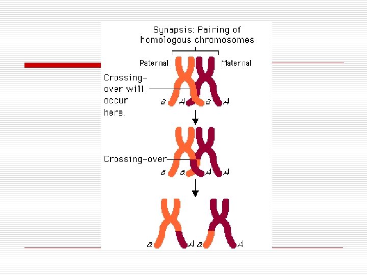
Crossing Over o Homologous chromosomes exchange portions of DNA. o A gene’s distance from the centromere is related to the frequency of cross over. o As meiosis continues this increases the differences between gametes and possible offspring.
M&M Comparison o T Chart o Comparison Activity with Fuzzy Sticks. n Draw only the cells required. n Increase genetic diversity during Step by adding Crossing Over. ACTUALLY cut your “chromosomes” and reattach to model crossing over. n Fertilization must occur with another group.
Mitosis vs. Meiosis Activity(20 min. ) Complete the activity on your own paper. Draw ONLY the cells required with the appropriately sized and colored chromosome. • When modeling Metaphase I demonstrate CROSSING OVER in one of the homologous pairs of your model. • Fertilization must be completed with another group at the same point in the activity. • Answer all questions.
Cell Cycle Lab Pt. II (online) 1. Go to Pearson Lab Bench and complete the Meiosis Tutorial with questions. 2. Data on your lab paper: Use the data from the Sordaria slide on the Analysis page to fill in Part II data for your lab sheet. 3. Take the lab quiz.
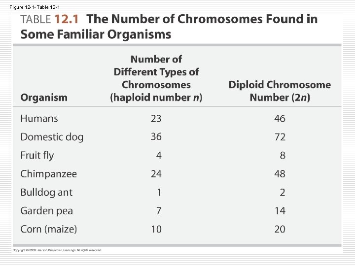
Figure 12 -1 -Table 12 -1
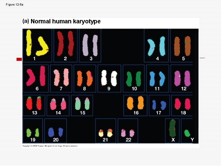
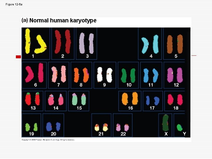
Figure 12 -6 a Normal human karyotype
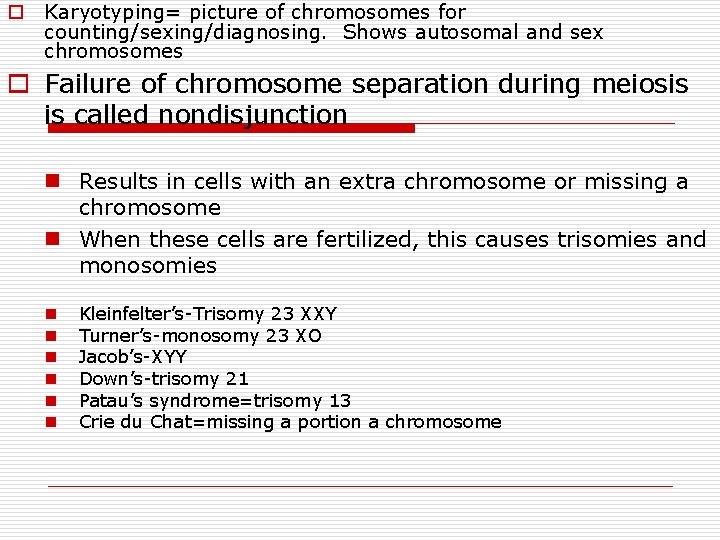
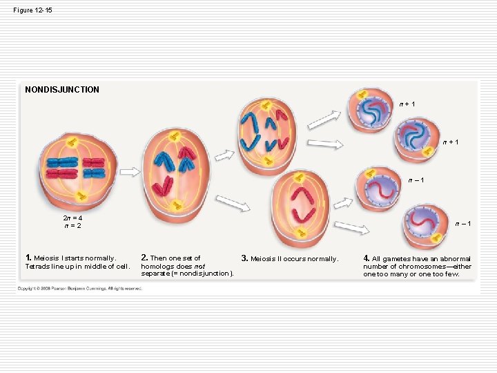
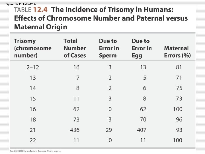
o Karyotyping= picture of chromosomes for counting/sexing/diagnosing. Shows autosomal and sex chromosomes o Failure of chromosome separation during meiosis is called nondisjunction n Results in cells with an extra chromosome or missing a chromosome n When these cells are fertilized, this causes trisomies and monosomies n n n Kleinfelter’s-Trisomy 23 XXY Turner’s-monosomy 23 XO Jacob’s-XYY Down’s-trisomy 21 Patau’s syndrome=trisomy 13 Crie du Chat=missing a portion a chromosome
Figure 12 -6 a Normal human karyotype
Figure 12 -15 NONDISJUNCTION n + 1 n – 1 2 n = 4 n = 2 1. Meiosis I starts normally. Tetrads line up in middle of cell. n – 1 2. Then one set of homologs does not separate (= nondisjunction). 3. Meiosis II occurs normally. 4. All gametes have an abnormal number of chromosomes—either one too many or one too few.
Figure 12 -15 -Table 12 -4
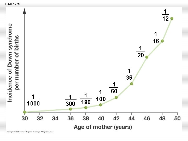
Figure 12 -16 1 12 1 16 1 20 1 36 1 1000 1 1 300 180 1 1 60 100
Genetics Review (heredity) o Notes o Sponge Bob: basic inheritance (complete dominance) o Incomplete, Codominance, Pollyallele o Sex-linked Traits o Dihybrids o At least two of each problem!
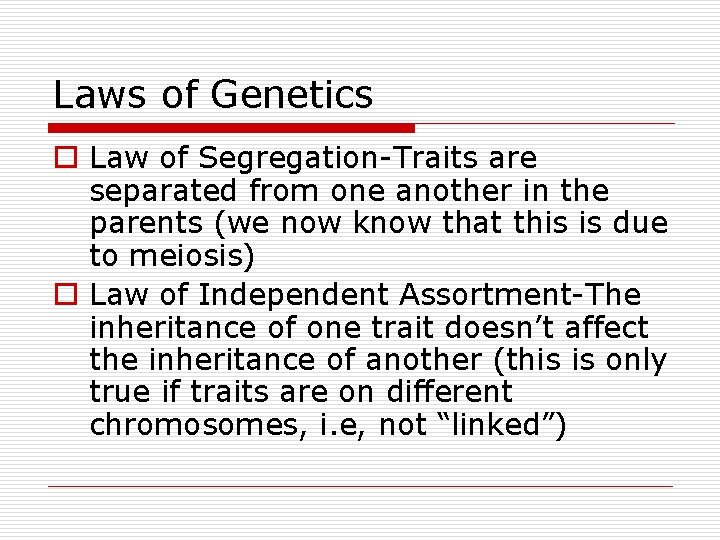
Laws of Genetics o Law of Segregation-Traits are separated from one another in the parents (we now know that this is due to meiosis) o Law of Independent Assortment-The inheritance of one trait doesn’t affect the inheritance of another (this is only true if traits are on different chromosomes, i. e, not “linked”)
Figure 13 -7 Rr parent Dominant allele for seed shape Recessive allele for seed shape Chromosomes replicate Meiosis I Alleles segregate Gametes Meiosis II Principle of segregation: Each gamete carries only one allele for seed shape, because the alleles have segregated during meiosis.
Figure 13 -8 R y y R r r Y R Rr Replicated chromosomes prior to meiosis Y r R R r Alleles for seed shape Alleles for seed color r Chromosomes can line up in two ways during meiosis I Y Y y y R Meiosis I R Y y y. Y Y r r R y y Y Meiosis I R Gametes Y Y 1/4 RY Y Meiosis II r R r Y y y Meiosis II R r r y y 1/4 ry r R R y y 1/4 Ry r Y Y 1/4 r. Y Principle of independent assortment: The genes for seed shape and seed color assort independently, because they are located on different chromosomes.
Mendel and Genetics o Mendel bred pea plants to determine the rules of heredity o Terms: n n n P=Parent generation F 1=First filial generation (offspring from P) F 2=Second filial generation (offspring from F 1) Allele=alternate form of a trait Dominant=always expressed Recessive=only expressed if dominant isn’t present Homozygous=2 copies of the same allele(pure/true breeding, ) Heterozygous=2 different alleles (carrier, hybrid) Genotype=Combination of genes (letters) Phenotype=Expression of the combination of genes Selfed/Self-fertizilation=crossed with same genotype Testcross=cross with homozygous recessive to determine unknown phenotype.
What Mendel figured out o Rarely blending of inheritance (purple x white yields either purple or white, not lavendar) o Alternate versions of each gene exist (2 alleles for each gene) o Each individual inherits 1 allele from each parent o Predictable ratios occur when crossing individuals o + 2 laws of inheritance
Types of crosses o Monohybrids-tracing inheritance of 1 trait at a time n Complete Dominance = 1 loci, 2 phenotypes are possible n Practice Problems #1, 6, 12,
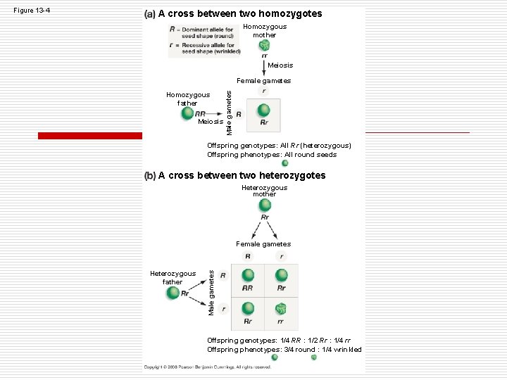
A cross between two homozygotes Homozygous mother Meiosis Homozygous father Meiosis Male gametes Female gametes Offspring genotypes: All Rr (heterozygous) Offspring phenotypes: All round seeds A cross between two heterozygotes Heterozygous mother Female gametes Heterozygous father Male gametes Figure 13 -4 Offspring genotypes: 1/4 RR : 1/2 Rr : 1/4 rr Offspring phenotypes: 3/4 round : 1/4 wrinkled
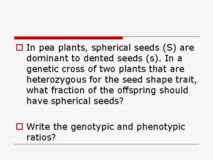
o In pea plants, spherical seeds (S) are dominant to dented seeds (s). In a genetic cross of two plants that are heterozygous for the seed shape trait, what fraction of the offspring should have spherical seeds? o Write the genotypic and phenotypic ratios?
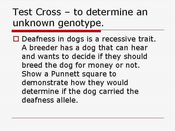
Test Cross – to determine an unknown genotype. o Deafness in dogs is a recessive trait. A breeder has a dog that can hear and wants to decide if they should breed the dog for money or not. Show a Punnett square to demonstrate how they would determine if the dog carried the deafness allele.
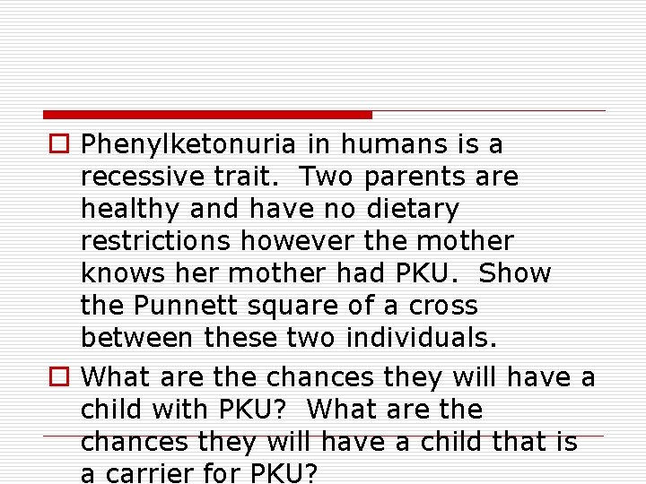
o Phenylketonuria in humans is a recessive trait. Two parents are healthy and have no dietary restrictions however the mother knows her mother had PKU. Show the Punnett square of a cross between these two individuals. o What are the chances they will have a child with PKU? What are the chances they will have a child that is a carrier for PKU?
Types of crosses o Dihybrids-tracing inheritance of 2 traits together at a time. n Complete Dominance = 1 loci, 2 phenotypes are possible n Practice Problems # 2, 10 and pg. 284 #7
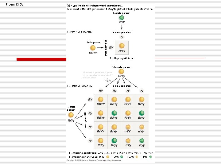
Hypothesis of independent assortment: Alleles of different genes don’t stay together when gametes form. Female parent F 1 PUNNET SQUARE Male gametes Male parent Female gametes F 1 offspring all Rr. Yy F 2 female parent Alleles at R gene and Y gene go to gametes independently of each other F 2 PUNNET SQUARE Female gametes F 2 male parent Male gametes Figure 13 -5 a F 2 offspring genotypes: 9/16 R–Y– : 3/16 R–yy : 3/16 rr. Y– : 1/16 rryy F 2 offspring phenotypes: 9/16 : 3/16 : 1/16
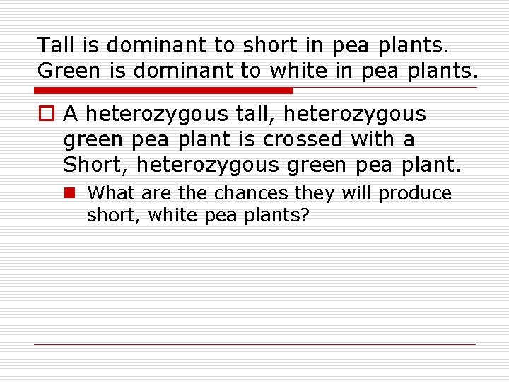
Tall is dominant to short in pea plants. Green is dominant to white in pea plants. o A heterozygous tall, heterozygous green pea plant is crossed with a Short, heterozygous green pea plant. n What are the chances they will produce short, white pea plants?
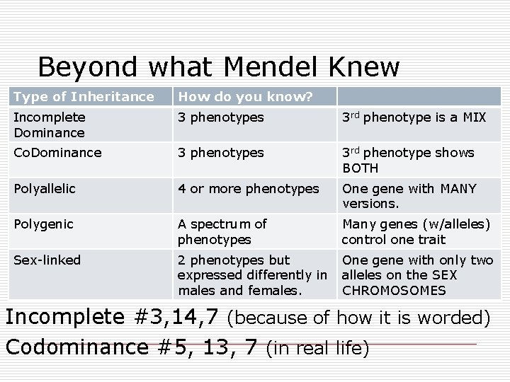
Beyond what Mendel Knew Type of Inheritance How do you know? Incomplete Dominance 3 phenotypes 3 rd phenotype is a MIX Co. Dominance 3 phenotypes 3 rd phenotype shows BOTH Polyallelic 4 or more phenotypes One gene with MANY versions. Polygenic A spectrum of phenotypes Many genes (w/alleles) control one trait Sex-linked 2 phenotypes but expressed differently in males and females. One gene with only two alleles on the SEX CHROMOSOMES Incomplete #3, 14, 7 (because of how it is worded) Codominance #5, 13, 7 (in real life)
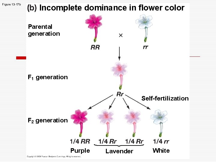
Figure 13 -17 b Incomplete dominance in flower color Parental generation F 1 generation Self-fertilization F 2 generation Purple Lavender White
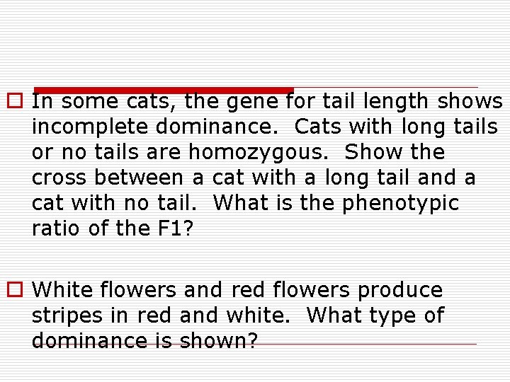
o In some cats, the gene for tail length shows incomplete dominance. Cats with long tails or no tails are homozygous. Show the cross between a cat with a long tail and a cat with no tail. What is the phenotypic ratio of the F 1? o White flowers and red flowers produce stripes in red and white. What type of dominance is shown?
Polyalleleic-one loci but many alleles. (complete and codominance) #4, 8 o Blood Typing: There are three alleles that create human blood types. Chart…
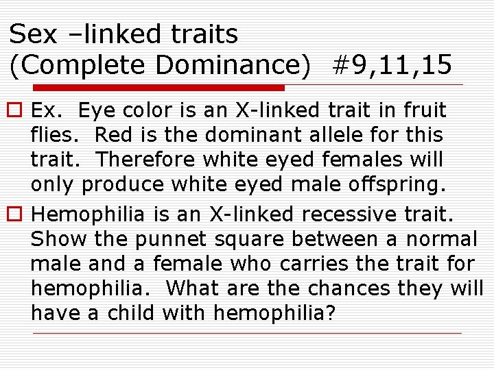
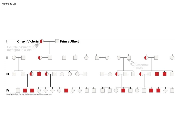
Sex –linked traits (Complete Dominance) #9, 11, 15 o Ex. Eye color is an X-linked trait in fruit flies. Red is the dominant allele for this trait. Therefore white eyed females will only produce white eyed male offspring. o Hemophilia is an X-linked recessive trait. Show the punnet square between a normal male and a female who carries the trait for hemophilia. What are the chances they will have a child with hemophilia?
Dihybrid Cross A = Axial Flowers, a = terminal flowers R = Round Seed, r = wrinkled seed o P Generation: A plant that is heterozygous for both traits crossed with a plant that is heterozygous for flower location and homozygous dominant for seed shape. o Textbook #7, 12 o Use these for multiple traits at a time OR polygenic traits. Example: Eye Color AABBCC or aabbcc
But what is dominant, really? o Tay-Sachs-inability to metabolize certain lipids because of a malfunctioning enzyme o Only children with 2 copies of the allele have the disease-at the organismal level, this is recessive o However, heterozygotes, biochemically appear to be the result of incomplete dominance-lipid metabolism levels are intermediate between those who don’t have Tay-Sachs and those who o Molecularly, heterozygotes produce equal levels of functional and non-functional enzymes-appearing to be co-dominant
Other single-gene locus influences o Multiple alleles n Ex: Human Blood type n I A, I B, i o Pleiotropy n When a single gene affects many traits n Ex: sickle cell disease and its multiple symptoms
Sex-linked Traits o X-chromosome inheritance n b/c males have only 1 copy, they’re most likely affected by these, but females can still inherit these n Pass from mother to children o Y-chromosome inheritance n Only males affected n Pass from father to sons
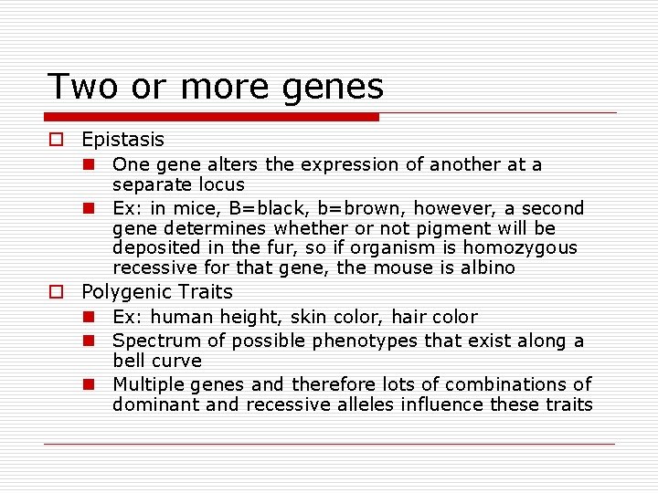
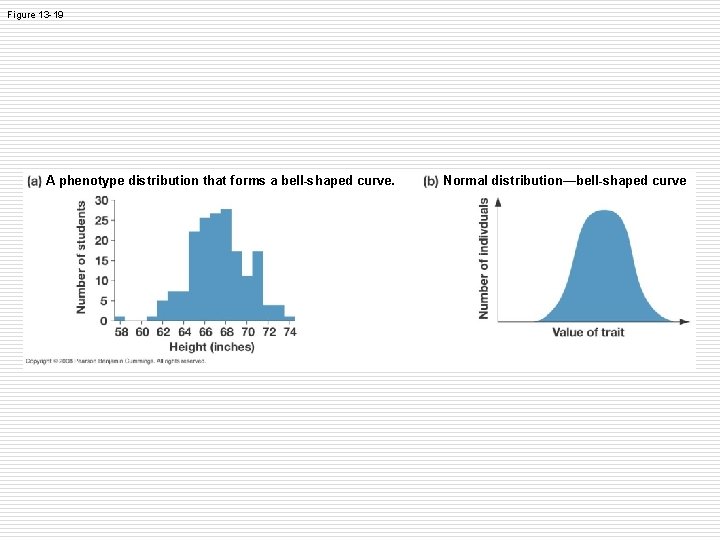
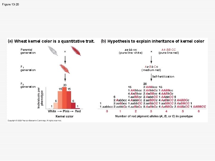
Two or more genes o Epistasis n One gene alters the expression of another at a separate locus n Ex: in mice, B=black, b=brown, however, a second gene determines whether or not pigment will be deposited in the fur, so if organism is homozygous recessive for that gene, the mouse is albino o Polygenic Traits n Ex: human height, skin color, hair color n Spectrum of possible phenotypes that exist along a bell curve n Multiple genes and therefore lots of combinations of dominant and recessive alleles influence these traits
Figure 13 -19 A phenotype distribution that forms a bell-shaped curve. Normal distribution—bell-shaped curve
Figure 13 -20 Wheat kernel color is a quantitative trait. Parental generation Hypothesis to explain inheritance of kernel color aa bb cc (pure-line white) F 1 generation AA BB CC (pure-line red) Aa Bb Cc (medium red) Self-fertilization F 2 generation 20 15 6 1
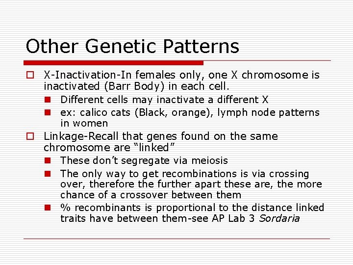
Other Genetic Patterns o X-Inactivation-In females only, one X chromosome is inactivated (Barr Body) in each cell. n Different cells may inactivate a different X n ex: calico cats (Black, orange), lymph node patterns in women o Linkage-Recall that genes found on the same chromosome are “linked” n These don’t segregate via meiosis n The only way to get recombinations is via crossing over, therefore the further apart these are, the more chance of a crossover between them n % recombinants is proportional to the distance linked traits have between them-see AP Lab 3 Sordaria
Does Genotype determine Phenotype? o The short answer is “no!” o Environmental influences also affect gene expression, giving us a norm of reaction for a phenotype (range of possible phenotypes)
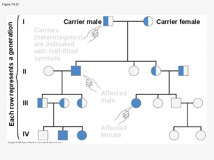
Pedigree Analysis o Pedigrees are family trees that use specific notations that geneticists use to predict the inheritance pattern of a trait
Each row represents a generation Figure 13 -21 I Carrier male Carriers (heterozygotes) are indicated with half-filled symbols II III Affected male IV Affected female Carrier female
Figure 13 -23 I Queen Victoria Prince Albert Female carrier of hemophilia allele II Affected male III IV
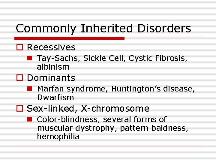
Commonly Inherited Disorders o Recessives n Tay-Sachs, Sickle Cell, Cystic Fibrosis, albinism o Dominants n Marfan syndrome, Huntington’s disease, Dwarfism o Sex-linked, X-chromosome n Color-blindness, several forms of muscular dystrophy, pattern baldness, hemophilia
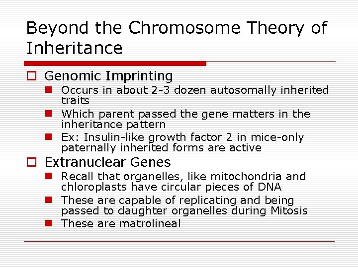
Beyond the Chromosome Theory of Inheritance o Genomic Imprinting n Occurs in about 2 -3 dozen autosomally inherited traits n Which parent passed the gene matters in the inheritance pattern n Ex: Insulin-like growth factor 2 in mice-only paternally inherited forms are active o Extranuclear Genes n Recall that organelles, like mitochondria and chloroplasts have circular pieces of DNA n These are capable of replicating and being passed to daughter organelles during Mitosis n These are matrolineal
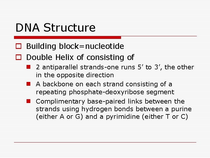
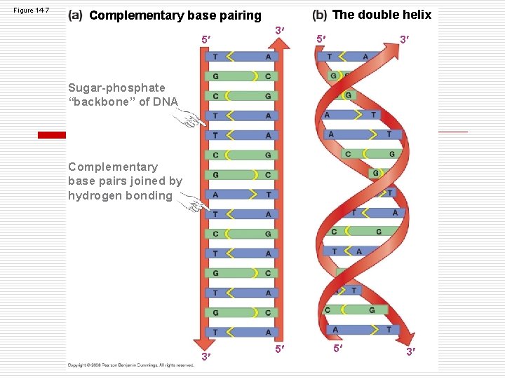
DNA Structure o Building block=nucleotide o Double Helix of consisting of n 2 antiparallel strands-one runs 5’ to 3’, the other in the opposite direction n A backbone on each strand consisting of a repeating phosphate-deoxyribose segment n Complimentary base-paired links between the strands using hydrogen bonds between a purine (either A or G) and a pyrimidine (either T or C)
Figure 14 -6 Structure of a deoxyribonucleotide Phosphate group attached to 5 carbon of the sugar Could be adenine (A), thymine (T), guanine (G) cytosine (C) 5 Hydroxyl (OH) group on 3 carbon of the sugar 3 Primary structure of DNA 5 end of strand Sugar-phosphate backbone of DNA strand Nitrogencontaining bases project from the backbone 5 Phosphodiester bond links deoxyribonucleotides 3 end of strand
Figure 14 -7 The double helix Complementary base pairing 5 3 Sugar-phosphate “backbone” of DNA Complementary base pairs joined by hydrogen bonding 3 5 5 3
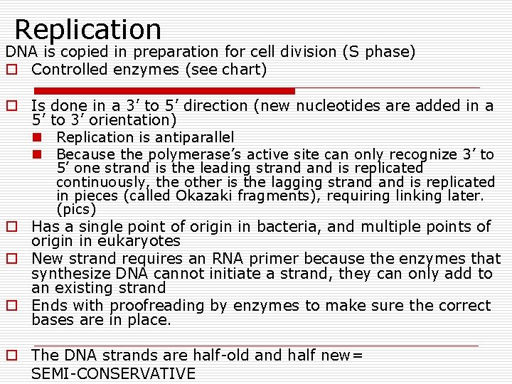
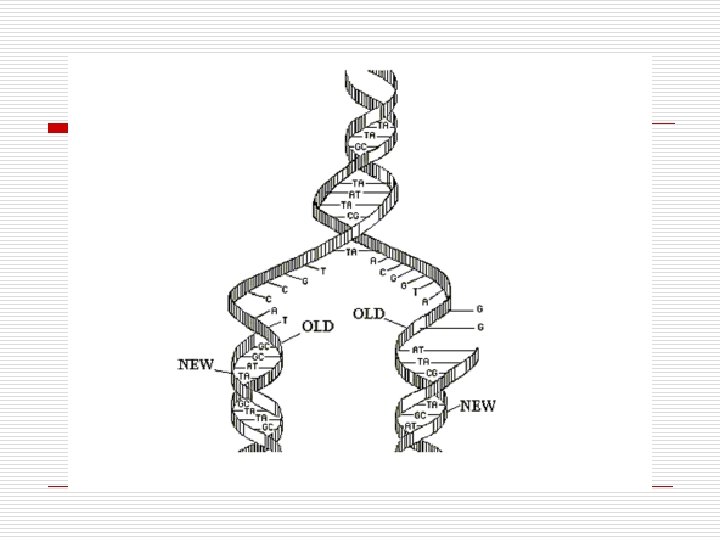
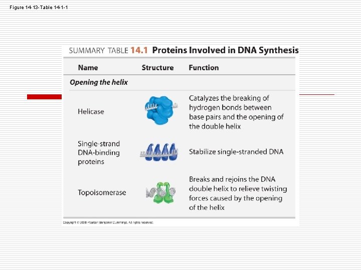
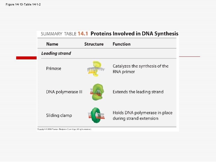
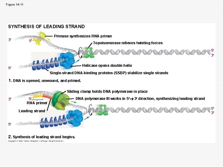
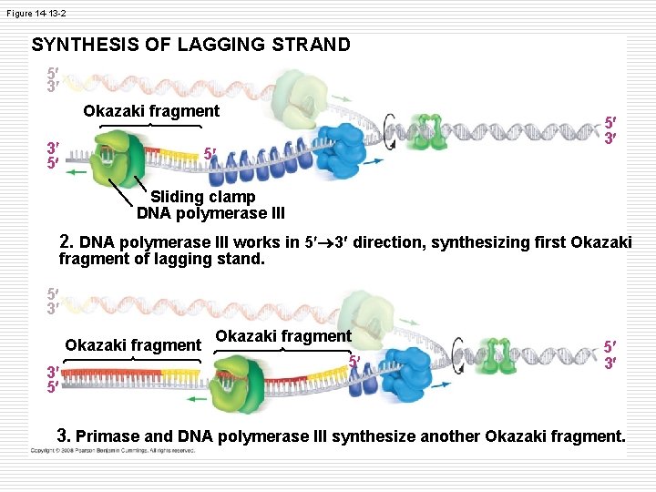
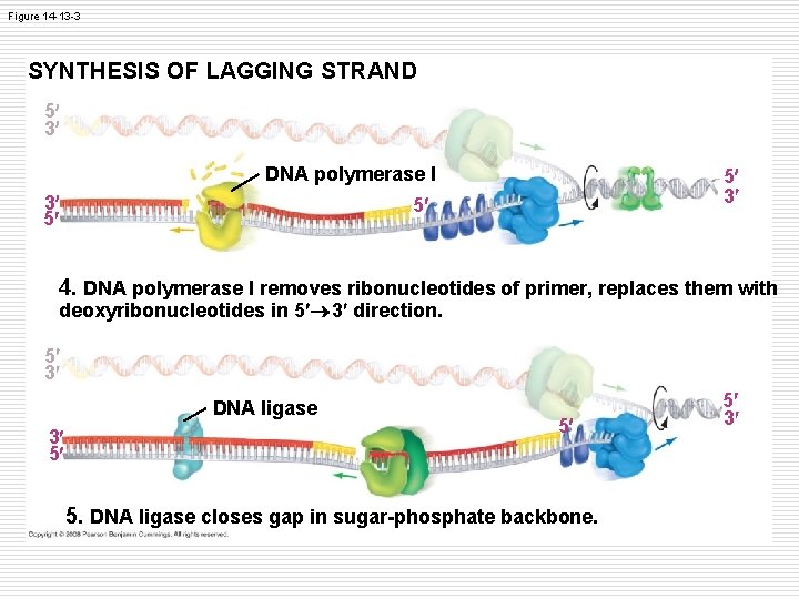
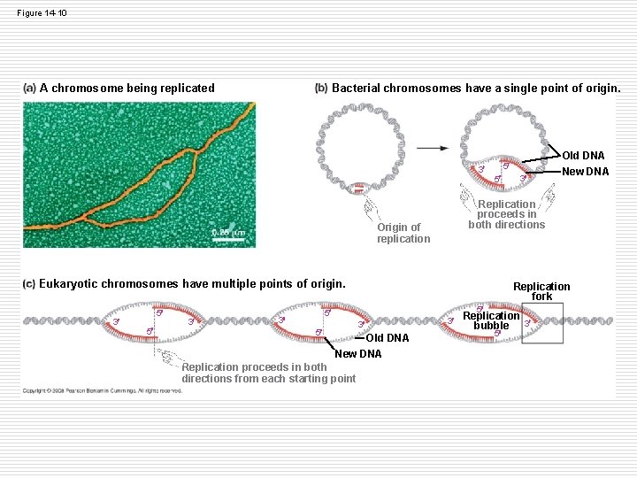
Replication DNA is copied in preparation for cell division (S phase) o Controlled enzymes (see chart) o Is 5’ n n done in a 3’ to 5’ direction (new nucleotides are added in a to 3’ orientation) Replication is antiparallel Because the polymerase’s active site can only recognize 3’ to 5’ one strand is the leading strand is replicated continuously, the other is the lagging strand is replicated in pieces (called Okazaki fragments), requiring linking later. (pics) o Has a single point of origin in bacteria, and multiple points of origin in eukaryotes o New strand requires an RNA primer because the enzymes that synthesize DNA cannot initiate a strand, they can only add to an existing strand o Ends with proofreading by enzymes to make sure the correct bases are in place. o The DNA strands are half-old and half new= SEMI-CONSERVATIVE
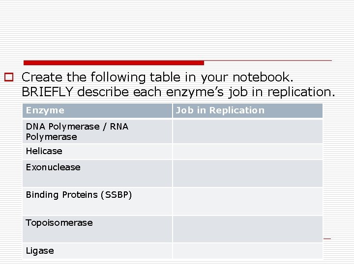
o Create the following table in your notebook. BRIEFLY describe each enzyme’s job in replication. Enzyme DNA Polymerase / RNA Polymerase Helicase Exonuclease Binding Proteins (SSBP) Topoisomerase Ligase Job in Replication
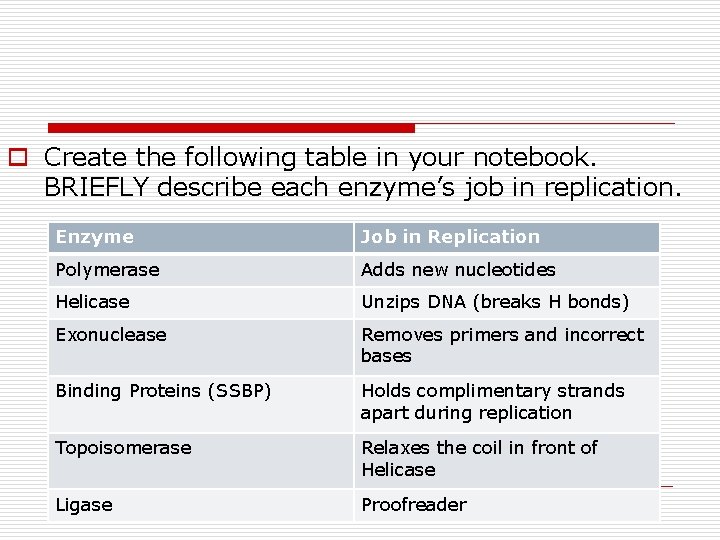
o Create the following table in your notebook. BRIEFLY describe each enzyme’s job in replication. Enzyme Job in Replication Polymerase Adds new nucleotides Helicase Unzips DNA (breaks H bonds) Exonuclease Removes primers and incorrect bases Binding Proteins (SSBP) Holds complimentary strands apart during replication Topoisomerase Relaxes the coil in front of Helicase Ligase Proofreader
What it really looks like… o Animation 1 o Animation 2
Coloring!
Figure 14 -13 -Table 14 -1 -1
Figure 14 -13 -Table 14 -1 -2
Figure 14 -11 SYNTHESIS OF LEADING STRAND 3 Primase synthesizes RNA primer 5 Topoisomerase relieves twisting forces 5 3 Helicase opens double helix 5 Single-strand DNA-binding proteins (SSBP) stabilize single strands 1. DNA is opened, unwound, and primed. Sliding clamp holds DNA polymerase in place 3 5 RNA primer Leading strand 5 2. Synthesis of leading strand begins. DNA polymerase III works in 5 3 direction, synthesizing leading strand 5 3
Figure 14 -13 -2 SYNTHESIS OF LAGGING STRAND 5 3 Okazaki fragment 3 5 5 3 5 Sliding clamp DNA polymerase III 2. DNA polymerase III works in 5 3 direction, synthesizing first Okazaki fragment of lagging stand. 5 3 Okazaki fragment 3 5 Okazaki fragment 5 5 3 3. Primase and DNA polymerase III synthesize another Okazaki fragment.
Figure 14 -13 -3 SYNTHESIS OF LAGGING STRAND 5 3 DNA polymerase I 3 5 5 3 5 4. DNA polymerase I removes ribonucleotides of primer, replaces them with deoxyribonucleotides in 5 3 direction. 5 3 DNA ligase 3 5 5 5. DNA ligase closes gap in sugar-phosphate backbone. 5 3
Figure 14 -10 Bacterial chromosomes have a single point of origin. A chromosome being replicated 3 5 5 Replication proceeds in both directions Origin of replication Eukaryotic chromosomes have multiple points of origin. 3 5 5 3 Replication fork 5 5 3 Old DNA New DNA Replication proceeds in both directions from each starting point 3 Old DNA New DNA 3 Replication bubble 3 5
Diagram Write TIME down the side of the paper. Draw an arrow at the top of the paper to show the direction of the replication fork. On each stage… o Color the original DNA RED. o Draw a GREEN circle where TOPOISOMERASE would be located. o Draw a BLACK circle where HELICASE would be located. o Highlight the RNA PRIMER in YELLOW. o Color all new DNA PURPLE. o Add a key at the bottom of your diagram.
RNA o Bring your DNA model pieces up to the front to create one large piece of DNA. o RNA section of your activity sheet! o Use the model pieces on the back table to create an RNA nucleotide. o 8 minutes.
Why RNA? o RNA, like DNA is made of nucleotides, however: n n RNA nucleotides use ribose, not deoxyribose Uracil, a base, replaces Thymine RNA is single-stranded RNA contains only 1 gene at a given time o RNA can leave the nucleus through the nuclear pores. o RNA polymerase does not need a backbone to start an RNA strand. *remember the primers* o RNA code is written as codons (3 -base segments) o Three types: m. RNA, t. RNA, r. RNA
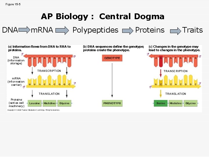
Figure 15 -5 AP Biology : Central Dogma DNA m. RNA Polypeptides DNA sequences define the genotype; proteins create the phenotype. Information flows from DNA to RNA to proteins. 3 DNA (information storage) Proteins 5 Changes in the genotype may lead to changes in the phenotype. 3 5 TRANSCRIPTION m. RNA (information carrier) 5 TRANSCRIPTION 3 3 5 TRANSLATION Proteins (active cell machinery) Traits TRANSLATION
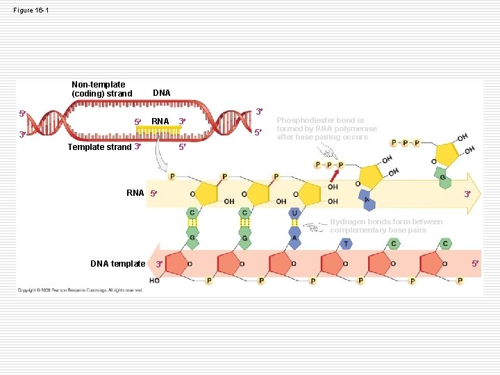
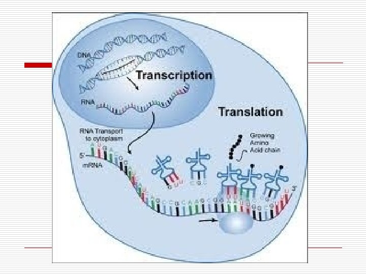
Transcription o This is the 1 st step of protein synthesis, but also makes any RNA molecule (m. RNA, t. RNA, r. RNA). o Begins at the promoter and ends at the terminator sequence. o Involves: n Opening DNA at one place only, and when complete, the DNA closes again – Thanks hydrogen bonding! n Making an RNA copy of a gene from DNA (only the leading strand) n More enzymes !
Figure 16 -1 Non-template (coding) strand 5 DNA 3 5 RNA 3 5 3 Template strand 3 Phosphodiester bond is formed by RNA polymerase after base pairing occurs 5 RNA 5 3 Hydrogen bonds form between complementary base pairs DNA template 3 5
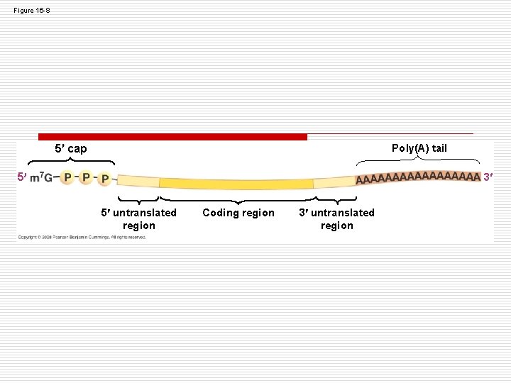
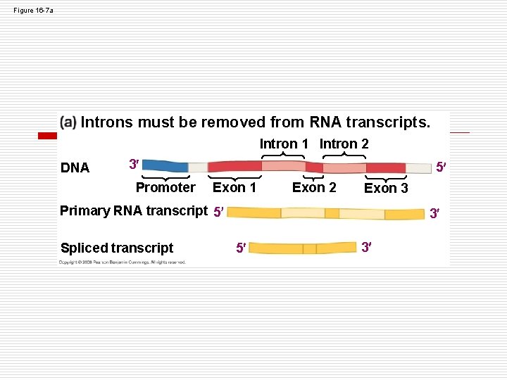
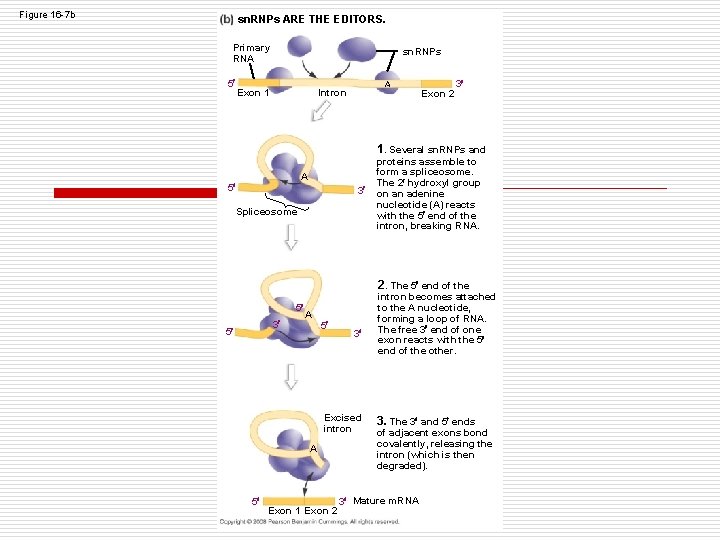
Post-transcription RNA modification o After transcription, the new RNA molecule is called pre-RNA because it is not yet translation-ready. o End-modification n 5’ end receives a cap with modified Guanine n 3’ end receives a poly-A tail (30 -250 Adenines) that help attach to the ribosome for translation o RNA splicing n RNA has large, noncoding sections that must be removed n Introns are noncoding regions between coding regions n Exons are regions that actually get translated (they exit the nucleus) n Small nuclear ribonucleoproteins (sn. RNPs-”snurps”) or spliceosomes cut out the introns and join their flanking exons together.
Figure 16 -8 5 cap Poly(A) tail 5 3 5 untranslated region Coding region 3 untranslated region
Figure 16 -7 a Introns must be removed from RNA transcripts. Intron 1 Intron 2 DNA 3 Promoter 5 Exon 1 Exon 2 Exon 3 Primary RNA transcript 5 Spliced transcript 3 5 3
Figure 16 -7 b sn. RNPs ARE THE EDITORS. Primary RNA 5 sn. RNPs Exon 1 A Intron Exon 2 3 1. Several sn. RNPs and A 5 3 Spliceosome proteins assemble to form a spliceosome. The 2 hydroxyl group on an adenine nucleotide (A) reacts with the 5 end of the intron, breaking RNA. 2. The 5 end of the 5 3 5 A 5 3 Excised intron A 5 Exon 1 Exon 2 intron becomes attached to the A nucleotide, forming a loop of RNA. The free 3 end of one exon reacts with the 5 end of the other. 3. The 3 and 5 ends of adjacent exons bond covalently, releasing the intron (which is then degraded). 3 Mature m. RNA
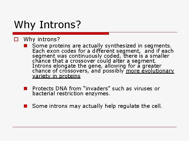
Why Introns? o Why introns? n Some proteins are actually synthesized in segments. Each exon codes for a different segment, and if each segment was continuously coded, there is a smaller chance that a crossover could alter a segment. Introns elongate the gene, allowing for a greater chance of crossovers, and possibly more evolutionary variety in proteins n Protects DNA from “invaders” such as viruses or bacterial restriction enzymes. n Some introns may actually help regulate the cell.
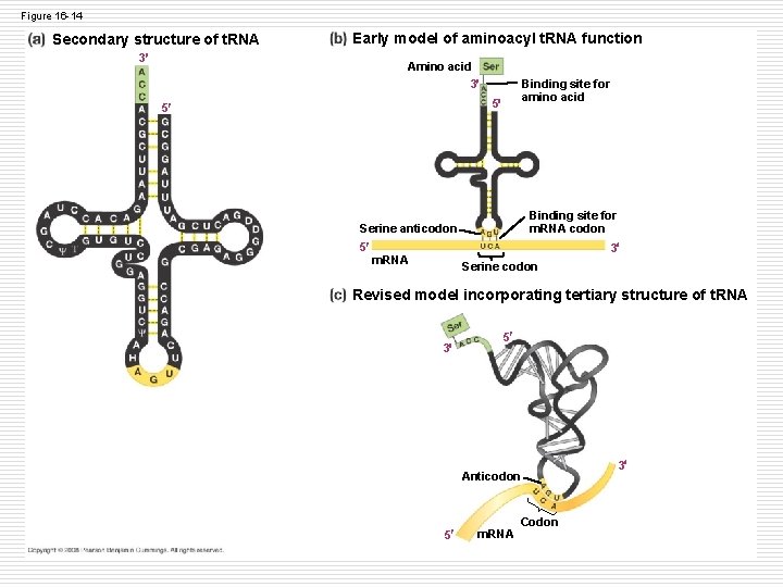
Translation o m. RNA goes to the ribosome to be “read” o t. RNA carries amino acids to the ribosome to be assembled. The anticodon matches with the m. RNA codon that is in place in the ribosome to check the code. o Initiators help this process by attaching at the start codon sequence (AUG)
Figure 16 -14 Secondary structure of t. RNA Early model of aminoacyl t. RNA function 3 Amino acid 3 Binding site for amino acid 5 5 Binding site for m. RNA codon Serine anticodon 5 3 m. RNA Serine codon Revised model incorporating tertiary structure of t. RNA 3 5 Anticodon 5 m. RNA Codon 3
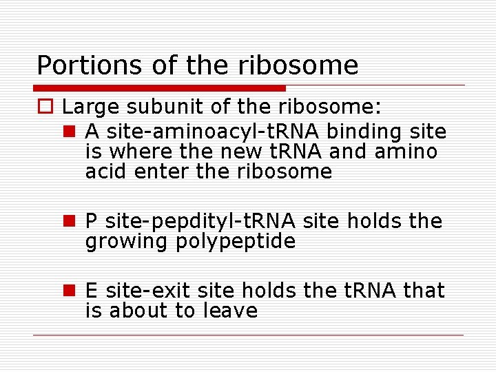
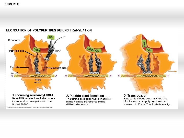
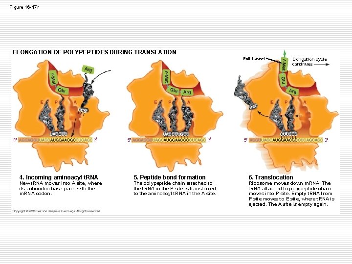
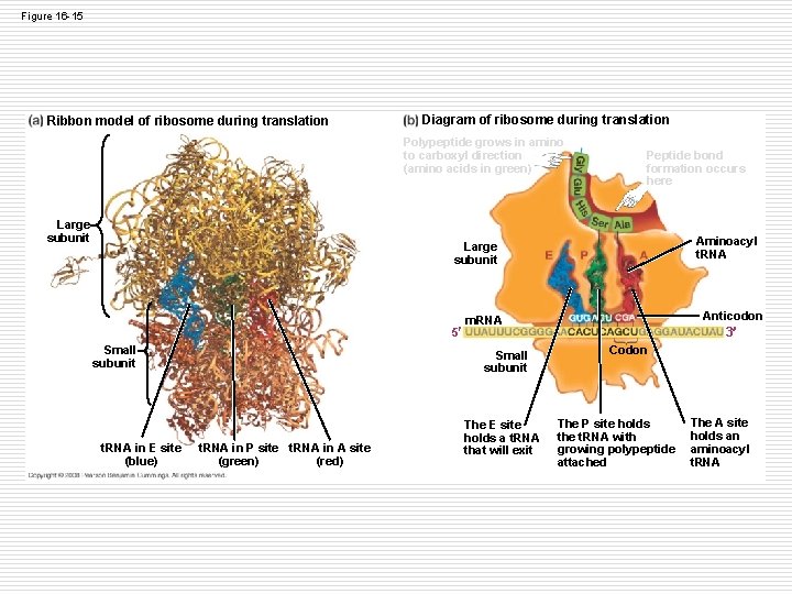
Portions of the ribosome o Large subunit of the ribosome: n A site-aminoacyl-t. RNA binding site is where the new t. RNA and amino acid enter the ribosome n P site-pepdityl-t. RNA site holds the growing polypeptide n E site-exit site holds the t. RNA that is about to leave
Figure 16 -17 l ELONGATION OF POLYPEPTIDES DURING TRANSLATION Ribosome t. RNA Peptidyl site Exit site Aminoacyl site m. RNA 5 3 Start codon 1. Incoming aminoacyl t. RNA New t. RNA moves into A site, where its anticodon base pairs with the m. RNA codon. 2. Peptide bond formation The amino acid attached to the t. RNA in the P site is transferred to the t. RNA in the A site. 3. Translocation Ribosome moves down m. RNA. The t. RNA attached to polypeptide chain moves into P site. The A site is empty.
Figure 16 -17 r ELONGATION OF POLYPEPTIDES DURING TRANSLATION Exit tunnel 5 3 4. Incoming aminoacyl t. RNA New t. RNA moves into A site, where its anticodon base pairs with the m. RNA codon. 3 5 5. Peptide bond formation The polypeptide chain attached to the t. RNA in the P site is transferred to the aminoacyl t. RNA in the A site. Elongation cycle continues 3 5 6. Translocation Ribosome moves down m. RNA. The t. RNA attached to polypeptide chain moves into P site. Empty t. RNA from P site moves to E site, where t. RNA is ejected. The A site is empty again.
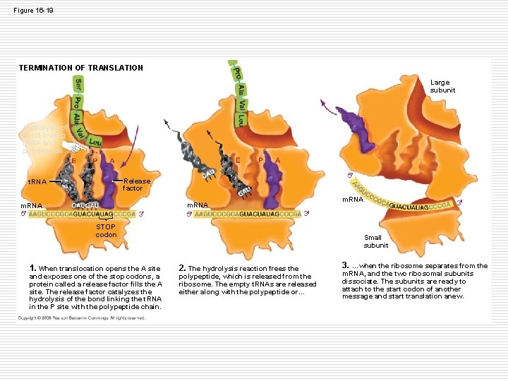
Figure 16 -19 TERMINATION OF TRANSLATION Large subunit Hydrolysis of bond linking t. RNA and polypeptide 5 Release factor t. RNA m. RNA 5 3 m. RNA 5 m. RNA 3 STOP codon 1. When translocation opens the A site and exposes one of the stop codons, a protein called a release factor fills the A site. The release factor catalyzes the hydrolysis of the bond linking the t. RNA in the P site with the polypeptide chain. 3 Small subunit 2. The hydrolysis reaction frees the polypeptide, which is released from the ribosome. The empty t. RNAs are released either along with the polypeptide or… 3. …when the ribosome separates from the m. RNA, and the two ribosomal subunits dissociate. The subunits are ready to attach to the start codon of another message and start translation anew.
Figure 16 -15 Ribbon model of ribosome during translation Diagram of ribosome during translation Polypeptide grows in amino to carboxyl direction (amino acids in green) Large subunit Peptide bond formation occurs here Aminoacyl t. RNA Large subunit 5 Small subunit t. RNA in E site (blue) Small subunit t. RNA in P site t. RNA in A site (green) (red) Anticodon m. RNA The E site holds a t. RNA that will exit 3 Codon The P site holds the t. RNA with growing polypeptide attached The A site holds an aminoacyl t. RNA
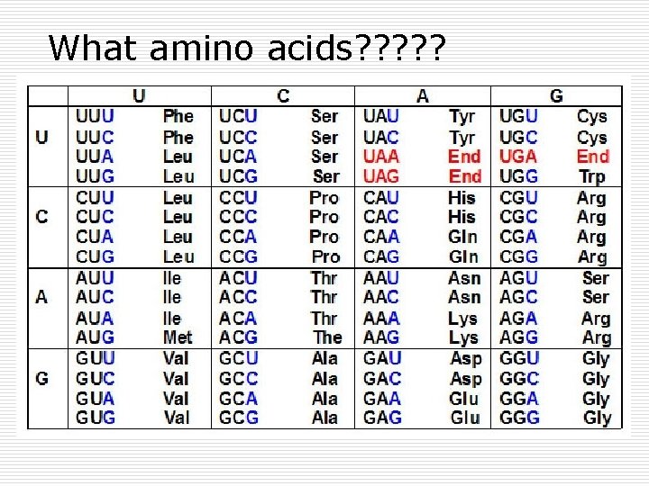
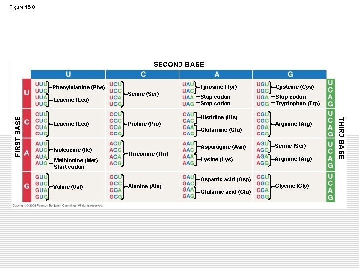
What amino acids? ? ?
Figure 15 -8 SECOND BASE Phenylalanine (Phe) Serine (Ser) Cysteine (Cys) Stop codon Tryptophan (Trp) Histidine (His) Leucine (Leu) Isoleucine (Ile) Proline (Pro) Threonine (Thr) Methionine (Met) Start codon Arginine (Arg) Glutamine (Glu) Asparagine (Asn) Serine (Ser) Lysine (Lys) Arginine (Arg) Aspartic acid (Asp) Valine (Val) Alanine (Ala) Glutamic acid (Glu) Glycine (Gly) THIRD BASE FIRST BASE Leucine (Leu) Tyrosine (Tyr)
Activities o Part A- 10 minutes IN YOUR NOTES o Protein Synthesis and Mutation n To be collected
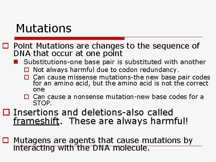
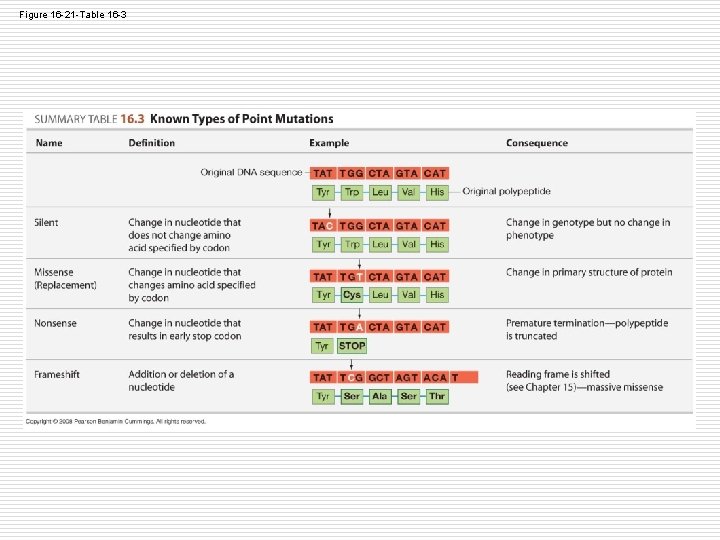
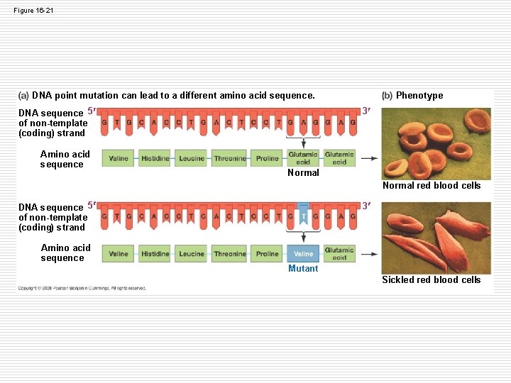
Mutations o Point Mutations are changes to the sequence of DNA that occur at one point n Substitutions-one base pair is substituted with another o Not always harmful due to codon redundancy. o Can cause missense mutations-the new base pair codes for an amino acid, but the amino acid is not the correct one o Can cause a nonsense mutation-new base codes for a STOP. o Insertions and deletions-also called frameshift. These are always harmful! o Mutagens are agents that cause mutations by interacting with the DNA molecule.
Figure 16 -21 -Table 16 -3
Figure 16 -21 DNA point mutation can lead to a different amino acid sequence. DNA sequence 5 of non-template (coding) strand Amino acid sequence Phenotype 3 Normal red blood cells DNA sequence 5 of non-template (coding) strand 3 Amino acid sequence Mutant Sickled red blood cells
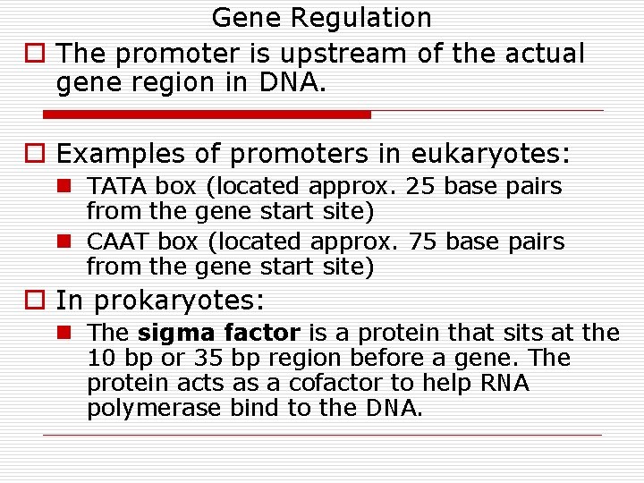
Gene Regulation o The promoter is upstream of the actual gene region in DNA. o Examples of promoters in eukaryotes: n TATA box (located approx. 25 base pairs from the gene start site) n CAAT box (located approx. 75 base pairs from the gene start site) o In prokaryotes: n The sigma factor is a protein that sits at the 10 bp or 35 bp region before a gene. The protein acts as a cofactor to help RNA polymerase bind to the DNA.
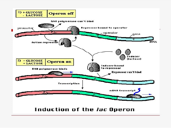
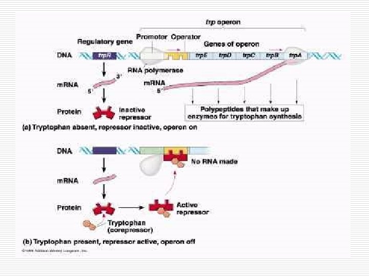
Gene Expression: Operons o An operon is a group of bacterial genes with a common promoter that act as a unit. o All genes in the operon are regulated together. n If one is transcribed they all are transcribed. o Operons can be regulated depending on the environment surrounding the cell. o Repressor molecules can bind to the operon to prohibit transcription = turning off the genes. o Reading and Diagrams: Would you describe the lac and trp operons as inducible or repressible?