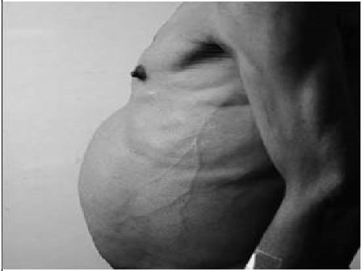
Ascites Definition fluid in the peritoneal cavity Causes


 – chylomicrons are present")


")


 Diuretics Aldosterone")


")
 Mortality is")

")


 Constructional apraxia (the patient is unable to")


- Slides: 42
Ascites
Definition: fluid in the peritoneal cavity
Causes of ascites divided according to the type of ascitic fluid: Straw-coloured Cirrhosis Congestive cardiac failure Constrictive pericarditis Meigs' syndrome Hepatic vein obstruction (Budd-Chiari syndrome) Hypoproteinemia, (e. g. nephrotic syndrome) Malignancy Infective: Tuberculosis, following perforation, SBP Chronic pancreatitis
Chylous Obstruction of main lymphatic duct (e. g. by carcinoma) – chylomicrons are present Cirrhosis (occasional) Hemorrhagic Malignancy Ruptured ectopic pregnancy Abdominal trauma Acute pancreatitis
Pathogenesis 1 - Sodium and water retention: Nitric oxide, atrial natriuretic peptide and prostaglandins are potent vasodilators that increase in liver cirrhosis. They produce peripheral arterial vasodilatation and consequent reduction in the effective blood volume.
2 - Portal hypertension : Exerts a local hydrostatic pressure increased hepatic and splanchnic production of lymph and transudation of fluid into the peritoneal cavity. 3 - Low serum albumin Further contributes by a reduction in plasma oncotic pressure.
Clinical features Abdominal swelling Rapidly accumulating ascites may be due to!!. Mild generalized abdominal pain are common but, if more severe !!.
Respiratory distress. Shifting dullness. Peripheral edema. Pleural effusion (usually on the right side)
Investigations Diagnostic aspiration: ■ Cell count: A neutrophil count above 250 cells /mm 3 is indicative SBP. ■ Bacterial stain and culture: for bacteria and acid-fast bacilli.
Protein: > 3 gm /d. L suggests an exudate. Cytology: for malignant cells. Amylase: to exclude pancreatic ascites. Abdominal Ultrasound
Portal hypertension Peritonial disease or heart failure or kidney disease
Treatment Treat the underlying disease Rest Salt restriction (< 2 gm /day) Diuretics Aldosterone inhibitor (spironolactone) Loop diuretic Paracentesis Peritoneal Ultrafilteration Shunts Liver transplantation
Bed rest alone will lead to a diuresis in a small proportion of people by improving renal perfusion. Dietary sodium restriction and maintain an adequate protein and calorie intake. Fluid restriction is probably not necessary unless the serum sodium is under 128 mmol /L
The diuretic of first choice is the aldosterone antagonist spironolactone, staring at 100 mg (up to 400) daily. S/E: Gynecomastia; (amiloride, 5 -15 mg daily, is then substituted) The aim of diuretic therapy should be to produce 0. 7 kg weight loss or 1. 0 kg in 24 hours if peripheral edema is present
A loop diuretic, such as furosemide 20 mg or bumetanide 1 mg daily, may be added if response is poor. S/E: hyponatraemia, hypokalaemia and volume depletion. Diuretics should be temporarily discontinued if there is a rise in serum creatinine, hyperkalaemia or precoma
Paracentesis Symptomatic tense ascites. In practice, up to 20 L can be removed over 4 -6 hours. Side Effects: *Hepatic encephalopathy. *Hypovolemia.
Shunts TIPS is useful for resistant ascites The use of a peritoneo-venous shunt has been abandoned in most centres as there is high rate of blockage.
Spontaneous bacterial peritonitis (SBP)
Infection of ascitic fluid Usually gram negative (E. Coli, Klebsiella and enterococci) Mortality is high Should be suspected in any patient with ascites with evidence of clinical deterioration Diagnosis: ascitic tap PMN >250 cell /cmm Treatment: third generation cephalosporin IV
Occurs in approximately 8% of cirrhotics with ascites. Recurrence is common (70% within a year) and an oral quinolone, e. g. norfloxacin, 400 mg twice daily is prescribed to prevent it.
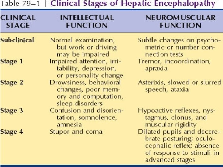
Hepatic Encephalopathy (Portosystemic encephalopathy )
Hepatic Encephalopathy Reversible impairment of neurological function secondary to liver disease Acute: seen with acute liver failure Acute on chronic: in established cirrhosis
Pathogenesis The mechanism is unknown but several factors are thought to play a part. In cirrhosis, the portal blood bypasses the liver via the collaterals and the 'toxic' metabolites pass directly to the brain to produce the encephalopathy.
Many 'toxic' substances are suggested as: Ammonia, Free fatty acids, Mercaptans False neurotransmitters Activation GABA system. Increased aromatic AA and reduced branched-chain AA (valine, leucine and isoleucine).
Factors precipitating portosystemic encephalopathy • • High dietary protein Gastrointestinal haemorrhage Constipation Infection, including spontaneous bacterial peritonitis
• • • Fluid and electrolyte disturbance Diuretic therapy or paracentesis Drugs (e. g. CNS depressants) Portosystemic shunt operations, Progressive liver damage Development of hepatocellular carcinoma
Clinical features Chronically, there is a disorder of personality, mood and intellect, with reversal of sleep rhythm. The patient is irritable, confused, disoriented and has slow slurred speech. Coma occurs as the encephalopathy becomes more marked, but there is hyperreflexia and increased tone.
Signs include: Fetor hepaticus Flapping tremor (asterixis) Constructional apraxia (the patient is unable to draw a five-pointed star) The ability to join numbers and letters with a pen within a certain time is prolonged
Drawing Tests
■ EEG: may show characterestic delta waves. ■ Visual evoked responses also detect subclinical encephalopathy. ■ Arterial blood ammonia is occasionally useful in the differential diagnosis of the cause of the coma and to follow the course of encephalopathy.
Management ■ Identify and remove the possible precipitating cause. ■ Restriction of protein intake. ■ Give purgation and enemas to empty the bowels of nitrogenous substances. ■ Lactulose (10 -30 m. L three times daily) is an osmotic purgative and reduces the colonic p. H and limits ammonia absorption.
■ Antibiotics for eradication of bacterial flora: Rifaximin or metronidazole. Neomycin is less commonly used. ■ L-ornithine L-aspartate: decrease blood amonia ■ Benzodiazepine antagonists e. g. flumazenil ■ Liver transplantation.
Hepatorenal Syndrome
The renal failure is described as 'functional'. The hepatorenal syndrome occurs typically in a patient with advanced cirrhosis with jaundice and ascites. Advanced cases may progress beyond the 'functional' stage to produce an acute tubular necrosis.
The initiating factor is thought to be extreme peripheral vasodilatation possibly due to nitric oxide, leading to decrease in the effective blood volume and hypotension. This activates the hemostatic mechanisms, causing a rise in plasma renin, aldosterone, norepinephrine and vasopressin, leading to vasoconstriction of the renal vasculature
Diuretic therapy should be stopped and intravascular hypovolemia corrected. IV vasoconstrictors Liver transplantation is the best option. The overall prognosis is poor.
Hepatopulmonary syndrome
Hypoxemia occurring in patients with advanced liver disease. It is due to intrapulmonary vascular dilatation with no evidence of primary pulmonary disease. The patients have features of cirrhosis with cyanosis, with more severe disease are breathless on standing (platypnea).
Transthoracic Echo may show intrapulmonary shunting CT pulmonary angiography Arterial blood gases confirm the arterial oxygen desaturation. These changes are improved with liver transplantation.