Articulation Articulation is the movement and placement of
![Articulation • Articulation is the movement and placement of the articulators to shape the Articulation • Articulation is the movement and placement of the articulators to shape the](https://slidetodoc.com/presentation_image_h/04546f097aab3246a5650f60aa7fd3ce/image-1.jpg)
![• Articulation sites Bilabials = Labiodental + Interdentals = Lingual alveolars = Palatals • Articulation sites Bilabials = Labiodental + Interdentals = Lingual alveolars = Palatals](https://slidetodoc.com/presentation_image_h/04546f097aab3246a5650f60aa7fd3ce/image-2.jpg)
![• Source-filter theory = explains how glottal tone is changed into speech sounds • Source-filter theory = explains how glottal tone is changed into speech sounds](https://slidetodoc.com/presentation_image_h/04546f097aab3246a5650f60aa7fd3ce/image-3.jpg)
![• THE ARTICULATORS • First we will examine the framework and muscles, then • THE ARTICULATORS • First we will examine the framework and muscles, then](https://slidetodoc.com/presentation_image_h/04546f097aab3246a5650f60aa7fd3ce/image-4.jpg)
![• Lateral view • Lateral view](https://slidetodoc.com/presentation_image_h/04546f097aab3246a5650f60aa7fd3ce/image-5.jpg)
![What sutures are shown? What sutures are shown?](https://slidetodoc.com/presentation_image_h/04546f097aab3246a5650f60aa7fd3ce/image-6.jpg)
![1. Mandible • Mental symphysis = two parts fused here • Mental protuberance = 1. Mandible • Mental symphysis = two parts fused here • Mental protuberance =](https://slidetodoc.com/presentation_image_h/04546f097aab3246a5650f60aa7fd3ce/image-7.jpg)
![](https://slidetodoc.com/presentation_image_h/04546f097aab3246a5650f60aa7fd3ce/image-8.jpg)
![](https://slidetodoc.com/presentation_image_h/04546f097aab3246a5650f60aa7fd3ce/image-9.jpg)
![Hemifacial microsomia Hemifacial microsomia](https://slidetodoc.com/presentation_image_h/04546f097aab3246a5650f60aa7fd3ce/image-10.jpg)
![Hemifacial Microsomia Characteristics • Hemi = half, although present in bilateral condition in up Hemifacial Microsomia Characteristics • Hemi = half, although present in bilateral condition in up](https://slidetodoc.com/presentation_image_h/04546f097aab3246a5650f60aa7fd3ce/image-11.jpg)
![Pierre Robin sequence shown below. Etiology is unknown. It can occur as one sign Pierre Robin sequence shown below. Etiology is unknown. It can occur as one sign](https://slidetodoc.com/presentation_image_h/04546f097aab3246a5650f60aa7fd3ce/image-12.jpg)
![2. Maxillae: two bones join to make up upper jaw (anterior ¾ of hard 2. Maxillae: two bones join to make up upper jaw (anterior ¾ of hard](https://slidetodoc.com/presentation_image_h/04546f097aab3246a5650f60aa7fd3ce/image-13.jpg)
![](https://slidetodoc.com/presentation_image_h/04546f097aab3246a5650f60aa7fd3ce/image-14.jpg)
![](https://slidetodoc.com/presentation_image_h/04546f097aab3246a5650f60aa7fd3ce/image-15.jpg)
![](https://slidetodoc.com/presentation_image_h/04546f097aab3246a5650f60aa7fd3ce/image-16.jpg)
![](https://slidetodoc.com/presentation_image_h/04546f097aab3246a5650f60aa7fd3ce/image-17.jpg)
![](https://slidetodoc.com/presentation_image_h/04546f097aab3246a5650f60aa7fd3ce/image-18.jpg)
![](https://slidetodoc.com/presentation_image_h/04546f097aab3246a5650f60aa7fd3ce/image-19.jpg)
![3. • Nasal bones: small oblong bones, form bridge of nose Articulate with frontal 3. • Nasal bones: small oblong bones, form bridge of nose Articulate with frontal](https://slidetodoc.com/presentation_image_h/04546f097aab3246a5650f60aa7fd3ce/image-20.jpg)
![4. Palatine bones: shaped like letter "L“ – Contribute to formation of 3 cavities: 4. Palatine bones: shaped like letter "L“ – Contribute to formation of 3 cavities:](https://slidetodoc.com/presentation_image_h/04546f097aab3246a5650f60aa7fd3ce/image-21.jpg)
![5. Lacrimal bones – – smallest facial bones form medial walls of orbital cavities 5. Lacrimal bones – – smallest facial bones form medial walls of orbital cavities](https://slidetodoc.com/presentation_image_h/04546f097aab3246a5650f60aa7fd3ce/image-22.jpg)
![6. Inferior nasal conchae – – Inferior nasal conchae (scroll-like) (inferior turbinates): forms part 6. Inferior nasal conchae – – Inferior nasal conchae (scroll-like) (inferior turbinates): forms part](https://slidetodoc.com/presentation_image_h/04546f097aab3246a5650f60aa7fd3ce/image-23.jpg)
![7. Vomer • • Inferior and posterior half of bony septum Name means plowshare 7. Vomer • • Inferior and posterior half of bony septum Name means plowshare](https://slidetodoc.com/presentation_image_h/04546f097aab3246a5650f60aa7fd3ce/image-24.jpg)
![8. Zygomatic bones OR Malar* bones – – forms zygomatic arch (OR cheekbones) with 8. Zygomatic bones OR Malar* bones – – forms zygomatic arch (OR cheekbones) with](https://slidetodoc.com/presentation_image_h/04546f097aab3246a5650f60aa7fd3ce/image-25.jpg)
![Treacher Collins syndrome: one feature is hypoplasia of the malar (zygomatic bones). Rib cartilages Treacher Collins syndrome: one feature is hypoplasia of the malar (zygomatic bones). Rib cartilages](https://slidetodoc.com/presentation_image_h/04546f097aab3246a5650f60aa7fd3ce/image-26.jpg)
![Father, daughter, granddaughter; all have TC Father, daughter, granddaughter; all have TC](https://slidetodoc.com/presentation_image_h/04546f097aab3246a5650f60aa7fd3ce/image-27.jpg)
![• http: //www. a 3 bs. com/3 d/A 291/index. cfm – Outstanding animations • http: //www. a 3 bs. com/3 d/A 291/index. cfm – Outstanding animations](https://slidetodoc.com/presentation_image_h/04546f097aab3246a5650f60aa7fd3ce/image-28.jpg)
![• http: //www. innerbody. com/htm/body. html – Select skeletal system • Skull views • http: //www. innerbody. com/htm/body. html – Select skeletal system • Skull views](https://slidetodoc.com/presentation_image_h/04546f097aab3246a5650f60aa7fd3ce/image-29.jpg)
![• Bones of Cranium • • • Ethmoid Frontal Parietal (2) Occipital Temporal • Bones of Cranium • • • Ethmoid Frontal Parietal (2) Occipital Temporal](https://slidetodoc.com/presentation_image_h/04546f097aab3246a5650f60aa7fd3ce/image-30.jpg)
![1. Ethmoid bone • Extremely complex (2 nd only to sphenoid), yet delicate (light 1. Ethmoid bone • Extremely complex (2 nd only to sphenoid), yet delicate (light](https://slidetodoc.com/presentation_image_h/04546f097aab3246a5650f60aa7fd3ce/image-31.jpg)
![http: //www. theodora. com/anatomy/ http: //www. theodora. com/anatomy/](https://slidetodoc.com/presentation_image_h/04546f097aab3246a5650f60aa7fd3ce/image-32.jpg)
![• http: //anatomy. med. umich. edu/nervous_system/ear. html – – – Scroll down and • http: //anatomy. med. umich. edu/nervous_system/ear. html – – – Scroll down and](https://slidetodoc.com/presentation_image_h/04546f097aab3246a5650f60aa7fd3ce/image-33.jpg)
![](https://slidetodoc.com/presentation_image_h/04546f097aab3246a5650f60aa7fd3ce/image-34.jpg)
![• Ethmoid: parts – A. Cribriform plate = separates cranial from nasal cavities • Ethmoid: parts – A. Cribriform plate = separates cranial from nasal cavities](https://slidetodoc.com/presentation_image_h/04546f097aab3246a5650f60aa7fd3ce/image-35.jpg)
![Arrow indicates falx cerebri Arrow indicates falx cerebri](https://slidetodoc.com/presentation_image_h/04546f097aab3246a5650f60aa7fd3ce/image-36.jpg)
![• Where is crista galli? • Where is crista galli?](https://slidetodoc.com/presentation_image_h/04546f097aab3246a5650f60aa7fd3ce/image-37.jpg)
![2. Frontal bone • Anterior cranial case, forehead, supraorbital region (roof of orbital cavity) 2. Frontal bone • Anterior cranial case, forehead, supraorbital region (roof of orbital cavity)](https://slidetodoc.com/presentation_image_h/04546f097aab3246a5650f60aa7fd3ce/image-38.jpg)
![Frontal bone: Parts – A. Superciliary arches = – B. Glabella = prominence above Frontal bone: Parts – A. Superciliary arches = – B. Glabella = prominence above](https://slidetodoc.com/presentation_image_h/04546f097aab3246a5650f60aa7fd3ce/image-39.jpg)
![Can you locate the superciliary arches? Can you locate the superciliary arches?](https://slidetodoc.com/presentation_image_h/04546f097aab3246a5650f60aa7fd3ce/image-40.jpg)
![• • 3. Parietal bones Roof of cranium Joined by sagittal suture Separated • • 3. Parietal bones Roof of cranium Joined by sagittal suture Separated](https://slidetodoc.com/presentation_image_h/04546f097aab3246a5650f60aa7fd3ce/image-41.jpg)
![• 4. Occipital bone • Lower and posterior portion of cranium • Parts: • 4. Occipital bone • Lower and posterior portion of cranium • Parts:](https://slidetodoc.com/presentation_image_h/04546f097aab3246a5650f60aa7fd3ce/image-42.jpg)
![](https://slidetodoc.com/presentation_image_h/04546f097aab3246a5650f60aa7fd3ce/image-43.jpg)
![• 5. Temporal bones • Lateral base and sides of cranium • Divided • 5. Temporal bones • Lateral base and sides of cranium • Divided](https://slidetodoc.com/presentation_image_h/04546f097aab3246a5650f60aa7fd3ce/image-44.jpg)
![](https://slidetodoc.com/presentation_image_h/04546f097aab3246a5650f60aa7fd3ce/image-45.jpg)
![• 6. Sphenoid bone • Most complex of cranial bones • Located posterior • 6. Sphenoid bone • Most complex of cranial bones • Located posterior](https://slidetodoc.com/presentation_image_h/04546f097aab3246a5650f60aa7fd3ce/image-46.jpg)
![• Sphenoid: Parts – A. Body or corpus (view superiorly) • Sella turcica • Sphenoid: Parts – A. Body or corpus (view superiorly) • Sella turcica](https://slidetodoc.com/presentation_image_h/04546f097aab3246a5650f60aa7fd3ce/image-47.jpg)
![Medial pterygoid plate Medial pterygoid plate](https://slidetodoc.com/presentation_image_h/04546f097aab3246a5650f60aa7fd3ce/image-48.jpg)
![](https://slidetodoc.com/presentation_image_h/04546f097aab3246a5650f60aa7fd3ce/image-49.jpg)
![• Dentition • Function: – Mastication – Surfaces for articulation of speech sounds • Dentition • Function: – Mastication – Surfaces for articulation of speech sounds](https://slidetodoc.com/presentation_image_h/04546f097aab3246a5650f60aa7fd3ce/image-50.jpg)
![](https://slidetodoc.com/presentation_image_h/04546f097aab3246a5650f60aa7fd3ce/image-51.jpg)
![• Surface terms – Midline (between central incisors – Buccal (toward cheek) – • Surface terms – Midline (between central incisors – Buccal (toward cheek) –](https://slidetodoc.com/presentation_image_h/04546f097aab3246a5650f60aa7fd3ce/image-52.jpg)
![• Occlusion = alignment of maxillary teeth with mandibular teeth. – Types of • Occlusion = alignment of maxillary teeth with mandibular teeth. – Types of](https://slidetodoc.com/presentation_image_h/04546f097aab3246a5650f60aa7fd3ce/image-53.jpg)
![• Class II malocclusion: the 1 st mandibular molar is retracted at least • Class II malocclusion: the 1 st mandibular molar is retracted at least](https://slidetodoc.com/presentation_image_h/04546f097aab3246a5650f60aa7fd3ce/image-54.jpg)
![• Class I malocclusion: • Other dental anomalies – Overbite—vertical dimension – Overjet—horizontal • Class I malocclusion: • Other dental anomalies – Overbite—vertical dimension – Overjet—horizontal](https://slidetodoc.com/presentation_image_h/04546f097aab3246a5650f60aa7fd3ce/image-55.jpg)
![Name the occlusion Name the occlusion](https://slidetodoc.com/presentation_image_h/04546f097aab3246a5650f60aa7fd3ce/image-56.jpg)
![](https://slidetodoc.com/presentation_image_h/04546f097aab3246a5650f60aa7fd3ce/image-57.jpg)
![A C B D E A C B D E](https://slidetodoc.com/presentation_image_h/04546f097aab3246a5650f60aa7fd3ce/image-58.jpg)
![• Cavities of Vocal Tract – Oral, pharyngeal, nasal • Cavities of Vocal Tract – Oral, pharyngeal, nasal](https://slidetodoc.com/presentation_image_h/04546f097aab3246a5650f60aa7fd3ce/image-59.jpg)
![– Oral tract/cavity Anterior faucial pillar Posterior faucial pillar Lingual frenum – Oral tract/cavity Anterior faucial pillar Posterior faucial pillar Lingual frenum](https://slidetodoc.com/presentation_image_h/04546f097aab3246a5650f60aa7fd3ce/image-60.jpg)
![Ankyloglossia Ankyloglossia](https://slidetodoc.com/presentation_image_h/04546f097aab3246a5650f60aa7fd3ce/image-61.jpg)
![In little children, the superior labial frenum is often attached between the two central In little children, the superior labial frenum is often attached between the two central](https://slidetodoc.com/presentation_image_h/04546f097aab3246a5650f60aa7fd3ce/image-62.jpg)
![– Pharyngeal cavity » Nasopharynx—space above soft palate (bounded anteriorly by nasal choanae) (choana – Pharyngeal cavity » Nasopharynx—space above soft palate (bounded anteriorly by nasal choanae) (choana](https://slidetodoc.com/presentation_image_h/04546f097aab3246a5650f60aa7fd3ce/image-63.jpg)
![• Facial muscles • Facial landmarks: – Cupids bow – Vermilion – Lips: • Facial muscles • Facial landmarks: – Cupids bow – Vermilion – Lips:](https://slidetodoc.com/presentation_image_h/04546f097aab3246a5650f60aa7fd3ce/image-64.jpg)
![Bifid uvula Bifid uvula](https://slidetodoc.com/presentation_image_h/04546f097aab3246a5650f60aa7fd3ce/image-65.jpg)
![• Orbicularis oris: principle muscle acting on lips – no origin or insertion • Orbicularis oris: principle muscle acting on lips – no origin or insertion](https://slidetodoc.com/presentation_image_h/04546f097aab3246a5650f60aa7fd3ce/image-66.jpg)
![• 2 transverse muscles • Buccinator muscle (transverse) (deep m) (bugler’s muscle) – • 2 transverse muscles • Buccinator muscle (transverse) (deep m) (bugler’s muscle) –](https://slidetodoc.com/presentation_image_h/04546f097aab3246a5650f60aa7fd3ce/image-67.jpg)
![](https://slidetodoc.com/presentation_image_h/04546f097aab3246a5650f60aa7fd3ce/image-68.jpg)
![• Risorius muscle (transverse) (L risus = laughter) – Runs parallel and superior • Risorius muscle (transverse) (L risus = laughter) – Runs parallel and superior](https://slidetodoc.com/presentation_image_h/04546f097aab3246a5650f60aa7fd3ce/image-69.jpg)
![• Angular muscles • Zygomatic Minor: – Origin = malar surface of zygomatic • Angular muscles • Zygomatic Minor: – Origin = malar surface of zygomatic](https://slidetodoc.com/presentation_image_h/04546f097aab3246a5650f60aa7fd3ce/image-70.jpg)
![• Levator Labii Superior: – Origin = lower border of orbit mainly (some • Levator Labii Superior: – Origin = lower border of orbit mainly (some](https://slidetodoc.com/presentation_image_h/04546f097aab3246a5650f60aa7fd3ce/image-71.jpg)
![• Levator Labaii Superior Alaeque Nasi M. : – origin = frontal process • Levator Labaii Superior Alaeque Nasi M. : – origin = frontal process](https://slidetodoc.com/presentation_image_h/04546f097aab3246a5650f60aa7fd3ce/image-72.jpg)
![• Depressor Labii Inferior: – origin = oblique line of mandible, inserts into • Depressor Labii Inferior: – origin = oblique line of mandible, inserts into](https://slidetodoc.com/presentation_image_h/04546f097aab3246a5650f60aa7fd3ce/image-73.jpg)
![• Vertical muscles • Mentalis: – origin = mental turberosity; inserts = some • Vertical muscles • Mentalis: – origin = mental turberosity; inserts = some](https://slidetodoc.com/presentation_image_h/04546f097aab3246a5650f60aa7fd3ce/image-74.jpg)
![• Depressor Anguli Oris = – superficial; – origin = oblique line; insertion • Depressor Anguli Oris = – superficial; – origin = oblique line; insertion](https://slidetodoc.com/presentation_image_h/04546f097aab3246a5650f60aa7fd3ce/image-75.jpg)
![• Levator Anguli Oris = – deep m. ; – origin = canine • Levator Anguli Oris = – deep m. ; – origin = canine](https://slidetodoc.com/presentation_image_h/04546f097aab3246a5650f60aa7fd3ce/image-76.jpg)
![FYI Anatomically, dimples may be caused by variations in the structure of the facial FYI Anatomically, dimples may be caused by variations in the structure of the facial](https://slidetodoc.com/presentation_image_h/04546f097aab3246a5650f60aa7fd3ce/image-77.jpg)
![• • http: //www. ivy-rose. co. uk/Topics/Facial. Muscles. htm The above cited website • • http: //www. ivy-rose. co. uk/Topics/Facial. Muscles. htm The above cited website](https://slidetodoc.com/presentation_image_h/04546f097aab3246a5650f60aa7fd3ce/image-78.jpg)
![• Regions of tongue: – tip—nearest central incisors – Blade—just below alveolar ridge • Regions of tongue: – tip—nearest central incisors – Blade—just below alveolar ridge](https://slidetodoc.com/presentation_image_h/04546f097aab3246a5650f60aa7fd3ce/image-79.jpg)
![• • Taste buds: – Anterior: sweet & salty (posterior = bitter) – • • Taste buds: – Anterior: sweet & salty (posterior = bitter) –](https://slidetodoc.com/presentation_image_h/04546f097aab3246a5650f60aa7fd3ce/image-80.jpg)
![• Plane/cut shown below? • Pix from: • http: //www. meddean. luc. edu/lumen/meded/grossana • Plane/cut shown below? • Pix from: • http: //www. meddean. luc. edu/lumen/meded/grossana](https://slidetodoc.com/presentation_image_h/04546f097aab3246a5650f60aa7fd3ce/image-81.jpg)
![• Intrinsic muscles • Overall function = shaping tongue – Superior longitudinal • • Intrinsic muscles • Overall function = shaping tongue – Superior longitudinal •](https://slidetodoc.com/presentation_image_h/04546f097aab3246a5650f60aa7fd3ce/image-82.jpg)
![• Tongue anomalies – Microglossia – Macroglossia (e. g. , Beckwith-Wiedemann syndrome – • Tongue anomalies – Microglossia – Macroglossia (e. g. , Beckwith-Wiedemann syndrome –](https://slidetodoc.com/presentation_image_h/04546f097aab3246a5650f60aa7fd3ce/image-83.jpg)
![• • 1. Extrinsic tongue muscles Function = general movement of tongue Genioglossus • • 1. Extrinsic tongue muscles Function = general movement of tongue Genioglossus](https://slidetodoc.com/presentation_image_h/04546f097aab3246a5650f60aa7fd3ce/image-84.jpg)
![2. Hyoglossus • Origin: hyoid; insertion: sides of tongue • Function: pulls sides of 2. Hyoglossus • Origin: hyoid; insertion: sides of tongue • Function: pulls sides of](https://slidetodoc.com/presentation_image_h/04546f097aab3246a5650f60aa7fd3ce/image-85.jpg)
![3. Styloglossus • • • Origin: styloid process of ? ; insertion: inferior sides 3. Styloglossus • • • Origin: styloid process of ? ; insertion: inferior sides](https://slidetodoc.com/presentation_image_h/04546f097aab3246a5650f60aa7fd3ce/image-86.jpg)
![• 4. palatoglossus – Commonly called anterior faucial pillar – Can be considered • 4. palatoglossus – Commonly called anterior faucial pillar – Can be considered](https://slidetodoc.com/presentation_image_h/04546f097aab3246a5650f60aa7fd3ce/image-87.jpg)
![• Muscles of mastication • • Function = elevate or depress mandible These • Muscles of mastication • • Function = elevate or depress mandible These](https://slidetodoc.com/presentation_image_h/04546f097aab3246a5650f60aa7fd3ce/image-88.jpg)
![2. Temporalis muscle • • • Fan-shaped muscle Origin = temporal fossa of temporal 2. Temporalis muscle • • • Fan-shaped muscle Origin = temporal fossa of temporal](https://slidetodoc.com/presentation_image_h/04546f097aab3246a5650f60aa7fd3ce/image-89.jpg)
![3. Medial (internal) Pterygoid muscle • Origin = pterygoid plate and fossa (sphenoid bone) 3. Medial (internal) Pterygoid muscle • Origin = pterygoid plate and fossa (sphenoid bone)](https://slidetodoc.com/presentation_image_h/04546f097aab3246a5650f60aa7fd3ce/image-90.jpg)
![Protruder 4. Lateral (external) Pterygoid muscle • Origin: Has two heads (sphenoid— pterygoid plate Protruder 4. Lateral (external) Pterygoid muscle • Origin: Has two heads (sphenoid— pterygoid plate](https://slidetodoc.com/presentation_image_h/04546f097aab3246a5650f60aa7fd3ce/image-91.jpg)
![Depressors 5. Digastricus • Has anterior and posterior belly, united by central tendon • Depressors 5. Digastricus • Has anterior and posterior belly, united by central tendon •](https://slidetodoc.com/presentation_image_h/04546f097aab3246a5650f60aa7fd3ce/image-92.jpg)
![6. Mylohyoid • • Origin = mylohyoid line Insertion = Median raphe and hyoid 6. Mylohyoid • • Origin = mylohyoid line Insertion = Median raphe and hyoid](https://slidetodoc.com/presentation_image_h/04546f097aab3246a5650f60aa7fd3ce/image-93.jpg)
![7. Geniohyoid • Origin = mental spine on inner aspect of mental symphysis (just 7. Geniohyoid • Origin = mental spine on inner aspect of mental symphysis (just](https://slidetodoc.com/presentation_image_h/04546f097aab3246a5650f60aa7fd3ce/image-94.jpg)
![8. Platysma • Considered a facial muscle (embryologically developed from same primordia from which 8. Platysma • Considered a facial muscle (embryologically developed from same primordia from which](https://slidetodoc.com/presentation_image_h/04546f097aab3246a5650f60aa7fd3ce/image-95.jpg)
![Muscles of the Velum • • • Levator veli palatini Musculus uvulae Tensor veli Muscles of the Velum • • • Levator veli palatini Musculus uvulae Tensor veli](https://slidetodoc.com/presentation_image_h/04546f097aab3246a5650f60aa7fd3ce/image-96.jpg)
![](https://slidetodoc.com/presentation_image_h/04546f097aab3246a5650f60aa7fd3ce/image-97.jpg)
![• 2. Musculus uvulae – Paired muscle (sometimes referred to as one muscle) • 2. Musculus uvulae – Paired muscle (sometimes referred to as one muscle)](https://slidetodoc.com/presentation_image_h/04546f097aab3246a5650f60aa7fd3ce/image-98.jpg)
![](https://slidetodoc.com/presentation_image_h/04546f097aab3246a5650f60aa7fd3ce/image-99.jpg)
![• 3. Tensor Veli Palatini Origin: sphenoid bone Insertion: converges on tendon; tendon • 3. Tensor Veli Palatini Origin: sphenoid bone Insertion: converges on tendon; tendon](https://slidetodoc.com/presentation_image_h/04546f097aab3246a5650f60aa7fd3ce/image-100.jpg)
![](https://slidetodoc.com/presentation_image_h/04546f097aab3246a5650f60aa7fd3ce/image-101.jpg)
![• 4. Palatoglossus (anterior faucial pillars) – Origin: anterior surface of soft palate--in • 4. Palatoglossus (anterior faucial pillars) – Origin: anterior surface of soft palate--in](https://slidetodoc.com/presentation_image_h/04546f097aab3246a5650f60aa7fd3ce/image-102.jpg)
![](https://slidetodoc.com/presentation_image_h/04546f097aab3246a5650f60aa7fd3ce/image-103.jpg)
![• 5. Palatopharyngeus (posterior faucial pillars) – Origin: Fan-shaped in soft palate – • 5. Palatopharyngeus (posterior faucial pillars) – Origin: Fan-shaped in soft palate –](https://slidetodoc.com/presentation_image_h/04546f097aab3246a5650f60aa7fd3ce/image-104.jpg)
- Slides: 104
![Articulation Articulation is the movement and placement of the articulators to shape the Articulation • Articulation is the movement and placement of the articulators to shape the](https://slidetodoc.com/presentation_image_h/04546f097aab3246a5650f60aa7fd3ce/image-1.jpg)
Articulation • Articulation is the movement and placement of the articulators to shape the vocal tract. • The buzz produced by the vibrating vocal folds is shaped into sounds or phonemes by the articulatory system. • As the laryngeal or glottal vibrations travel superiorly through the vocal tract, they are selectively attenuated so that certain frequencies are transmitted more strongly than others. – Vocal tract = pharyngeal, nasal, & oral cavities
![Articulation sites Bilabials Labiodental Interdentals Lingual alveolars Palatals • Articulation sites Bilabials = Labiodental + Interdentals = Lingual alveolars = Palatals](https://slidetodoc.com/presentation_image_h/04546f097aab3246a5650f60aa7fd3ce/image-2.jpg)
• Articulation sites Bilabials = Labiodental + Interdentals = Lingual alveolars = Palatals = Velars = Glottal =
![Sourcefilter theory explains how glottal tone is changed into speech sounds • Source-filter theory = explains how glottal tone is changed into speech sounds](https://slidetodoc.com/presentation_image_h/04546f097aab3246a5650f60aa7fd3ce/image-3.jpg)
• Source-filter theory = explains how glottal tone is changed into speech sounds by the vocal tract. – The three cavities of the vocal tract can change shapes due to moveable articulators – The changes in shape cause resonant frequencies to change. • Resonant frequency = frequency that cavity responds to most effectively • Example: bottle with water* • /s/ vs /sh/ Which has a lower frequency? • http: //www. uiowa. edu/~acadtech/phonetics/# • See articulation anatomy on the website above. – With consonants, sources of sound can include turbulence of frication with or without voicing. – Articulators are movable or not movable
![THE ARTICULATORS First we will examine the framework and muscles then • THE ARTICULATORS • First we will examine the framework and muscles, then](https://slidetodoc.com/presentation_image_h/04546f097aab3246a5650f60aa7fd3ce/image-4.jpg)
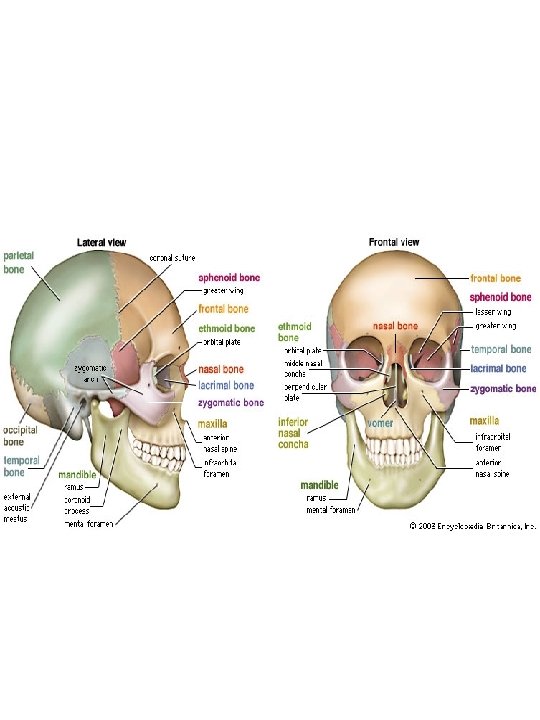
• THE ARTICULATORS • First we will examine the framework and muscles, then relate structures to speech production • SKULL = supportive framework – Composed of 22 bones. – All except mandible are rigidly joined together by sutures* – Main sutures = coronal. sagittal, lambdoidal, and squamosal suture • Coronal = divides skull into front and back; between frontal and parietal bones • Sagittal = divides skull in L and R; between the two parietal bones • Lambdoidal = between parietal and occipital bones; shaped like Greek letter • Squamosal = between parietal and temporal bones – Identify these sutures on the next two slides • Sutures = joints that are immoveable • Lambdoidal = occipital
![Lateral view • Lateral view](https://slidetodoc.com/presentation_image_h/04546f097aab3246a5650f60aa7fd3ce/image-5.jpg)
• Lateral view
![What sutures are shown What sutures are shown?](https://slidetodoc.com/presentation_image_h/04546f097aab3246a5650f60aa7fd3ce/image-6.jpg)
What sutures are shown?
![1 Mandible Mental symphysis two parts fused here Mental protuberance 1. Mandible • Mental symphysis = two parts fused here • Mental protuberance =](https://slidetodoc.com/presentation_image_h/04546f097aab3246a5650f60aa7fd3ce/image-7.jpg)
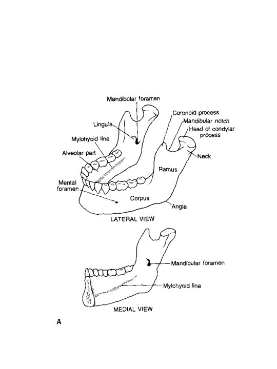
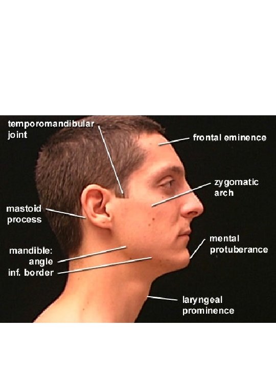
1. Mandible • Mental symphysis = two parts fused here • Mental protuberance = midline; creates triangular eminence (prominence) • Mental foramen • Corpus • Angle • Ramus • Coronoid process • Condylar process (articulates with temporal bone, allowing rotation of mandible) • Mandibular foramen (inner aspect) (branch of Trigeminal, CV, goes through here to provide sensory innervation to teeth and gums) • Mylohyoid line (mylohyoid muscle inserts here) • Mandibular hypoplasia and micrognathia • May cause some instances of cleft palate • Can be corrected surgically or with distraction osteogenesis
![](https://slidetodoc.com/presentation_image_h/04546f097aab3246a5650f60aa7fd3ce/image-8.jpg)
![](https://slidetodoc.com/presentation_image_h/04546f097aab3246a5650f60aa7fd3ce/image-9.jpg)
![Hemifacial microsomia Hemifacial microsomia](https://slidetodoc.com/presentation_image_h/04546f097aab3246a5650f60aa7fd3ce/image-10.jpg)
Hemifacial microsomia
![Hemifacial Microsomia Characteristics Hemi half although present in bilateral condition in up Hemifacial Microsomia Characteristics • Hemi = half, although present in bilateral condition in up](https://slidetodoc.com/presentation_image_h/04546f097aab3246a5650f60aa7fd3ce/image-11.jpg)
Hemifacial Microsomia Characteristics • Hemi = half, although present in bilateral condition in up to 15%-16% of cases • Unilateral or bilateral underdev of mandible • Unilateral or bilateral underdev/malformations of ear (microtia, ear tags, etc) • Unilateral or bilateral reduction in size and flattening of maxilla • Narrowing of the opening of the eye Causes: thought caused by hemorrhage from artery that produces hematoma in area of branchial arches (head, neck area dev from branchial arches). Occurs during first 6 -8 weeks of pregnancy. • Inheritance: sporadic; reports of familial cases. May be auto dominant, auto recessive, or multifactorial.
![Pierre Robin sequence shown below Etiology is unknown It can occur as one sign Pierre Robin sequence shown below. Etiology is unknown. It can occur as one sign](https://slidetodoc.com/presentation_image_h/04546f097aab3246a5650f60aa7fd3ce/image-12.jpg)
Pierre Robin sequence shown below. Etiology is unknown. It can occur as one sign of some syndromes. Note the micrognathia of the infant. Young child has distractor in place and trach.
![2 Maxillae two bones join to make up upper jaw anterior ¾ of hard 2. Maxillae: two bones join to make up upper jaw (anterior ¾ of hard](https://slidetodoc.com/presentation_image_h/04546f097aab3246a5650f60aa7fd3ce/image-13.jpg)
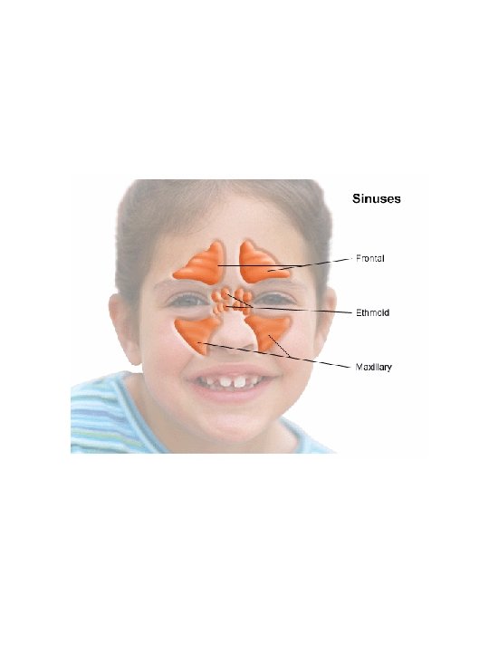
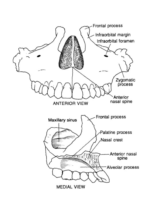
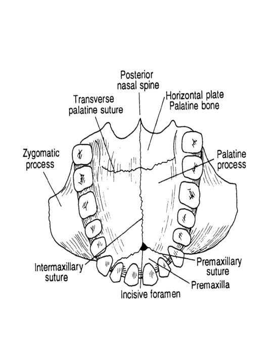
2. Maxillae: two bones join to make up upper jaw (anterior ¾ of hard palate) – – – – – Frontal process (superior-most part of maxillae); maxilla articulates with frontal bone Zygomatic arch = articulates with zygomatic bone Infraorbital foramen (passageway for branch of CV (Trigeminal) that supplies sensation to lower eyelid, upper lip, and nasal alae) Anterior nasal spine Alveolar process = Maxillary sinuses (see next slide) Palatine process = articulates with palatine bone Incisive foramen = nerve passageway for nasal mucosa Premaxillary suture = line that divides primary palate OR premaxilla from secondary palate. • Premaxilla can be clefted as either unilateral or bilateral Intermaxillary suture = cleft of hard palate follows this suture
![](https://slidetodoc.com/presentation_image_h/04546f097aab3246a5650f60aa7fd3ce/image-14.jpg)
![](https://slidetodoc.com/presentation_image_h/04546f097aab3246a5650f60aa7fd3ce/image-15.jpg)
![](https://slidetodoc.com/presentation_image_h/04546f097aab3246a5650f60aa7fd3ce/image-16.jpg)
![](https://slidetodoc.com/presentation_image_h/04546f097aab3246a5650f60aa7fd3ce/image-17.jpg)
![](https://slidetodoc.com/presentation_image_h/04546f097aab3246a5650f60aa7fd3ce/image-18.jpg)
![](https://slidetodoc.com/presentation_image_h/04546f097aab3246a5650f60aa7fd3ce/image-19.jpg)
![3 Nasal bones small oblong bones form bridge of nose Articulate with frontal 3. • Nasal bones: small oblong bones, form bridge of nose Articulate with frontal](https://slidetodoc.com/presentation_image_h/04546f097aab3246a5650f60aa7fd3ce/image-20.jpg)
3. • Nasal bones: small oblong bones, form bridge of nose Articulate with frontal bone (above), perpendicular plate of ethmoid, and nasal bone from opposite side, and maxilla.
![4 Palatine bones shaped like letter L Contribute to formation of 3 cavities 4. Palatine bones: shaped like letter "L“ – Contribute to formation of 3 cavities:](https://slidetodoc.com/presentation_image_h/04546f097aab3246a5650f60aa7fd3ce/image-21.jpg)
4. Palatine bones: shaped like letter "L“ – Contribute to formation of 3 cavities: floor and lateral wall of nasal cavity, roof of mouth (posterior 1/4), floor of orbital cavity – – – Posterior nasal spine Horizontal plate = posterior ¼ of hard palate Orbital process = sm portion of floor of orbital cavity
![5 Lacrimal bones smallest facial bones form medial walls of orbital cavities 5. Lacrimal bones – – smallest facial bones form medial walls of orbital cavities](https://slidetodoc.com/presentation_image_h/04546f097aab3246a5650f60aa7fd3ce/image-22.jpg)
5. Lacrimal bones – – smallest facial bones form medial walls of orbital cavities
![6 Inferior nasal conchae Inferior nasal conchae scrolllike inferior turbinates forms part 6. Inferior nasal conchae – – Inferior nasal conchae (scroll-like) (inferior turbinates): forms part](https://slidetodoc.com/presentation_image_h/04546f097aab3246a5650f60aa7fd3ce/image-23.jpg)
6. Inferior nasal conchae – – Inferior nasal conchae (scroll-like) (inferior turbinates): forms part of lateral nasal wall Note: medial and superior nasal conchae are processes of the ethmoid bone (to be discussed later) Mucosal lining overlying the nasal conchae is thickest of the nose and is richly invested with vascular supply. Air is warmed and humidified as passes through the conchae
![7 Vomer Inferior and posterior half of bony septum Name means plowshare 7. Vomer • • Inferior and posterior half of bony septum Name means plowshare](https://slidetodoc.com/presentation_image_h/04546f097aab3246a5650f60aa7fd3ce/image-24.jpg)
7. Vomer • • Inferior and posterior half of bony septum Name means plowshare
![8 Zygomatic bones OR Malar bones forms zygomatic arch OR cheekbones with 8. Zygomatic bones OR Malar* bones – – forms zygomatic arch (OR cheekbones) with](https://slidetodoc.com/presentation_image_h/04546f097aab3246a5650f60aa7fd3ce/image-25.jpg)
8. Zygomatic bones OR Malar* bones – – forms zygomatic arch (OR cheekbones) with maxilla and temporal bones Makes up lateral orbit Processes names for bones with which zygomatic bones articulates Imp. muscles of articulation and mastication attach to the zygomatic bone.
![Treacher Collins syndrome one feature is hypoplasia of the malar zygomatic bones Rib cartilages Treacher Collins syndrome: one feature is hypoplasia of the malar (zygomatic bones). Rib cartilages](https://slidetodoc.com/presentation_image_h/04546f097aab3246a5650f60aa7fd3ce/image-26.jpg)
Treacher Collins syndrome: one feature is hypoplasia of the malar (zygomatic bones). Rib cartilages may be used to restructure the cheekbone. Single gene syndrome that is inherited in a dominant pattern.
![Father daughter granddaughter all have TC Father, daughter, granddaughter; all have TC](https://slidetodoc.com/presentation_image_h/04546f097aab3246a5650f60aa7fd3ce/image-27.jpg)
Father, daughter, granddaughter; all have TC
![http www a 3 bs com3 dA 291index cfm Outstanding animations • http: //www. a 3 bs. com/3 d/A 291/index. cfm – Outstanding animations](https://slidetodoc.com/presentation_image_h/04546f097aab3246a5650f60aa7fd3ce/image-28.jpg)
• http: //www. a 3 bs. com/3 d/A 291/index. cfm – Outstanding animations of all facial and cranial bones! • http: //www. gwc. maricopa. edu/class/bio 201/sku ll/skulltt. htm – Allows you to click on various bones of the skull and see the name and exact dimensions/location of the bone. • http: //www. meddean. luc. edu/lumen/meded/gro ssanatomy/dissector/index. html – Select Head and Neck – Select Muscles – Select muscles discussed in this section – Select Exam – Select Skull Practical – See if you can name the bones and parts discussed in the notes.
![http www innerbody comhtmbody html Select skeletal system Skull views • http: //www. innerbody. com/htm/body. html – Select skeletal system • Skull views](https://slidetodoc.com/presentation_image_h/04546f097aab3246a5650f60aa7fd3ce/image-29.jpg)
• http: //www. innerbody. com/htm/body. html – Select skeletal system • Skull views – Select muscular system • Select a facial muscle and see where it’s located and read the function. • http: //www. innerbody. com/anim/nasal. html
![Bones of Cranium Ethmoid Frontal Parietal 2 Occipital Temporal • Bones of Cranium • • • Ethmoid Frontal Parietal (2) Occipital Temporal](https://slidetodoc.com/presentation_image_h/04546f097aab3246a5650f60aa7fd3ce/image-30.jpg)
• Bones of Cranium • • • Ethmoid Frontal Parietal (2) Occipital Temporal (2) Sphenoid
![1 Ethmoid bone Extremely complex 2 nd only to sphenoid yet delicate light 1. Ethmoid bone • Extremely complex (2 nd only to sphenoid), yet delicate (light](https://slidetodoc.com/presentation_image_h/04546f097aab3246a5650f60aa7fd3ce/image-31.jpg)
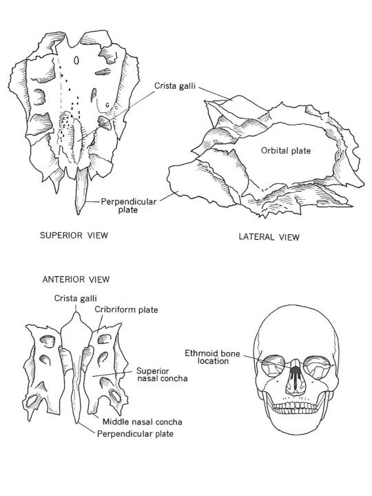
1. Ethmoid bone • Extremely complex (2 nd only to sphenoid), yet delicate (light weight); filled with air spaces Projects down from between the orbital plates, dividing nasal cavities (along with vomer bone) Contributes of walls of orbital and nasal cavities • •
![http www theodora comanatomy http: //www. theodora. com/anatomy/](https://slidetodoc.com/presentation_image_h/04546f097aab3246a5650f60aa7fd3ce/image-32.jpg)
http: //www. theodora. com/anatomy/
![http anatomy med umich edunervoussystemear html Scroll down and • http: //anatomy. med. umich. edu/nervous_system/ear. html – – – Scroll down and](https://slidetodoc.com/presentation_image_h/04546f097aab3246a5650f60aa7fd3ce/image-33.jpg)
• http: //anatomy. med. umich. edu/nervous_system/ear. html – – – Scroll down and click on skull structures Shows CT of head; ethmoid and maxillary sinuses displayed Can you name all of the bones shown below?
![](https://slidetodoc.com/presentation_image_h/04546f097aab3246a5650f60aa7fd3ce/image-34.jpg)
![Ethmoid parts A Cribriform plate separates cranial from nasal cavities • Ethmoid: parts – A. Cribriform plate = separates cranial from nasal cavities](https://slidetodoc.com/presentation_image_h/04546f097aab3246a5650f60aa7fd3ce/image-35.jpg)
• Ethmoid: parts – A. Cribriform plate = separates cranial from nasal cavities • Forms roof of nasal cavities • Houses olfactory bulbs • Pix below from csuchico website – B. Crista galli = projects upward from cribiform plate • Serves as attachment for falx cerebri – C. Middle and superior nasal conchae – D. Perpendicular plate = projects inferiorly • Superior bony part of nasal septum – E. Orbital plate
![Arrow indicates falx cerebri Arrow indicates falx cerebri](https://slidetodoc.com/presentation_image_h/04546f097aab3246a5650f60aa7fd3ce/image-36.jpg)
Arrow indicates falx cerebri
![Where is crista galli • Where is crista galli?](https://slidetodoc.com/presentation_image_h/04546f097aab3246a5650f60aa7fd3ce/image-37.jpg)
• Where is crista galli?
![2 Frontal bone Anterior cranial case forehead supraorbital region roof of orbital cavity 2. Frontal bone • Anterior cranial case, forehead, supraorbital region (roof of orbital cavity)](https://slidetodoc.com/presentation_image_h/04546f097aab3246a5650f60aa7fd3ce/image-38.jpg)
2. Frontal bone • Anterior cranial case, forehead, supraorbital region (roof of orbital cavity) In infancy, frontal bone was divided in two, with the metopic suture separating the halves KNOW this figure and the bones shown. From: www. upstate. edu/cdb/grossanat/hnskullant. shtml • •
![Frontal bone Parts A Superciliary arches B Glabella prominence above Frontal bone: Parts – A. Superciliary arches = – B. Glabella = prominence above](https://slidetodoc.com/presentation_image_h/04546f097aab3246a5650f60aa7fd3ce/image-39.jpg)
Frontal bone: Parts – A. Superciliary arches = – B. Glabella = prominence above nasal notch – C. Frontal sinuses = located behind superciliary arches glabella
![Can you locate the superciliary arches Can you locate the superciliary arches?](https://slidetodoc.com/presentation_image_h/04546f097aab3246a5650f60aa7fd3ce/image-40.jpg)
Can you locate the superciliary arches?
![3 Parietal bones Roof of cranium Joined by sagittal suture Separated • • 3. Parietal bones Roof of cranium Joined by sagittal suture Separated](https://slidetodoc.com/presentation_image_h/04546f097aab3246a5650f60aa7fd3ce/image-41.jpg)
• • 3. Parietal bones Roof of cranium Joined by sagittal suture Separated from occipital bone by lambdoidal suture • Laterally, separated from the temporal bones by the squamosal suture • Superior and inferior temporal lines • Pix from www. upstate. edu/cdb/grossanat/hnskullant. s html
![4 Occipital bone Lower and posterior portion of cranium Parts • 4. Occipital bone • Lower and posterior portion of cranium • Parts:](https://slidetodoc.com/presentation_image_h/04546f097aab3246a5650f60aa7fd3ce/image-42.jpg)
• 4. Occipital bone • Lower and posterior portion of cranium • Parts: – A. Foramen magnum = medulla oblongata passes through (from superior to inferior) and becomes spinal cord – B. Occipital protuberance = external midline projection that is visible from behind – C. Cerebellar fossa (internal structure); houses cerebellum – D. Condyles = articulate with C 1 Occipital protuberance
![](https://slidetodoc.com/presentation_image_h/04546f097aab3246a5650f60aa7fd3ce/image-43.jpg)
![5 Temporal bones Lateral base and sides of cranium Divided • 5. Temporal bones • Lateral base and sides of cranium • Divided](https://slidetodoc.com/presentation_image_h/04546f097aab3246a5650f60aa7fd3ce/image-44.jpg)
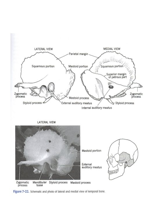
• 5. Temporal bones • Lateral base and sides of cranium • Divided into 4 parts • Parts: – A. Squamous: • zygomatic process (forms part of zygomatic arch • attachment for bulk of masseter muscle (muscle of mastication) • External auditory meatus • Mandibular fossa = forms TMJ with condyle of mandible – B. Mastoid • Attachment for neck muscles – C. Petrous = houses organs of hearing and equilibrium (see internal pix, next slide) – D. Styloid process (freq. missing in specimens); origin for several muscles
![](https://slidetodoc.com/presentation_image_h/04546f097aab3246a5650f60aa7fd3ce/image-45.jpg)
![6 Sphenoid bone Most complex of cranial bones Located posterior • 6. Sphenoid bone • Most complex of cranial bones • Located posterior](https://slidetodoc.com/presentation_image_h/04546f097aab3246a5650f60aa7fd3ce/image-46.jpg)
• 6. Sphenoid bone • Most complex of cranial bones • Located posterior to ethmoid anterior to foramen magnum • Likened to bar or butterfly
![Sphenoid Parts A Body or corpus view superiorly Sella turcica • Sphenoid: Parts – A. Body or corpus (view superiorly) • Sella turcica](https://slidetodoc.com/presentation_image_h/04546f097aab3246a5650f60aa7fd3ce/image-47.jpg)
• Sphenoid: Parts – A. Body or corpus (view superiorly) • Sella turcica (hypophyseal fossa)= houses pituitary gland • Optic nerve passes through body • Sphenoid sinuses located in body (air filled spaces) • Optic chiasm (directly anterior to hypophyseal fossa) located in chiasmatic groove. – B. Lesser and greater wings; comprise part of the orbital cavity – C. Lateral and medial pterygoid plates • Hamulus projects from each medial pterygoid plate; tendon of tensor veli palatini muscle rounds the hamulus as tensor changes direction from superior to horizontal orientation. • http: //www. a 3 bs. com/3 d/A 291/index. cfm
![Medial pterygoid plate Medial pterygoid plate](https://slidetodoc.com/presentation_image_h/04546f097aab3246a5650f60aa7fd3ce/image-48.jpg)
Medial pterygoid plate
![](https://slidetodoc.com/presentation_image_h/04546f097aab3246a5650f60aa7fd3ce/image-49.jpg)
![Dentition Function Mastication Surfaces for articulation of speech sounds • Dentition • Function: – Mastication – Surfaces for articulation of speech sounds](https://slidetodoc.com/presentation_image_h/04546f097aab3246a5650f60aa7fd3ce/image-50.jpg)
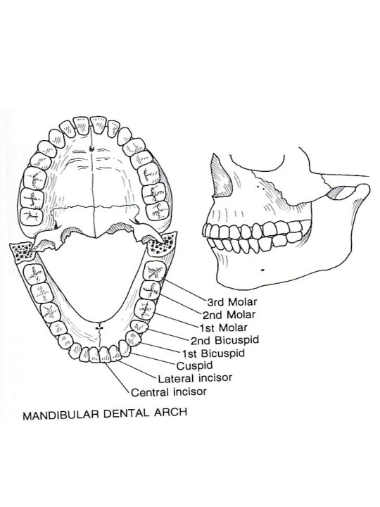
• Dentition • Function: – Mastication – Surfaces for articulation of speech sounds • Four types of teeth/number in permanent dental arch (mandible or maxilla) – Incisors (cutting) (4) – Cuspids/Canine (tearing) (2) – Bicuspids (two cusps) (4) – Molars (grinders) (6); 3 rd molars sometimes called wisdom teeth – Total # of permanent teeth: – Total # of deciduous teeth: – Teeth in the maxilla are larger than those in the mandible. – Maxillary teeth overlap mandibular teeth
![](https://slidetodoc.com/presentation_image_h/04546f097aab3246a5650f60aa7fd3ce/image-51.jpg)
![Surface terms Midline between central incisors Buccal toward cheek • Surface terms – Midline (between central incisors – Buccal (toward cheek) –](https://slidetodoc.com/presentation_image_h/04546f097aab3246a5650f60aa7fd3ce/image-52.jpg)
• Surface terms – Midline (between central incisors – Buccal (toward cheek) – Distal (away from central incisors) – Lingual (towards tongue) – Medial (towards midline) • Development – Deciduous teeth: • Lose incisors between 6 & 9 • Lose cuspids and molars 9 -12 • Lose 2 nd molars @ 10 • Teeth not in deciduous arch: • Teeth Anomalies (a few examples) – Supernumerary teeth – Microdontia (teeth smaller than normal)
![Occlusion alignment of maxillary teeth with mandibular teeth Types of • Occlusion = alignment of maxillary teeth with mandibular teeth. – Types of](https://slidetodoc.com/presentation_image_h/04546f097aab3246a5650f60aa7fd3ce/image-53.jpg)
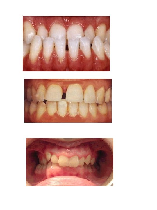
• Occlusion = alignment of maxillary teeth with mandibular teeth. – Types of occlusion-determined by alignment of 1 st molar in each arch. • Class I occlusion: (neutroclusion) cusp of the maxillary first molar occludes in the buccal groove of the mandibular molar. – Book definition = 1 st mandibular molar is ½ tooth advanced as compared to maxillary 1 st molar. – Max incisors overlap mandibular by a few millimeters – Considered to be the normal relationship between molars in the arches.
![Class II malocclusion the 1 st mandibular molar is retracted at least • Class II malocclusion: the 1 st mandibular molar is retracted at least](https://slidetodoc.com/presentation_image_h/04546f097aab3246a5650f60aa7fd3ce/image-54.jpg)
• Class II malocclusion: the 1 st mandibular molar is retracted at least one tooth from the 1 st maxillary molar • Class III malocclusion: 1 st mandibular molar is advanced farther than one tooth from 1 st mandibular molar.
![Class I malocclusion Other dental anomalies Overbitevertical dimension Overjethorizontal • Class I malocclusion: • Other dental anomalies – Overbite—vertical dimension – Overjet—horizontal](https://slidetodoc.com/presentation_image_h/04546f097aab3246a5650f60aa7fd3ce/image-55.jpg)
• Class I malocclusion: • Other dental anomalies – Overbite—vertical dimension – Overjet—horizontal dimension; refers to front teeth only – Diastema – Crossbite – Closed bite – Supraversion – Infraversion – Speech effects
![Name the occlusion Name the occlusion](https://slidetodoc.com/presentation_image_h/04546f097aab3246a5650f60aa7fd3ce/image-56.jpg)
Name the occlusion
![](https://slidetodoc.com/presentation_image_h/04546f097aab3246a5650f60aa7fd3ce/image-57.jpg)
![A C B D E A C B D E](https://slidetodoc.com/presentation_image_h/04546f097aab3246a5650f60aa7fd3ce/image-58.jpg)
A C B D E
![Cavities of Vocal Tract Oral pharyngeal nasal • Cavities of Vocal Tract – Oral, pharyngeal, nasal](https://slidetodoc.com/presentation_image_h/04546f097aab3246a5650f60aa7fd3ce/image-59.jpg)
• Cavities of Vocal Tract – Oral, pharyngeal, nasal
![Oral tractcavity Anterior faucial pillar Posterior faucial pillar Lingual frenum – Oral tract/cavity Anterior faucial pillar Posterior faucial pillar Lingual frenum](https://slidetodoc.com/presentation_image_h/04546f097aab3246a5650f60aa7fd3ce/image-60.jpg)
– Oral tract/cavity Anterior faucial pillar Posterior faucial pillar Lingual frenum
![Ankyloglossia Ankyloglossia](https://slidetodoc.com/presentation_image_h/04546f097aab3246a5650f60aa7fd3ce/image-61.jpg)
Ankyloglossia
![In little children the superior labial frenum is often attached between the two central In little children, the superior labial frenum is often attached between the two central](https://slidetodoc.com/presentation_image_h/04546f097aab3246a5650f60aa7fd3ce/image-62.jpg)
In little children, the superior labial frenum is often attached between the two central teeth. In most cases, this attachment migrates away from the teeth as the child approaches the age of 8 or 9.
![Pharyngeal cavity Nasopharynxspace above soft palate bounded anteriorly by nasal choanae choana – Pharyngeal cavity » Nasopharynx—space above soft palate (bounded anteriorly by nasal choanae) (choana](https://slidetodoc.com/presentation_image_h/04546f097aab3246a5650f60aa7fd3ce/image-63.jpg)
– Pharyngeal cavity » Nasopharynx—space above soft palate (bounded anteriorly by nasal choanae) (choana = funnel) » Association between removal of adenoids and VPI » Oropharynx (bounded anteriorly by fauces/faucial pillars; superiorly by ; inferiorly by hyoid bone » Laryngopharynx (bounded inferiorly by esophagus Pharyngeal tonsil or adenoids Eustachian tube orifice laryngopharynx
![Facial muscles Facial landmarks Cupids bow Vermilion Lips • Facial muscles • Facial landmarks: – Cupids bow – Vermilion – Lips:](https://slidetodoc.com/presentation_image_h/04546f097aab3246a5650f60aa7fd3ce/image-64.jpg)
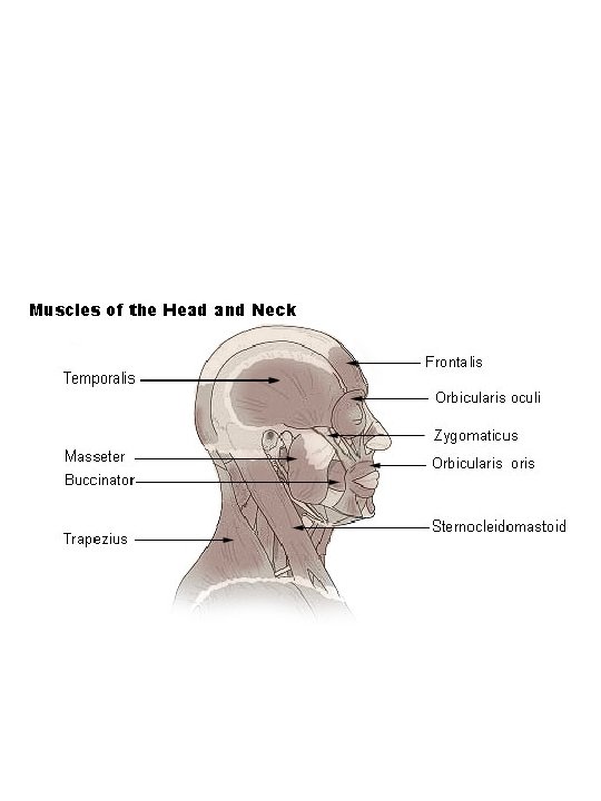
• Facial muscles • Facial landmarks: – Cupids bow – Vermilion – Lips: many facial muscles insert into lips • Important for articulation of bilabials and labiodentals philtrum
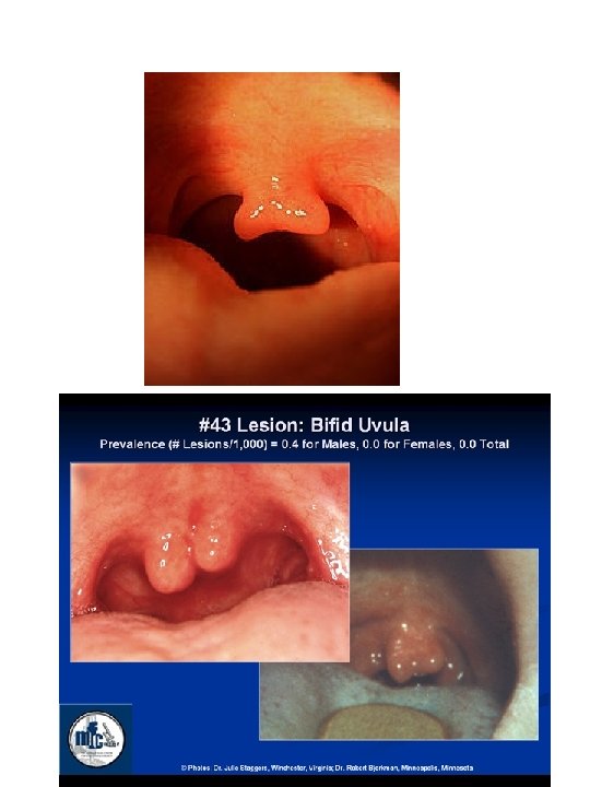
![Bifid uvula Bifid uvula](https://slidetodoc.com/presentation_image_h/04546f097aab3246a5650f60aa7fd3ce/image-65.jpg)
Bifid uvula
![Orbicularis oris principle muscle acting on lips no origin or insertion • Orbicularis oris: principle muscle acting on lips – no origin or insertion](https://slidetodoc.com/presentation_image_h/04546f097aab3246a5650f60aa7fd3ce/image-66.jpg)
• Orbicularis oris: principle muscle acting on lips – no origin or insertion – Main function is closing mouth, puckering lips – Many muscles insert into the orbicularis oris
![2 transverse muscles Buccinator muscle transverse deep m buglers muscle • 2 transverse muscles • Buccinator muscle (transverse) (deep m) (bugler’s muscle) –](https://slidetodoc.com/presentation_image_h/04546f097aab3246a5650f60aa7fd3ce/image-67.jpg)
• 2 transverse muscles • Buccinator muscle (transverse) (deep m) (bugler’s muscle) – Principle muscle of cheek – Action: compresses cheek, draws corner of mouth laterally – Masticatory func: keeps foods from slipping out between teeth while tongue works to keep food between grinding surfaces. – #37
![](https://slidetodoc.com/presentation_image_h/04546f097aab3246a5650f60aa7fd3ce/image-68.jpg)
![Risorius muscle transverse L risus laughter Runs parallel and superior • Risorius muscle (transverse) (L risus = laughter) – Runs parallel and superior](https://slidetodoc.com/presentation_image_h/04546f097aab3246a5650f60aa7fd3ce/image-69.jpg)
• Risorius muscle (transverse) (L risus = laughter) – Runs parallel and superior to buccinator – Action: retracts angle of mouth; – Action: when contracting in conjunction with zygomatic muscle results in smile – #38
![Angular muscles Zygomatic Minor Origin malar surface of zygomatic • Angular muscles • Zygomatic Minor: – Origin = malar surface of zygomatic](https://slidetodoc.com/presentation_image_h/04546f097aab3246a5650f60aa7fd3ce/image-70.jpg)
• Angular muscles • Zygomatic Minor: – Origin = malar surface of zygomatic bone-courses downward and medially – Function: elevation of upper lip • Zygomatic Major: – Origin = lateral to zygomatic minor on malar surface of zygomatic bone--inserts into corner of mouth – ACTION = draws corner of mouth up and backward = grinning, smiling Zygomatic minor
![Levator Labii Superior Origin lower border of orbit mainly some • Levator Labii Superior: – Origin = lower border of orbit mainly (some](https://slidetodoc.com/presentation_image_h/04546f097aab3246a5650f60aa7fd3ce/image-71.jpg)
• Levator Labii Superior: – Origin = lower border of orbit mainly (some fibers from frontal process of maxilla & some from zygomatic bone). Inserts near median of upper lip. – ACTION = elevator of upper lip & may evert it slightly L labii superior Zygomatic minor risorius Zygomatic major
![Levator Labaii Superior Alaeque Nasi M origin frontal process • Levator Labaii Superior Alaeque Nasi M. : – origin = frontal process](https://slidetodoc.com/presentation_image_h/04546f097aab3246a5650f60aa7fd3ce/image-72.jpg)
• Levator Labaii Superior Alaeque Nasi M. : – origin = frontal process of maxilla & infraorbital margin; courses downward & slightly laterally; 2 insertions = lateral portion of nose & o. oris – ACTION = elevator of upper lip and dilator of nostrils L l superior alaque nasi m
![Depressor Labii Inferior origin oblique line of mandible inserts into • Depressor Labii Inferior: – origin = oblique line of mandible, inserts into](https://slidetodoc.com/presentation_image_h/04546f097aab3246a5650f60aa7fd3ce/image-73.jpg)
• Depressor Labii Inferior: – origin = oblique line of mandible, inserts into lower lip lateral to midline. – ACTION = depresses lower lip D labii inferior
![Vertical muscles Mentalis origin mental turberosity inserts some • Vertical muscles • Mentalis: – origin = mental turberosity; inserts = some](https://slidetodoc.com/presentation_image_h/04546f097aab3246a5650f60aa7fd3ce/image-74.jpg)
• Vertical muscles • Mentalis: – origin = mental turberosity; inserts = some fibers insert into contralateral Mentalis, some into skin of chin, some into o. oris. – ACTION = wrinkles chin, everts lower lip mentalis
![Depressor Anguli Oris superficial origin oblique line insertion • Depressor Anguli Oris = – superficial; – origin = oblique line; insertion](https://slidetodoc.com/presentation_image_h/04546f097aab3246a5650f60aa7fd3ce/image-75.jpg)
• Depressor Anguli Oris = – superficial; – origin = oblique line; insertion = corner of mouth; – ACTION = depressor of angle of mouth, compresses lips by pulling upper lip downward onto lower lip D anguli oris
![Levator Anguli Oris deep m origin canine • Levator Anguli Oris = – deep m. ; – origin = canine](https://slidetodoc.com/presentation_image_h/04546f097aab3246a5650f60aa7fd3ce/image-76.jpg)
• Levator Anguli Oris = – deep m. ; – origin = canine fossa (pit or hollow just lateral to canine eminence); insertion = corner of mouth at upper lip; – ACTION = draws corners of mouth up and medially. L anguli oris
![FYI Anatomically dimples may be caused by variations in the structure of the facial FYI Anatomically, dimples may be caused by variations in the structure of the facial](https://slidetodoc.com/presentation_image_h/04546f097aab3246a5650f60aa7fd3ce/image-77.jpg)
FYI Anatomically, dimples may be caused by variations in the structure of the facial muscle known as zygomaticus major. Specifically, the presence of a double or bifid zygomaticus major muscle may explain the formation of cheek dimples. [2] This bifid variation of the muscle originates as a single structure from the zygomatic bone. As it travels anteriorly, it then divides with a superior bundle that inserts in the typical position above the corner of the mouth. An inferior bundle inserts below the corner of the mouth.
![http www ivyrose co ukTopicsFacial Muscles htm The above cited website • • http: //www. ivy-rose. co. uk/Topics/Facial. Muscles. htm The above cited website](https://slidetodoc.com/presentation_image_h/04546f097aab3246a5650f60aa7fd3ce/image-78.jpg)
• • http: //www. ivy-rose. co. uk/Topics/Facial. Muscles. htm The above cited website provides a good test of your knowledge about facial muscles. • Tongue = biol. func. = taste (complemented by smell), mastication (moves food between teeth for grinding, forms bolus, and propels bolus into pharynx), & deglutition (swallowing) • Most imp and most active articulator • Capable of rapid and subtle movements due to high innervation and complex arrangement of muscle fibers. • All muscles innervated by C XII; hypoglossal cranial nerve
![Regions of tongue tipnearest central incisors Bladejust below alveolar ridge • Regions of tongue: – tip—nearest central incisors – Blade—just below alveolar ridge](https://slidetodoc.com/presentation_image_h/04546f097aab3246a5650f60aa7fd3ce/image-79.jpg)
• Regions of tongue: – tip—nearest central incisors – Blade—just below alveolar ridge – Front—below hard palate (dorsum) – Back—below soft palate
![Taste buds Anterior sweet salty posterior bitter • • Taste buds: – Anterior: sweet & salty (posterior = bitter) –](https://slidetodoc.com/presentation_image_h/04546f097aab3246a5650f60aa7fd3ce/image-80.jpg)
• • Taste buds: – Anterior: sweet & salty (posterior = bitter) – Sides: sour – Terminal sulcus Foramen cecum = pit Sulcus terminalis = separates anterior 2/3 from posterior 1/3 Vallate papillae: V-shaped row, anterior to foramen cecum ?
![Planecut shown below Pix from http www meddean luc edulumenmededgrossana • Plane/cut shown below? • Pix from: • http: //www. meddean. luc. edu/lumen/meded/grossana](https://slidetodoc.com/presentation_image_h/04546f097aab3246a5650f60aa7fd3ce/image-81.jpg)
• Plane/cut shown below? • Pix from: • http: //www. meddean. luc. edu/lumen/meded/grossana tomy/dissector/ Try the Exams, Skull Practical. Only focus on structures that are named in the notes.
![Intrinsic muscles Overall function shaping tongue Superior longitudinal • Intrinsic muscles • Overall function = shaping tongue – Superior longitudinal •](https://slidetodoc.com/presentation_image_h/04546f097aab3246a5650f60aa7fd3ce/image-82.jpg)
• Intrinsic muscles • Overall function = shaping tongue – Superior longitudinal • Upper layer of tongue • Originates near epiglottis; inserts into tongue apex and lateral margins • Action: elevate tip – Inferior longitudinal • Origin: root of tongue & corpus of hyoid; inserts into tongue apex • Lower tongue base; absent in medial tongue base (genioglossus occupies medial area) • Action: tongue turns downward – Transverse • Action: narrow tongue – Vertical -- depression of tongue dorsum • Flatten tongue
![Tongue anomalies Microglossia Macroglossia e g BeckwithWiedemann syndrome • Tongue anomalies – Microglossia – Macroglossia (e. g. , Beckwith-Wiedemann syndrome –](https://slidetodoc.com/presentation_image_h/04546f097aab3246a5650f60aa7fd3ce/image-83.jpg)
• Tongue anomalies – Microglossia – Macroglossia (e. g. , Beckwith-Wiedemann syndrome – Bifid tongue – Pseudomacroglossia (associated with micrognathia)
![1 Extrinsic tongue muscles Function general movement of tongue Genioglossus • • 1. Extrinsic tongue muscles Function = general movement of tongue Genioglossus](https://slidetodoc.com/presentation_image_h/04546f097aab3246a5650f60aa7fd3ce/image-84.jpg)
• • 1. Extrinsic tongue muscles Function = general movement of tongue Genioglossus (pix www. answers. com) • Prime mover of tongue • Comprises bulk of tongue tissue • Strongest and largest of extrinsic muscles • Flat, triangular in shape • Origin at symphysis; fans out; inserts into tip and dorsum of tongue • Function: anterior portion retracts tongue; posterior fibers protrude tongue; together fibers depress tongue
![2 Hyoglossus Origin hyoid insertion sides of tongue Function pulls sides of 2. Hyoglossus • Origin: hyoid; insertion: sides of tongue • Function: pulls sides of](https://slidetodoc.com/presentation_image_h/04546f097aab3246a5650f60aa7fd3ce/image-85.jpg)
2. Hyoglossus • Origin: hyoid; insertion: sides of tongue • Function: pulls sides of tongue down; retracts and depresses tongue; raises hyoid • I’m considering the chondroglossus m. part of the hyoglossus. Chondro m. arises from lesser cornu
![3 Styloglossus Origin styloid process of insertion inferior sides 3. Styloglossus • • • Origin: styloid process of ? ; insertion: inferior sides](https://slidetodoc.com/presentation_image_h/04546f097aab3246a5650f60aa7fd3ce/image-86.jpg)
3. Styloglossus • • • Origin: styloid process of ? ; insertion: inferior sides of tongue Action: draws tongue back and up Pix: http: //webpages. charter. net/reinerwt/themodel. htm
![4 palatoglossus Commonly called anterior faucial pillar Can be considered • 4. palatoglossus – Commonly called anterior faucial pillar – Can be considered](https://slidetodoc.com/presentation_image_h/04546f097aab3246a5650f60aa7fd3ce/image-87.jpg)
• 4. palatoglossus – Commonly called anterior faucial pillar – Can be considered as a muscle of tongue or palate – Origin: anterior surface of soft palate; Insertion: sides of tongue – Function: elevates back of tongue, closes oropharyngeal isthmus and aids initiation of swallowing ; depresses soft palate – Motor innervation: C XI: Accessory
![Muscles of mastication Function elevate or depress mandible These • Muscles of mastication • • Function = elevate or depress mandible These](https://slidetodoc.com/presentation_image_h/04546f097aab3246a5650f60aa7fd3ce/image-88.jpg)
• Muscles of mastication • • Function = elevate or depress mandible These muscles are one of the most strongest in the body • http: //www. dentalwisdom. com/animationstudio/flashpla yer 28. html – View film on TMJ Elevators 1. Masseter • Most superficial muscle of mastication • Origin = zygomatic arch; insertion = ramus of mandible • Action = elevates mandible
![2 Temporalis muscle Fanshaped muscle Origin temporal fossa of temporal 2. Temporalis muscle • • • Fan-shaped muscle Origin = temporal fossa of temporal](https://slidetodoc.com/presentation_image_h/04546f097aab3246a5650f60aa7fd3ce/image-89.jpg)
2. Temporalis muscle • • • Fan-shaped muscle Origin = temporal fossa of temporal and parietal bones Insertion = coronoid process and ramus Function = elevates mandible and draws it back if protruded It is considered a snapping muscle, built for speed (tearing food)
![3 Medial internal Pterygoid muscle Origin pterygoid plate and fossa sphenoid bone 3. Medial (internal) Pterygoid muscle • Origin = pterygoid plate and fossa (sphenoid bone)](https://slidetodoc.com/presentation_image_h/04546f097aab3246a5650f60aa7fd3ce/image-90.jpg)
3. Medial (internal) Pterygoid muscle • Origin = pterygoid plate and fossa (sphenoid bone) • Forms mandibular sling with mandible (angle of mandible rests in the sling) • Function = elevates mandible
![Protruder 4 Lateral external Pterygoid muscle Origin Has two heads sphenoid pterygoid plate Protruder 4. Lateral (external) Pterygoid muscle • Origin: Has two heads (sphenoid— pterygoid plate](https://slidetodoc.com/presentation_image_h/04546f097aab3246a5650f60aa7fd3ce/image-91.jpg)
Protruder 4. Lateral (external) Pterygoid muscle • Origin: Has two heads (sphenoid— pterygoid plate and greater wing); insertion = mandible • Function = protrudes mandible
![Depressors 5 Digastricus Has anterior and posterior belly united by central tendon Depressors 5. Digastricus • Has anterior and posterior belly, united by central tendon •](https://slidetodoc.com/presentation_image_h/04546f097aab3246a5650f60aa7fd3ce/image-92.jpg)
Depressors 5. Digastricus • Has anterior and posterior belly, united by central tendon • • • Posterior belly originates from mastoid process and inserts onto corpus of hyoid bone where it joins with the anterior belly by means of the central tendon. Anterior belly originates from mandibular symphysis, goes to central tendon, Function • • Anterior = elevates hyoid and pulls it forward; depresses mandible when contracts in conjunction with posterior belly Posterior = pulls hyoid backward and upward; depresses mandible when contracts with anterior belly
![6 Mylohyoid Origin mylohyoid line Insertion Median raphe and hyoid 6. Mylohyoid • • Origin = mylohyoid line Insertion = Median raphe and hyoid](https://slidetodoc.com/presentation_image_h/04546f097aab3246a5650f60aa7fd3ce/image-93.jpg)
6. Mylohyoid • • Origin = mylohyoid line Insertion = Median raphe and hyoid Function = depresses mandible Forms floor of the mouth
![7 Geniohyoid Origin mental spine on inner aspect of mental symphysis just 7. Geniohyoid • Origin = mental spine on inner aspect of mental symphysis (just](https://slidetodoc.com/presentation_image_h/04546f097aab3246a5650f60aa7fd3ce/image-94.jpg)
7. Geniohyoid • Origin = mental spine on inner aspect of mental symphysis (just superior to mylohyoid m) • Insertion = hyoid bone • Action/Function = assist in elevating larynx or depressing mandible
![8 Platysma Considered a facial muscle embryologically developed from same primordia from which 8. Platysma • Considered a facial muscle (embryologically developed from same primordia from which](https://slidetodoc.com/presentation_image_h/04546f097aab3246a5650f60aa7fd3ce/image-95.jpg)
8. Platysma • Considered a facial muscle (embryologically developed from same primordia from which facial muscles originated) • Covers most of the lateral and anterior regions of neck • Action = mandibular depressor • Go to www. innerbody. com
![Muscles of the Velum Levator veli palatini Musculus uvulae Tensor veli Muscles of the Velum • • • Levator veli palatini Musculus uvulae Tensor veli](https://slidetodoc.com/presentation_image_h/04546f097aab3246a5650f60aa7fd3ce/image-96.jpg)
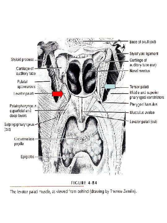
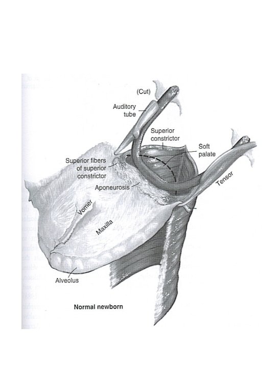
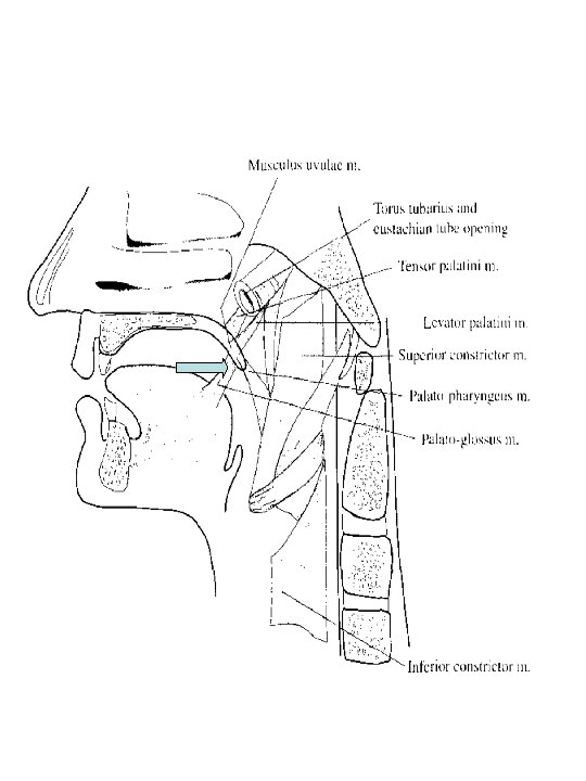
Muscles of the Velum • • • Levator veli palatini Musculus uvulae Tensor veli palatini Palatoglossus Palatopharyngeus • 1. levator veli palatini – Function: elevates and retracts soft palate or velum – Origin = petrous portion of temporal bone and wall of Eustachian tube. – Courses= downward and forward – Inserts into member from other side – Innervation= C XI (accessory) and CX (vagus)
![](https://slidetodoc.com/presentation_image_h/04546f097aab3246a5650f60aa7fd3ce/image-97.jpg)
![2 Musculus uvulae Paired muscle sometimes referred to as one muscle • 2. Musculus uvulae – Paired muscle (sometimes referred to as one muscle)](https://slidetodoc.com/presentation_image_h/04546f097aab3246a5650f60aa7fd3ce/image-98.jpg)
• 2. Musculus uvulae – Paired muscle (sometimes referred to as one muscle) – Function: • Shortens soft palate, creating bunching or bulk on nasal side that aids in velopharygeal closure, • and helps to elevate the soft palate. – Origin: posterior nasal spines of ? and from palatal aponeurosis. – Courses: near midline in soft palate Insertion: near base of uvula (midline pendulous structure) – Bifid uvula occurs in 1/75 people
![](https://slidetodoc.com/presentation_image_h/04546f097aab3246a5650f60aa7fd3ce/image-99.jpg)
![3 Tensor Veli Palatini Origin sphenoid bone Insertion converges on tendon tendon • 3. Tensor Veli Palatini Origin: sphenoid bone Insertion: converges on tendon; tendon](https://slidetodoc.com/presentation_image_h/04546f097aab3246a5650f60aa7fd3ce/image-100.jpg)
• 3. Tensor Veli Palatini Origin: sphenoid bone Insertion: converges on tendon; tendon rounds hamulus and attaches to palatine bones and inserts into palatine aponeurosis Function: acts on palatine aponeurosis to produce taut palate--TVP is active during inspiration, producing a taut palate; taut palate is more desirable for unimpeded airflow; A floppy palate would be more prone to make pharyngeal wall contact, esp. in the supine position; opens e. tube permitting press. equalization within middle ear cavity. Innervation: CN V, the trigeminal nerve
![](https://slidetodoc.com/presentation_image_h/04546f097aab3246a5650f60aa7fd3ce/image-101.jpg)
![4 Palatoglossus anterior faucial pillars Origin anterior surface of soft palatein • 4. Palatoglossus (anterior faucial pillars) – Origin: anterior surface of soft palate--in](https://slidetodoc.com/presentation_image_h/04546f097aab3246a5650f60aa7fd3ce/image-102.jpg)
• 4. Palatoglossus (anterior faucial pillars) – Origin: anterior surface of soft palate--in most individuals, attachments nearer uvula than rim of hard palate (suggests may have very limited ability to elevate tongue) – Insertion: dorsum and side of tongue – Function: velar lowering, i. e. drawing velum anteriorly; tongue upward and posterior movement, and constriction of anterior faucial pillars (helps propel food bolus); speech function remains unresolved; may be important in terms of opposing gravity (i. e. sleeping in the supine position). The elasticity of the palatoglossus would tend to keep the airway patent without m. contraction. Changes in elastic fibers occur with age, they get more lax. The result may be more mouth breathing and snoring in older individuals.
![](https://slidetodoc.com/presentation_image_h/04546f097aab3246a5650f60aa7fd3ce/image-103.jpg)
![5 Palatopharyngeus posterior faucial pillars Origin Fanshaped in soft palate • 5. Palatopharyngeus (posterior faucial pillars) – Origin: Fan-shaped in soft palate –](https://slidetodoc.com/presentation_image_h/04546f097aab3246a5650f60aa7fd3ce/image-104.jpg)
• 5. Palatopharyngeus (posterior faucial pillars) – Origin: Fan-shaped in soft palate – Insertion: Tapers to termination in posterior border of thyroid cartilage of larynx and into pharynx – Function: Active during orally produced, velum elevated speech sounds (although anatomically situated to lower the velum)-may function to make subtle adjustments of velar height when velum in elevated state; lowers the velum vertically; decrease distance between faucial pillars (action vigorous during swallowing and gagging). – Innervation: CN XI and X
Ellipsoidal joint example
What is a non movement area
What are the example of axial movement
What are the 4 processes of speech production?
Balanced articulation
Phonation and articulation
Articulation points and biconnected components
Hình ảnh bộ gõ cơ thể búng tay
Slidetodoc
Bổ thể
Tỉ lệ cơ thể trẻ em
Gấu đi như thế nào
Tư thế worms-breton
Chúa yêu trần thế
Các môn thể thao bắt đầu bằng từ đua
Thế nào là hệ số cao nhất
Các châu lục và đại dương trên thế giới
Cong thức tính động năng
Trời xanh đây là của chúng ta thể thơ
Mật thư tọa độ 5x5
Phép trừ bù
Phản ứng thế ankan
Các châu lục và đại dương trên thế giới
Thể thơ truyền thống
Quá trình desamine hóa có thể tạo ra
Một số thể thơ truyền thống
Cái miệng nó xinh thế
Vẽ hình chiếu vuông góc của vật thể sau
Biện pháp chống mỏi cơ
đặc điểm cơ thể của người tối cổ
Ví dụ giọng cùng tên
Vẽ hình chiếu đứng bằng cạnh của vật thể
Phối cảnh
Thẻ vin
đại từ thay thế
điện thế nghỉ
Tư thế ngồi viết
Diễn thế sinh thái là
Dot
Số nguyên tố là số gì
Tư thế ngồi viết
Lời thề hippocrates
Thiếu nhi thế giới liên hoan
ưu thế lai là gì
Sự nuôi và dạy con của hổ
Khi nào hổ con có thể sống độc lập
Hệ hô hấp
Từ ngữ thể hiện lòng nhân hậu
Thế nào là mạng điện lắp đặt kiểu nổi
Double articulation
What is this language
Vdoe vertical articulation
Articulation costo corporéale
Phonemes in spanish
Language
Pure vowels
Co-articulation
Language
Place of articulation consonants
Ischial spine
Quoll pronunciation
Indiana math framework
Manner of articulation
Fricative sounds
Manner of articulation definition
Ligament jugal
Double articulation
Articulation du lisfranc
Articulation in solution design document
Vertical articulation example
Co-articulation
Laminal sounds
Consonants place of articulation
Articulation gynglime
Rib vertebrae articulation
Schyndilèse
Vertical articulation template
Articulation interchondrale
Bones of the forehead
Lappareil locomoteur
They are called gliding vowels
Nasal voice
Kość klinowa budowa
Steep incisal guidance
The arrangement of the element of curriculum
What is an articulation point on a bone
Arrangement of curriculum
Caap articulation test
Cubiti
Articulation sports med
Manner of articulation
Tongue articulation
Articulation mechanism
Mode of articulation
Flexion du pied
Manner of articulation
Balanced articulation
Oacac articulation
M. articularis cubiti
Language
Bowen screener
Auscultatory triangle
Les os du pied humain
Voiceless sounds