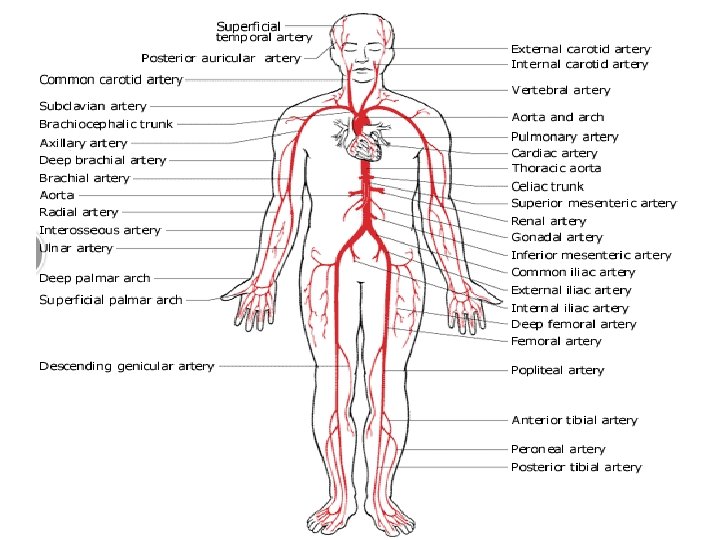
Arteries of lower extremity Superior gluteal artery Branch

Arteries of lower extremity ØSuperior gluteal artery: • Branch of posterior div. of internal iliac artery • Enters the gluteal region through greater sciatic foramen passing above the piriformis. • In the foramen it divides into superficial & deep branches. • Superficial branch supplies gluteus max. • Deep branch subdivides into sup. & inf. branches

Arteries of gluteal region Ø Inferior gluteal artery • Branch of ant. Div. of internal iliac artery • Enters gluteal region by passing through greter sciatic foramen below the piriformis. It Supplies • Musular branches • Cutaneous branches to buttock & back of thigh • Articular branch to hip joint • Artery to sciatic nerve • Cruciate anastamotic branch • Coccygeal branch

Arteries of gluteal region Ø Internal pudendal artery • Branch of ant. Div of internal iliac artey • Enters gluteal region through greater sciatic foramen

Trochanteric anastomosis This anastomosis is a channel of communication between internal iliac and femoral arteries. Situated near the trochanteric fossa • Supplies branches to head of femur • Inf. Div. of deep branch of sup. Gluteal artery • Ascending branch of medial circumflex femoral artery • Ascending branch of lateral circumflex femoral artery • Often the inf. Gluteal artery

Cruciate anastomosis • Situated at the level of lesser trochanter of femur Formed by • Inferior gluteal artery • Medial femoral circumflex artery • Lateral femoral circumflex artery • First perforating artery, a branch of profunda femoris artery
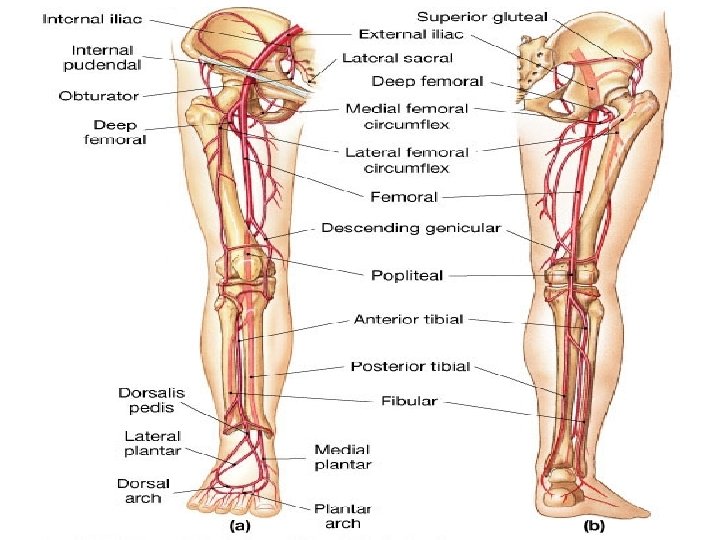
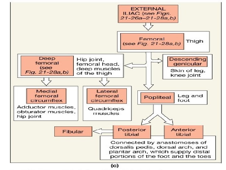
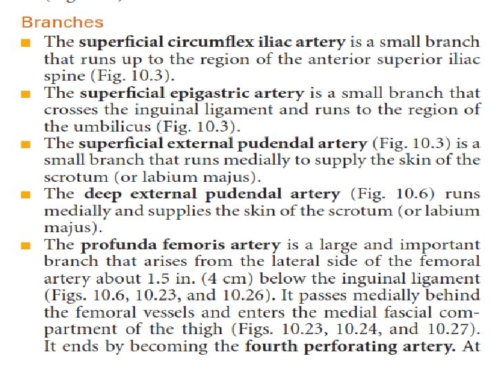
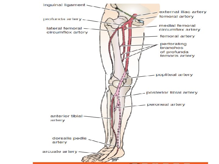

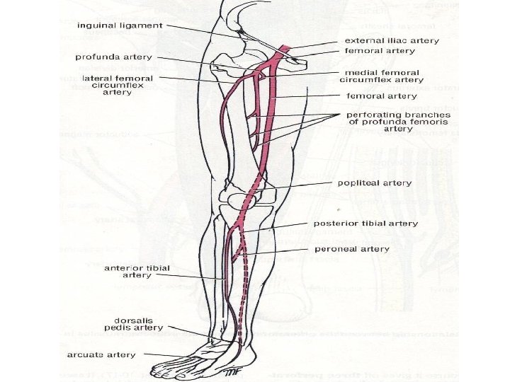
Profunda femoris • Largest branch of femoral artery • Main artery of all three compartments of thigh • Arises from lateral side of femoral artery 4 cm below the inguinal ligament • Branches: • Medial circumflex femoral artery • Lateral circumflex femoral artery • 4 perforating arteries

Profunda femoris & its branches

Popliteal artery • Continuation of femoral artery • Begins at the opening in the adductor magnus • Runs downwards &laterally to reach lower border of popliteus • At that point it terminates by dividing into ant. &post. Tibial arteries • Branches • Muscular : supply adductor magnus & hamstrings , terminate by anastomosing with 4 th perforating artery

Popliteal artery • Cutaneous: accompany small saphanous vein • Genicular: 5 in number • 2 superior: medial & lateral superior genicular arteries • 2 inferior: medial & lateral inferior genicular arteries • 1 middle

Anterior tibial artery • Smaller terminal branch • Passes into ant. Compartment by passing through gap in the upper part of interosseous membrane • Deep in the upper part of course • Supply adjacent muscles • Superficial in the lower part • passes behind the extensor retinaculum, has ext. hallucis longus tendon on its medial side and deep peroneal nerve and extensor digitorum longus on its lateral side • On entering the ankle region it becomes dorsalis pedis artery

Anterior tibial artery • Branches • Muscular branches • Anastomotic branches with branches of other arteries around knee and ankle joint • Vanea commitantes of anterior tibial artery joins with those of posterior tibial artery in the popliteal fossa to form popliteal vein

Dorsalis pedis artery • It terminates by passing downward into the sole between the two heads of 1 st dorsal interosseous muscle • Joins lateral planter artery to form planter arch • Branches 1) Lateral tarsal artery 2) Arcuate artery (gives off dorsal metatarsal artery) 3) 1 st dorsal metatarsal artery

Posterior tibial artery • • • Branches Peroneal artery Muscular branch Nutrient artery to tibia Anastomotic branch circumflex fibular branch malleolar branch calcaneal branch terminal branches Medial & lateral planter arteries
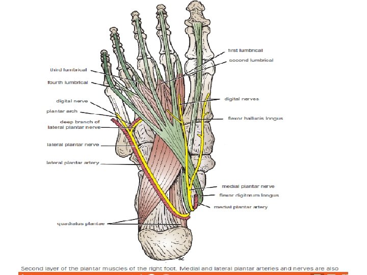

Ø Medial planter artery gives off muscular to the adjoining muscles 3 small superficial digital branches Ø lateral planter artery larger terminal branch ends at the base of 5 th metatarsal bone by continuing with planter arch branches 1) muscular branches 2)superficial branches supplying skin & 3)anastomotic branches 4) Calcaneal branch
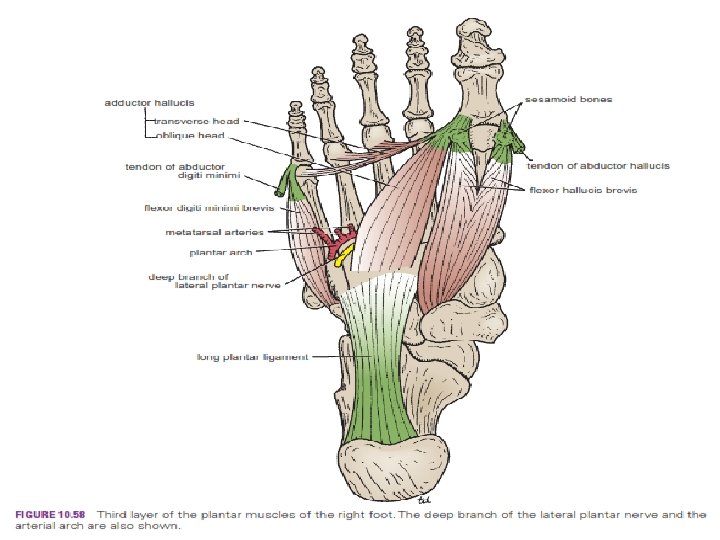

Planter arch • Formed by direct continuation of lateral planter artery. • Completed medially by dorsalis pedis artery • Extends from base of 5 th metatarsal bone to the proximal part of 1 st intermetatarsal space • Lies between 3 rd & 4 th layer of sole • Accompanied by vanea commitantes & deep branch of lateral planter nerve

Planter arch Branches 4 planter metatarsal arteries 3 proximal perforating arteries Communicate with the dorsal metatarsal arteries (branches of arcuate artery) • Distal end of each planter metatarsal artery gives of distal perforating artery • •
- Slides: 25