ARTERIES AND VEINS OF SCALP Venous sinuses 1


 Confluence of")


- Slides: 37
ARTERIES AND VEINS OF SCALP
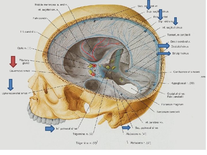
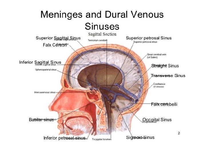
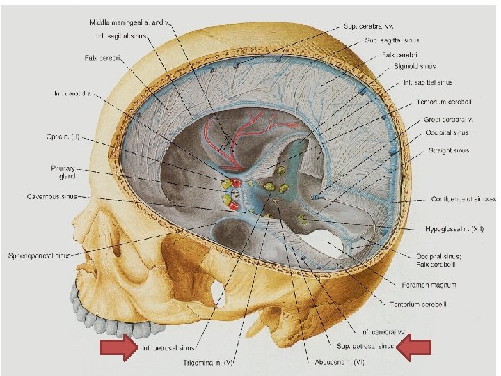
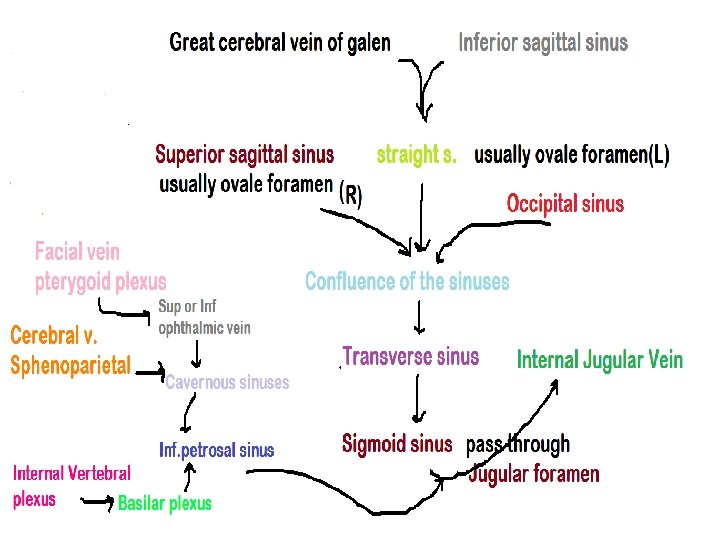
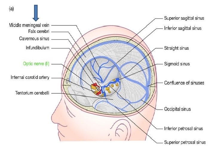
Venous sinuses • • • 1 -Sup. sagital sinus 2 -Inf. sagital sinus 3 -Sigmoid sinuses 4 -Occipital sinus 5 -Transverse sinuses 6 -Straight sinus 7 -Cavernous sinuses 8 -Intercavernous sinuses 9 -Sphenoparietal sinuses 10 -Sup. petrosal sinuses 11 -Inf. petrosal sinuses
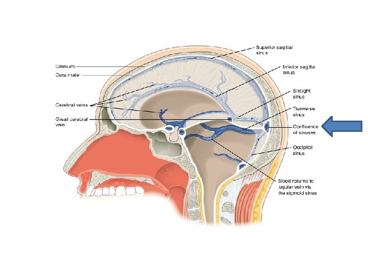
A little more about venous sinuses Rectus sinus(in base of falx cerebri) Confluence of transverse sinus(made of rectus sinus +sup. sagital sinus) Occipital sinus(located in the place that falx cerebelli and internal occipital crest linked together) and finally goes to confluence of sinuses
Confluence of sinuses A place that sup. sagital sinus, rectus sinus, occipital sinus, right and left taransverse sinuses join together. This dilatation is located near by internal occipital protuberance.
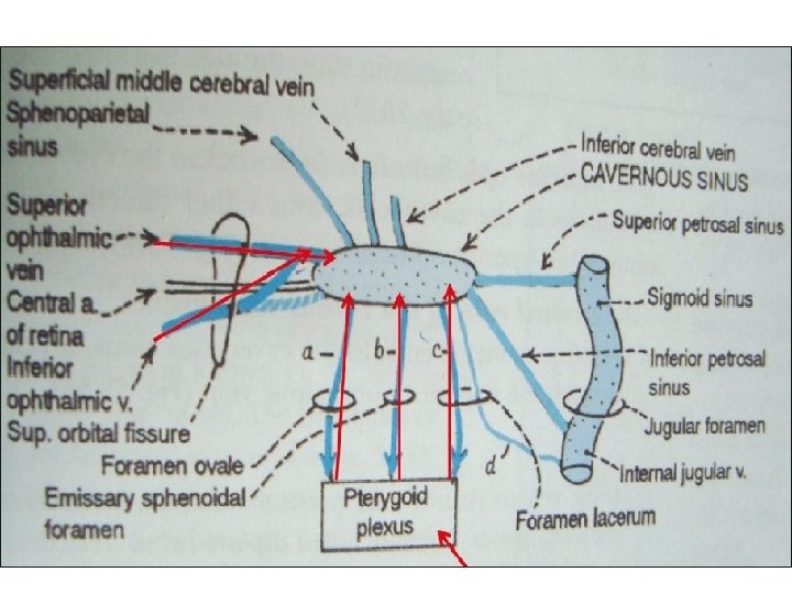
Inter cavernous • Intercavernous: 2 little vein in forward and backward of pituitary gland that connect cavernous sinuses together.
Superior and inferior petrosal sinuses • Sup. petrosal sinus: on the superior border of petros, started from cavernous sinus and ended in junction of transverse and sigmoid sinuses. • Inf. petrosal sinus: located in petrooccipital fissure. this sinus connect cavernous sinus to superior dilatation of internal jugular vein. right and left inf. petrosal sinus connect together by basilar plexus.
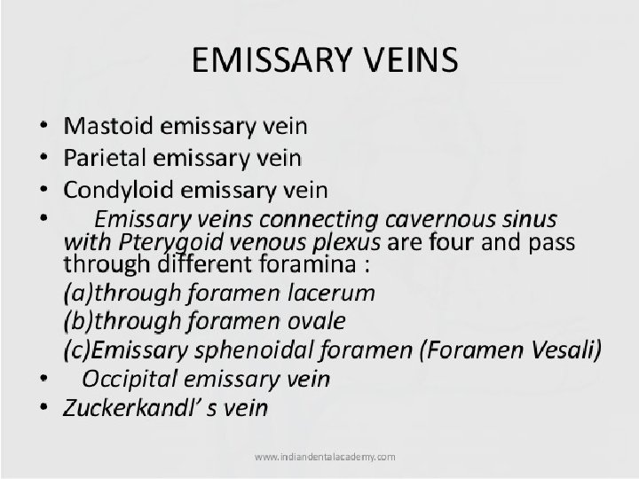
Emissary veins Connect the extracranial venous system with the intracranial venous system. it means they connect veins in outside of cranium to veins inside the cranium. There also emissary veins passing through the foramen ovale, jugular foramen, foramen lacerum Because the emissary veins are valveless they are an important part in selective brain cooling bidirectional flow of cooler blood from evaporat. Ing surface of the head. in general blood flow is from external to internal but the flow can be altered by increased intracranial pressure.
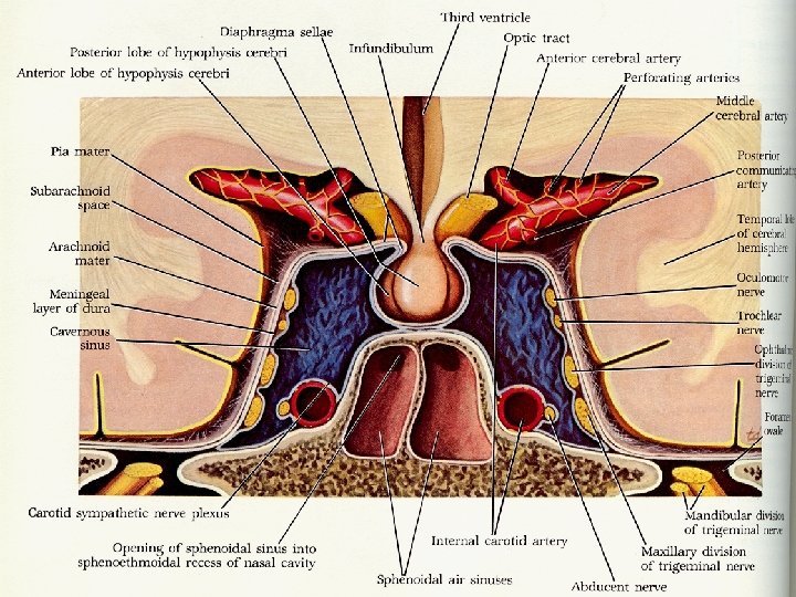
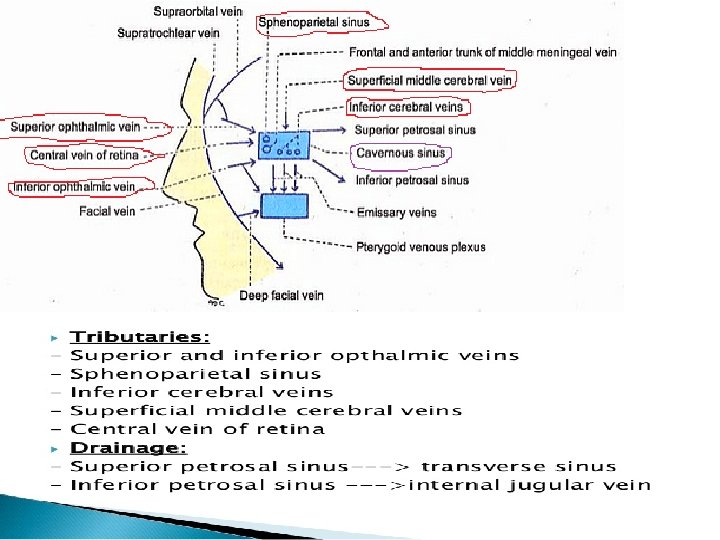
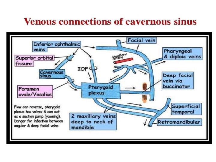
CAVERNOUS SINUSES • Pair sinuses, located in carotid groove in lateral side of body of sphenoid.
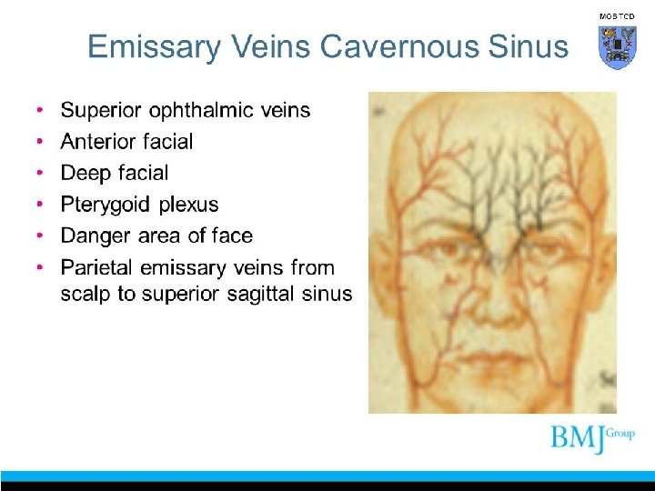
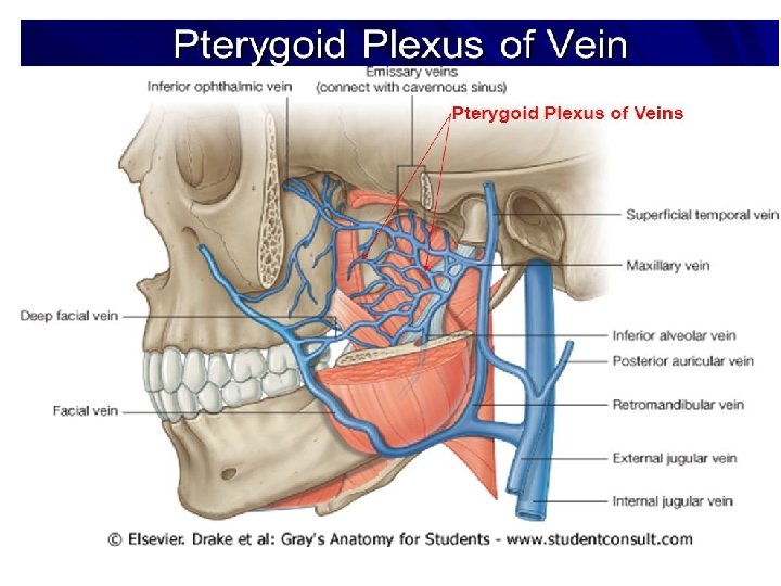
PTERYGOID PLEXUS • This plexus situated between mandibles ramus and pterygoid muscles, and is situated between the temporalis muscle and lateral pterygoid muscle. most of this veins receive blood from deep parts and one relative branch connect this plexus to facial vein. emissary veins or inferior ophthalmic vein can connect this plexus to cavernous sinus. • Because of these valveless veins blood pressure is bidirectional in head and face and can transfer to each other easily.
Tributaries received
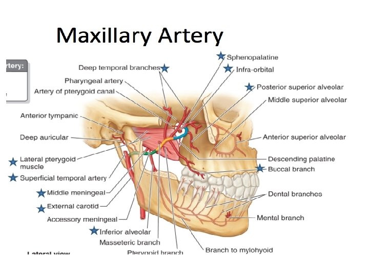
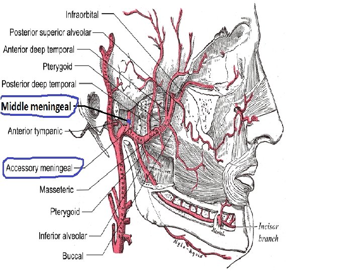
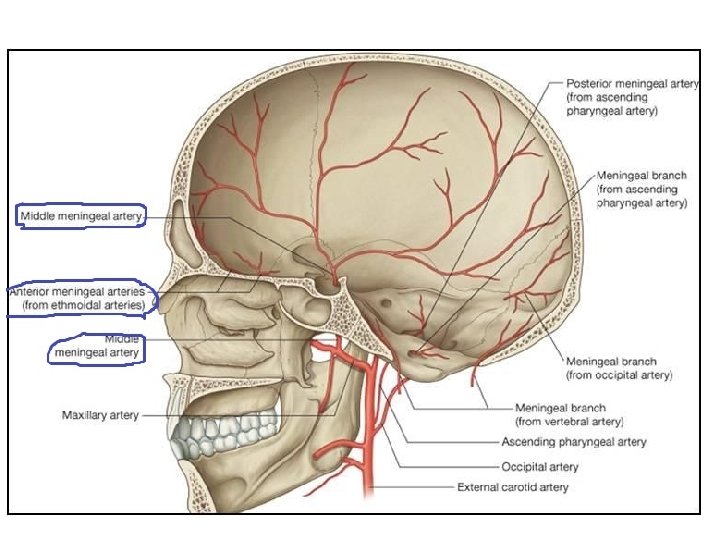
MENINGEAL ARTERIES • Anterior meningeal branch of anterior ethmoidal artery • Accessory meningeal artery • Middle meningeal artery • Posterior meningeal artery(ascending pharyngeal , vertebral and occipital)
MIDDLE MENINGEAL ARTERY • Is typically the 3 rd branch of first part (retromandibular part)of the maxillary artery • One of the 2 terminal branches of external carotid artery. after branching off the maxillary artery in infratemporal fossa, it runs through the spinosum to supply the dura matter. this artery is the largest of the 3 arteries that supply the meninges.
POSTERIOR MENINGEAL ARTERIES • These are quite similar to 3 branches. • Posterior meningeal artery originate from the vertebral opposite the foramen magnum, ramifies between the bone and dura matter in the cerebllar fossa, and supplies the falx cerebri.
MENINGEAL VEINS • Ant. meningeal V: goes to venous sinuses of anterior and posterior cranial fossa. • Mid. meningeal V: goes to sphenoidal sinus or pterygoid plexus. • Post. meningeal V: goes to venous sinuses of anterior and posterior cranial fossa.
ANTERIOR MENINGEAL VEIN