ARTERIAL SUPPLY OF BRAIN BY DR manjula vastrad

ARTERIAL SUPPLY OF BRAIN BY: DR. manjula vastrad asst. prof Dept of rachana shareera Smvvs rkm amc vijayapur

INTRODUCTION The brain is one of the most metabolically active organs in the body, receiving 17% of the total cardiac output. Brain accounts 2. 5% of body weight, but it receives 1/6 th of cardiac output.

INTRODUCTION �Amount of blood cerebral circulation carries – Cerebral Blood Flow �In an adult, Cerebral blood flow is typically 750 millilitres per minute. �The brain receives it’s blood from two pairs of arteries, the carotid and vertebral arteries which are interconnected in the cranial cavity – Arterial circle – Circle of Willis. �About 80% of the brain’s blood supply comes from the carotid, and the remaining 20% from the vertebral artery.

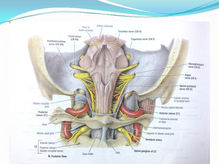
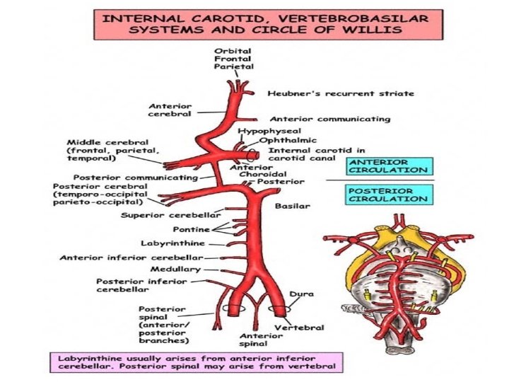
The Vertebro-basilar System The vertebral artery is a branch of the first part of subclavian artery. It enters the subarachnoid space in the upper part of vertebral canal after piercing the Dura & Arachnoid mater. It unite with its fellow at lower border of Pons and forms Basilar artery.
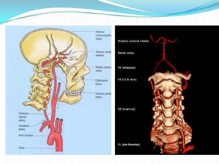

BRANCHES OF VERTEBRAL ARTERY 1. Posterior spinal artery 2. Posterior inferior cerebellar artery 3. Anterior spinal artery 4. Medullary branches 5. Meningeal branches

BRANCHES OF VERTEBRAL ARTERY

BASILAR ARTERY �Formed by the union of two vertebral arteries at the lower border of Pons. �Lies in the median groove of Pons in Cisterna pontis. �It ends by dividing into two Posterior Cerebral Arteries at the upper border of pons.

COURSE

Branches of basilar artery 1. Anterior inferior cerebellar artery 2. Labrynthine artery 3. Pontine branches 4. Superior cerebellar artery 5. Posterior cerebral artery


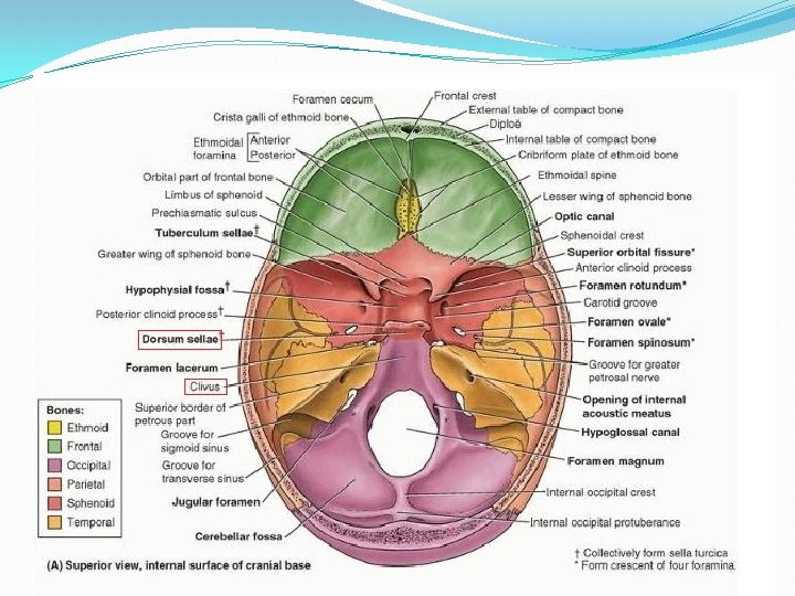
Internal carotid artery The internal carotid arteries arise in the neck from common carotid arteries. Internal carotid arteries enters middle cranial fossa through the carotid canal at the base of skull.

Course of ICA

Branches of ica 1. 2. 3. 4. 5. 6. Posterior communicating artery Ophthalmic artery Hypophysial artery Anterior choroidal artery Anterior cerebral artery Middle cerebral artery

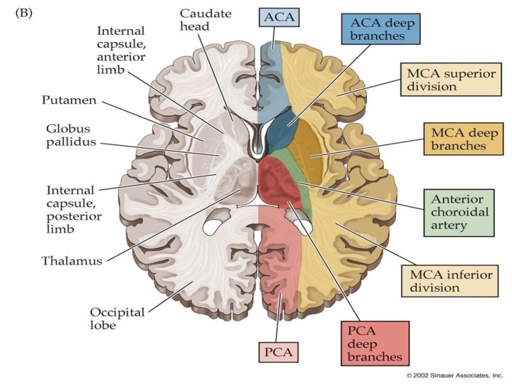
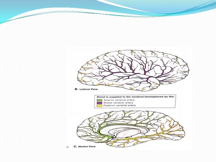
CEREBRAL ARTERIES 1. Anterior cerebral artery supply most of the medial and superior surfaces of the brain and the frontal pole. 2. Middle cerebral artery supply the lateral surface of the brain and the temporal pole. 3. Posterior cerebral artery supply the inferior surface of the brain and the occipital pole

CEREBRAL ARTERIES

Posterior cerebral artery These are the two terminal branches of Basilar Artery. Diverge at superior border of Pons. On the anterior surface of midbrain, each posterior cerebral artery receives posterior communicating branch from corresponding ICA.

BRANCHES OF PCA Posteromedial central branches 2. Posterolateral central branches 3. Posterior choroidal branches 4. Cortical branches 1.

CORTICAL BRANCHES OF PCA

Posterior Cerebral Artery & Important Areas Of Cortical Distribution LOBE AREA OCCIPITAL VISUAL TEMPORAL OLFACTORY

Anterior cerebral artery �It arises from the internal carotid artery below the anterior perforated substance, lateral to the optic chiasma. �Here it runs forwards and medially crossing above the optic nerve to reach the longitudinal fissure separating the two cerebral hemispheres.

Anterior cerebral artery v The arteries of the two sides lie close together and are united to each other by the anterior communicating artery. v Now turns sharply to reach the genu of the corpus callosum then body and ends in posterior part.

BRANCHES OF ACA

CORTICAL BRANCHES OF ACA

MIDDLE CEREBRAL ARTERY Large terminal branch of internal carotid artery which lies in line with it. It runs laterally in the stem of lateral sulcus where it branches and projects to many parts of lateral cerebral cortex. It supply blood to the anterior temporal lobes and insular cortices.

MIDDLE CEREBRAL ARTERY

MIDDLE CEREBRAL ARTERY

Choroid plexuses � The Anterior Choroidal A: from the Internal Carotid A supply the choroid plexus of the lateral ventricle. � The Posterior Choroidal A. : from the Posterior cerebral artery to supply the choroid plexus of the lateral ventricle and 3 rd ventricle. �The Choroidal Branch of Posterior inferior Cerebellar A – to supply choroidal plexuses of 4 th venticle

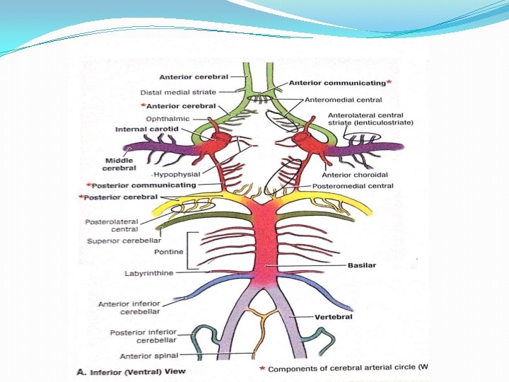
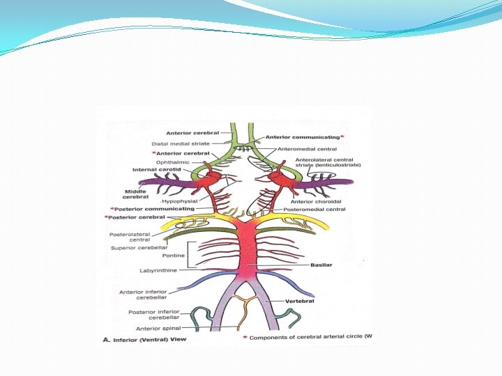
Circle Of Willis Site: at the base of the brain around interpeduncular fossa. • Function: Important anastomosis between the 2 internal carotid arteries in front & Vertebro-basilar system behind. • It attempts to equalize the flow of blood to different parts of brain and provides a collateral circulation in the event of obstruction of one of its components. • Arteries forming it: Rt. & Lt. internal carotid artery. Rt. & Lt. anterior cerebral arteries. Rt. & Lt. posterior communicating arteries. Anterior communicating artery.

CIRCLE OF WILLIS

APPLIED ANATOMY

STROKE ü Stroke or cerebrovascular accident: - Blockage in the artery – cerebral infarction • Carotid artery • Basilar artery ü Bleeding within the brain – Intracerebral haemorrhage • Aneurysm • Subarachnoid haemorrhage • Hypertension

Area of oxygendeprives brain Blockage Thrombus Plaque


MOYA DISEASE • This disease occurs when the large arteries carrying nutrients to the brain become blocked, causing fine blood vessels to develop in the surrounding area to compensate for the lack of blood flow. • • The name "moya disease" comes from the fact that these collateral vessels have the appearance of a puff of smoke ("moya" in Japanese).

HEMIPLEGIA q. Hemiplegia is a common condition. It is an upper motor neuron type of paralysis of one half of the body, including the face. It is usually due to an internal capsule lesion caused by thrombosis of one of the lenticulostriate branches of middle cerebral artery. q. One of the lenticulostriate branches is most frequently ruptured , it is known as charcot’s artery of cerebral hemorrhage. This lesion also produces hemiplegia with deep coma, and is ultimately fatal.

THANK YOU

Resin cast of the arterial supply of the brain

Arteries to specific brain areas Corpus striatum Internal capsule Medial and lateral striate branches Anterior and middle cerebral arteries Thalamus P communicating A , Basilar A. Posterior cerebral artery Midbrain Posterior cerebral Artery, Sup cerebellar Artery, Basilar Artery Pons Pontine branches of Basilar, Anterior inferior cerebellar, superior cerebellar Artery Medulla oblangata Vertebral, posterior inferior cerebellar artery, Anterior inferior cerebellar Artery Cerebellum Superior cerebellar, Anterior inferior cerebellar Artery, Posterior inferior cerebellar Artery

ACA – central branches Territory supplied by branches of the anterior and middle cerebral arteries is shown in red. Territory supplied by the anterior choroidal artery is shown in green
- Slides: 47