Arterial Pressure Monitoring Components of an Arterial Pressure


 Waveform")


 70 -90 mm Hg Cardiac Index (CI)-")


 • Monitoring after discontinuing a PA catheter or")

- Slides: 62
Arterial Pressure Monitoring
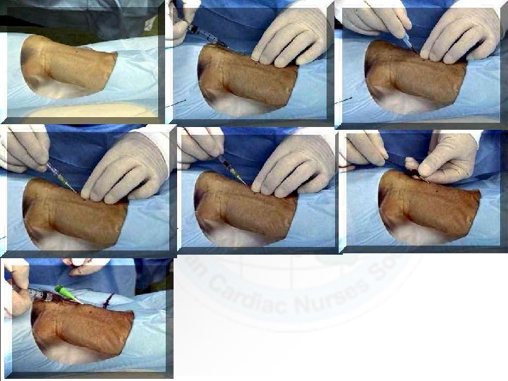
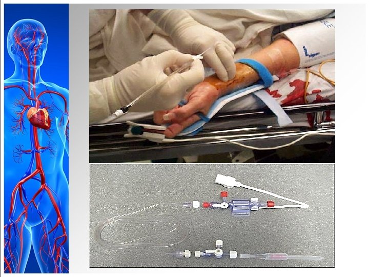
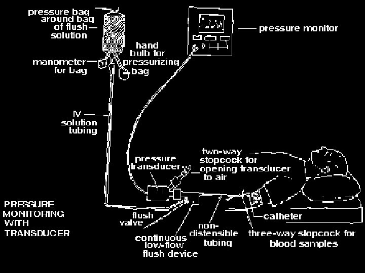
Components of an Arterial Pressure Monitoring System
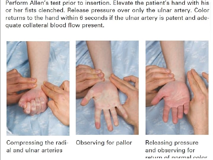
Arterial Pressure Monitoring • High- and low-pressure alarms based on patient’s status • Risks: üHemorrhage, infection, thrombus formation, neurovascular impairment, loss of limb (Assess 5 P’s)
• Allen’s test • May be incorrect as many as 14% of patients
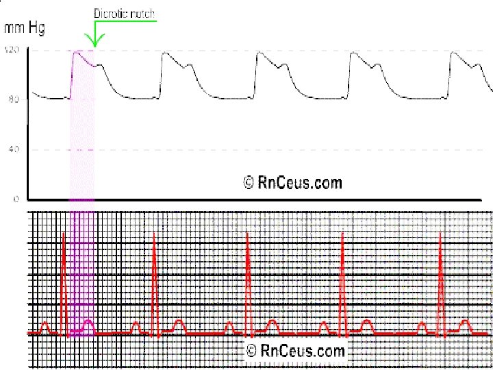
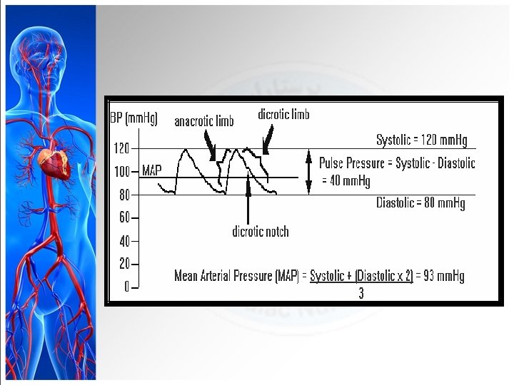
Arterial Pressure Tracing
Dicrotic notch signifies the closure of the aortic valve.
Arterial Pressure Monitoring
Arterial Pressure Monitoring
Arterial Pressure Monitoring
Arterial Pressure Monitoring
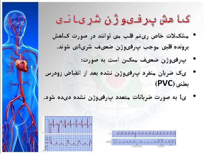
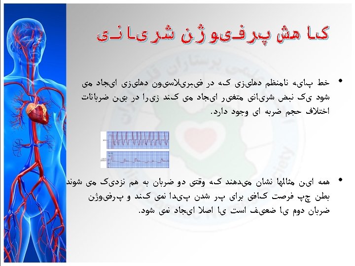
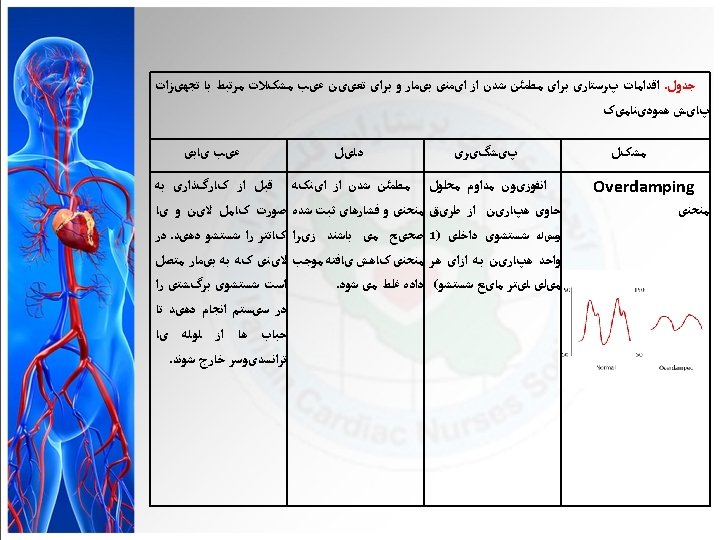
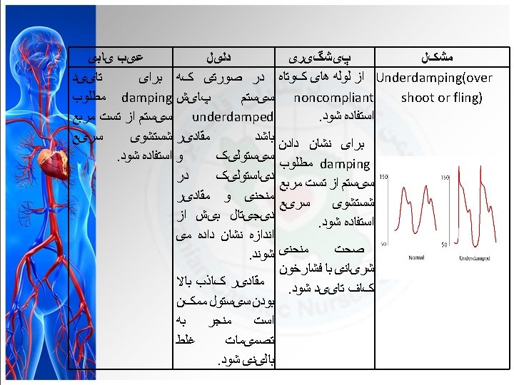
The over-damped arterial line waveform The over-damped trace will lose its dicrotic notch, and there wont be more than one oscillation. This happens when there is clot in the catheter tip, or an air bubble in the tubing.
The under-damped arterial line waveform The under-damped trace will overestimate the systolic, and there will be many post-flush oscillations.
• Dampened trace
• Resonant trace
Pulmonary Artery Pressure Monitoring
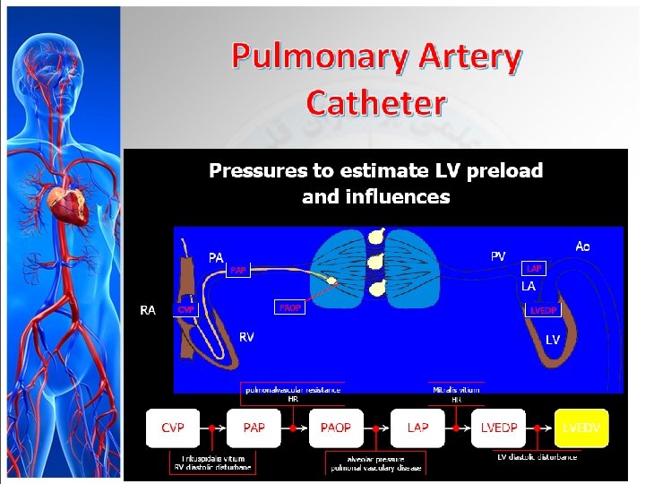
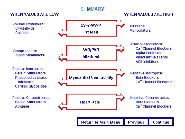
Pulmonary Artery Pressure Monitoring • Guides management of patients with complicated cardiac, pulmonary, and intravascular volume problems – PA diastolic (PAD) pressure and PAWP: Indicators of cardiac function and fluid volume status – Monitoring PA pressures allows for therapeutic manipulation of preload
Werner Forssmann The Nobel Prize in Physiology or Medicine 1956 “…develop a technique for the catheterization of the heart. This he did by inserting a cannula into his own antecubital vein, through which he passed a catheter for 65 cm and then walked to the X-ray department, where a photograph was taken of the catheter lying in his right auricle. ” -The Nobel Foundation 1956
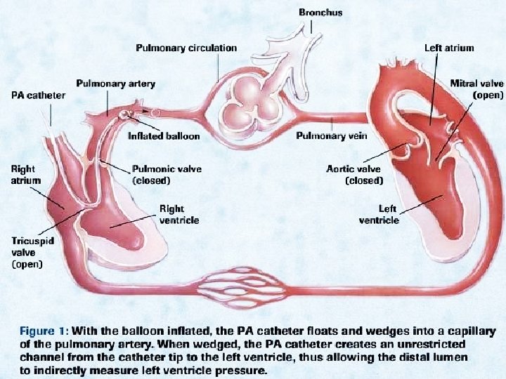
Pulmonary Artery Pressure Monitoring § PA flow-directed catheter § Distal lumen port in PA § Samples mixed venous blood § Thermistor lumen port near distal tip § Monitors core temperature § Thermo dilution method measuring CO
Pulmonary Artery Pressure Monitoring
Pulmonary Artery Pressure Monitoring § Proximal -Right atrium port Measurement of CVP, Injection of fluid, for CO measurement, Blood sampling, Administer medications
Purpose Measuring of pulmonary artery pressure § § § § Measuring of right atrium pressure. Measuring of right ventricle pressure. Measuring of cardiac out put. indirect measurement of the pressure in the left atrium heart atrium Assessing the status of the mitral valve.
Pulmonary Artery Catheter
“Floating a Swan”
Pulmonary Artery Catheter
RA WAVEFORM • • • Normal Value 0 -8 mm. Hg RAP = CVP Wave Fluctuations Due To Contractions
RV WAVEFORM • Normal Value 15 -25/0 -8 mm. Hg • Catheter In RV May Cause Ventricular Ectopy • Swan Tip May Drift From PA to RV
PA WAVEFORM • Normal Value 8 -15 mm. Hg • Dicrotic Notch Represents PV Closure • PAD Approximates PAWP (LVEDP) (in absence of lung or MV disease)
PAWP WAVEFORM • • Normal Value 8 -12 mm. Hg Balloon Floats and Wedges in Pulmonary Artery PAWP = LAP = LVEDP Wedging Can Cause Capillary Rupture
PA CATHETER WAVEFORMS A wave - due to atrial contraction. Absent in atrial fibrillation. Enlarged in tricuspid stenosis, pulmonary stenosis and pulmonary hypertension. C wave - due to bulging of tricuspid valve into the right atrium or possibly transmitted pulsations from the carotid artery. X descent - due to atrial relaxation. V wave - due to the rise in atrial pressure before the tricuspid valve opens. Enlarged in tricuspid regurgitation. Y descent - due to atrial emptying as blood enters the ventricle. Canon waves - large waves not corresponding to a, v or c waves. Due to complete heart block or junctional arrhythmias.
PA INSERTION WAVEFORMS • • A B C D A = RA (CVP) Waveform B = RV Waveform C = PA Waveform D = PAWP Waveform
PAWP WAVEFORM
PAWP WAVEFORM
PAWP WAVEFORM
PAWP WAVEFORM
PAWP WAVEFORM
PAWP WAVEFORM
PAWP WAVEFORM
PAWP WAVEFORM
PA Waveforms during Insertion
Hemodynamics: Normal value Mean Arterial Pressure (MAP) 70 -90 mm Hg Cardiac Index (CI)- 2. 2 -4. 0 L/min/m 2 Cardiac Output (CO)- 4 -8 L/min Central Venous Pressure (CVP) (also known as Right Atrial Pressure (RA)) 2 -8 mm. Hg Pulmonary Artery Pressure (PA) Systolic 20 -30 mm. Hg (PAS) Diastolic 4 -12 mm. Hg (PAD) Mean 15 -25 mm. Hg Pulmonary Capillary Wedge Pressure (PWCP) 6 -12 mm. Hg Systemic Vascular Resistance(SVR) 800 -1200
Measuring Cardiac Output • SVR, SVRI, SV, and SVI can calculated when CO is measured – ↑ SVR • Vasoconstriction from shock • Hypertension • ↑ Release or administration of epinephrine or other vasoactive inotropes • Left ventricular failure
Complications with PA Catheters § Infection and sepsis § Asepsis for insertion and maintenance of catheter and tubing mandatory § Change flush bag, pressure tubing, transducer, and stopcock every 96 hours § Air embolus (e. g. , disconnection)
Complications with PA Catheters § Ventricular dysrhythmias § During PA catheter insertion or removal § If tip migrates back from PA to right ventricle § PA catheter cannot be wedged § May need repositioning
Complications with PA Catheters § Pulmonary infarction or PA rupture § Balloon rupture (e. g. , overinflation) § Prolonged inflation § Spontaneous wedging § Thrombus/embolus formation
Noninvasive Hemodynamic Monitoring § Major indications • Early signs and symptoms of pulmonary or cardiac dysfunction • Differentiation of cardiac or pulmonary cause of shortness of breath • Evaluation of etiology and management of hypotension
Noninvasive Hemodynamic Monitoring § Major indications(cont’d) • Monitoring after discontinuing a PA catheter or justification for insertion of a PA catheter • Evaluation of pharmacotherapy • Diagnosis of rejection following cardiac transplantation
QUESTION?