Arterial Blood Gas Interpretation Associate Professor Dr Samah

Arterial Blood Gas Interpretation Associate Professor Dr. Samah Shehata

OBJECTIVES ABG Sampling Interpretation of ABG Oxygenation status Acid Base status Case Scenarios

ABG – Procedure and Precautions Ideally - Pre-heparinised ABG syringes - Syringe should be FLUSHED with 0. 5 ml of 1: 1000 Heparin solution and emptied. DO NOT LEAVE EXCESSIVE HEPARIN IN THE SYRINGE HEPARIN DILUTIONAL EFFECT HCO 3 PCO 2 Only small 0. 5 ml Heparin for flushing and discard it Syringes must have > 50% blood. Use only 2 ml or less syringe.
 • Brachial artery")
Sites for obtaining ABG • Radial artery ( most common ) • Brachial artery • Femoral artery Radial is the most preferable site used because: • It is easy to access • It is not a deep artery which facilitate palpation, stabilization and puncturing • The artery has a collateral blood circulation 4 A. Y. T

ALLEN’S TEST It is a test done to determine that collateral circulation is present from the ulnar artery 5

Ensure No Air Bubbles. Syringe must be sealed immediately after withdrawing sample. ◦ Contact with AIR BUBBLES Air bubble = PO 2 150 mm Hg , PCO 2 0 mm Hg Air Bubble + Blood = PO 2 PCO 2 ABG Syringe must be transported at the earliest to the laboratory for EARLY analysis via COLD CHAIN

Interpretation of ABG OXYGENATION ACID BASE

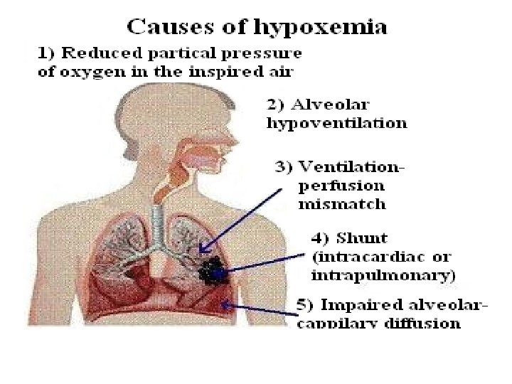
O X Y G E N A T I O N Determination of Pa. O 2 is dependant upon As Age, Fi. O 2, Patm the expected Pa. O 2 • Pa. O 2 = 109 - 0. 4 (Age) As Fi. O 2 the expected Pa. O 2 • Alveolar Gas Equation: • P AO 2= (PB-P h 2 o) x Fi. O 2 - p. CO 2/R P AO 2 = partial pressure of oxygen in alveolar gas, PB = barometric pressure (760 mm. Hg), Ph 2 o = water vapor pressure (47 mm Hg), Fi. O 2 = fraction of inspired oxygen, PCO 2 = partial pressure of CO 2 in the ABG, R = respiratory quotient (0. 8)

Determination of the Pa. O 2 / Fi. O 2 ratio Inspired Air Fi. O 2 = 21% Pi. O 2 = 150 mm. Hg Palv. O 2 = 100 mm. Hg CO 2 Pa. O 2 = 95 mm. Hg O 2
 Gives understanding that")
PO 2/ Fi. O 2 ratio ( P: F Ratio ) Gives understanding that the patients OXYGENATION with respect to OXYGEN delivered is more important than simply the PO 2 value. Example, Patient 1 On Room Air Patient 2 On MV PO 2 68 90 Fi. O 2 21% (0. 21) 50% (0. 50) P: F Ratio 324 180

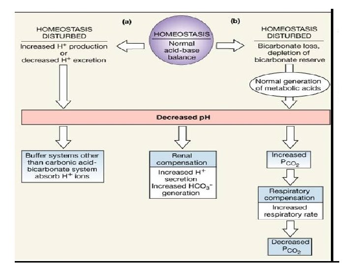
A C I D Acid Base Balance H+ ion concentration in the body is precisely regulated The body understands the importance of H+ and hence devised DEFENCES against any change in its concentration- B A S E BICARBONATE BUFFER SYSTEM Acts in few seconds RESPIRATORY REGULATION RENAL REGULATION Acts in few minutes Acts in hours to days
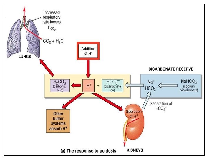
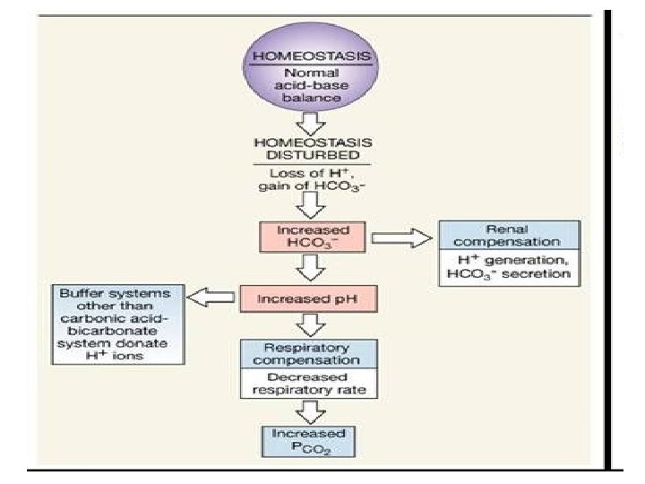
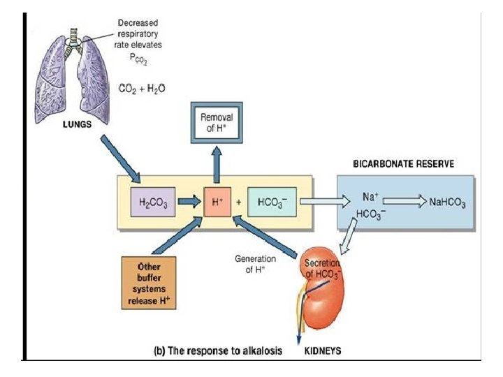

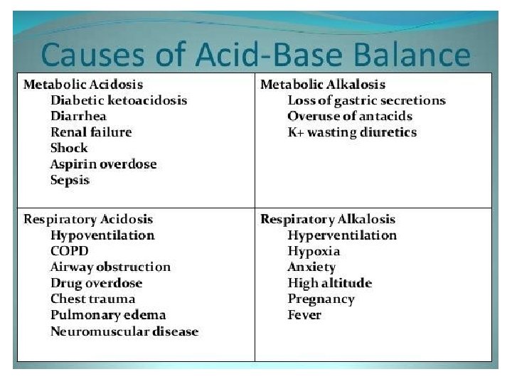
Assessment of ACID BASE Balance �Definitions and Terminology ACIDOSIS – presence of a process which tends to p. H by virtue of gain of H + or loss of HCO 3 ALKALOSIS – presence of a process which tends to p. H by virtue of loss of H+ or gain of HCO 3 - If these changes, change p. H, suffix ‘emia’ is added ACIDEMIA – reduction in arterial p. H (p. H<7. 35) ALKALEMIA – increase in arterial p. H (p. H>7. 45)
![If PCO 2 & [HCO 3] move in opposite directions](http://slidetodoc.com/presentation_image_h2/17383971d63b03f94df20e1ced4adca7/image-22.jpg "If PCO 2 & [HCO 3] move in opposite directions")
If PCO 2 & [HCO 3] move in opposite directions

Mixed disturbance

STEP WISE APPROACH to Interpretation Of ABG reports

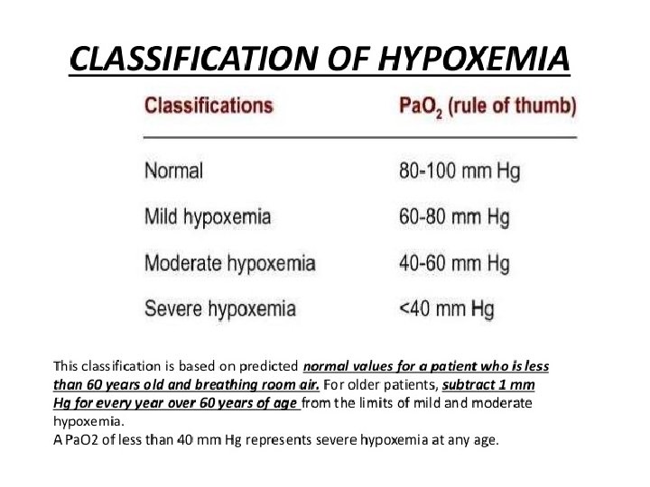
Normal Values ANALYTE Normal Value Units p. H 7. 35 - 7. 45 PCO 2 35 - 45 mm Hg PO 2 80 – 100 mm Hg` [HCO 3] 22 – 26 meq/L Sa. O 2 95 -100 % Anion Gap 5 -11 meq/L ∆HCO 3 +2 to -2 meq/L

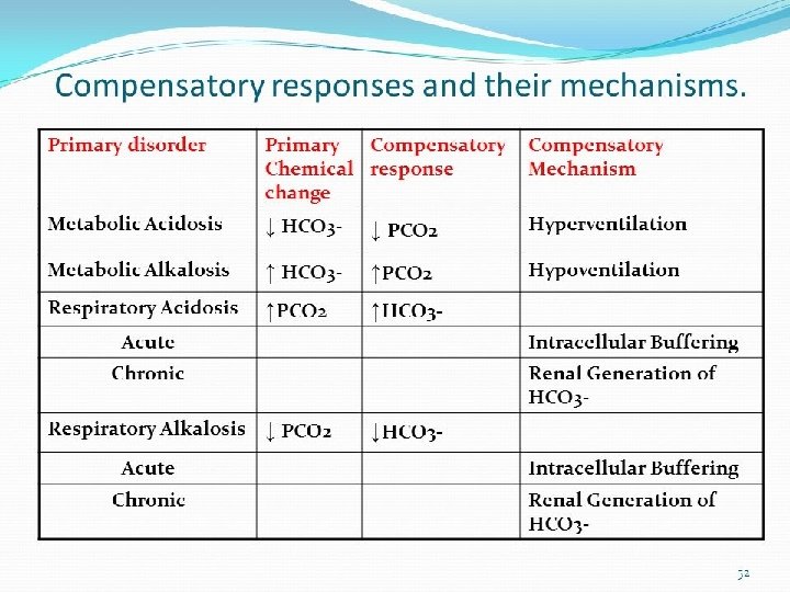
STEP 1 STEP 2 • ACIDEMIA or ALKALEMIA? • RESPIRATORY or METABOLIC? STEP 3 • If Respiratory – Compensated or Not? • If METABOLIC – ANION GAP? STEP 4

STEP 1 ACIDEMIA OR ALKALEMIA? Look at p. H <7. 35 - acidemia >7. 45 – alkalemia

STEP 2 RESPIRATORY or METABOLIC? IS PRIMARY DISTURBANCE RESPIRATORY OR METABOLIC? p. H PCO 2 or p. H PCO 2 METABOLIC p. H PCO 2 or p. H PCO 2 RESPIRATORY

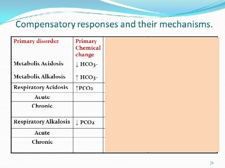
STEP 3 RESPIRATORYCompensated/decompensated?

a. Respiratory acidosis Phase PH Pa. CO 2 HCO 3 UNCOMPENSATED ↓ ↑ ------ Because there is no response from the kidneys yet to acidosis the HCO 3 will remain normal Phase PH Pa. CO 2 HCO 3 PARTIAL COMPENSATED ↓ ↑ ↑ The kidneys start to respond to the acidosis by increasing the amount of circulating HCO 3 Phase PH Pa. CO 2 HCO 3 FULL COMPENSATED N ↑ ↑ PH return to normal Pa. CO 2 & HCO 3 levels are still high to correct acidosis A. Y. T 31

B. Respiratory alkalosis Phase PH Pa. CO 2 HCO 3 UNCOMPENSATED ↑ ↓ ------ Because there is no response from the kidneys yet to acidosis the HCO 3 will remain normal Phase PH Pa. CO 2 HCO 3 PARTIAL COMPENSATED ↑ ↓ ↓ The kidneys start to respond to the alkalosis by decreasing the amount of circulating HCO 3 Phase PH Pa. CO 2 HCO 3 FULL COMPENSATED N ↓ ↓ PH return to normal Pa. CO 2 & HCO 3 levels are still low to correct alkalosis A. Y. T 32

STEP 4 • If METABOLIC – ANION GAP?

Electrochemical Balance in Blood 100% 90% 80% 70% 60% 50% 40% 30% 20% 10% 0% UC Na UA HCO 3 Cl CATIONS ANIONS Sulfate Phosphate Mg- OA K - Proteins Ca-HCO 3 Na- Cl

Anion Gap AG based on principle of electroneutrality: Total Serum Cations = Total Serum Anions M cations + U cations = M anions + U anions �Na + K + (Ca + Mg) = HCO 3 + Cl + (PO 4 + SO 4 + Protein + Organic Acids) Na + K + UC = HCO 3 + Cl + UA But in Blood there is a relative abundance of Anions, hence Anions > Cations Na + K – (HCO 3 + Cl) = UA – UC Na + K – (HCO 3 + Cl) = Anion Gap
 =")
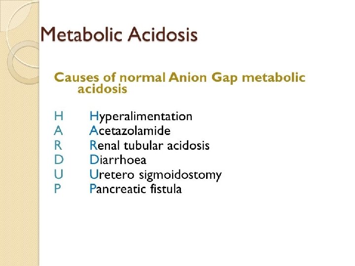
METABOLIC ACIDOSISANION GAP? IN METABOLIC ACIDOSIS WHAT IS THE ANION GAP? ANION GAP(AG) = (Na + K) – (HCO 3 + Cl) Normal Value =11 - 5 mmol /L High Anion Gap Metabolic Acidosis Normal Anion Gap Acidosis

High Anion Gap Metabolic Acidosis M METHANOL U UREMIA D P I L E ARF/CRF DIABETIC KETOACIDOSIS & other KETOSIS PARALDEHYDE, PROPYLENE GLYCOL ISONIAZIDE, IRON LACTIC ACIDOSIS ETHANOL, ETHYLENE GLYCOL S SALICYLATE

Clinical CASE SCENARIOS

CASE 1 62 years old Male patient COPD Breathlessness, progressively increased , aggravated on exertion, 2 days Chronic smoker expiratory rhonchi 22/7/2011 7: 30 am p. H 7. 20 PCO 2 92 mm. Hg PO 2 76 mm. Hg Actual HCO 3 SO 2 28. 00 mmol/l Fi. O 2 37% 89

STEP 1 – ACIDEMIA STEP 2 – p. H PCO 2 Respiratory STEP 3 Primary Respiratory Acidosis, partially compensated STEP 4 Mild hypoxemia

CASE 2 31/7/2011 11: 30 pm 63 years old , Male patient p. H 7. 18 PCO 2 21. 00 CRF PO 2 Breathlessness Actual Decreased Urine Otpt. 2 days HCO 3 Vomiting 10 -15 Base Excess 82 7. 80 -18. 80 SO 2 95 Na 140. 6 Chloride 102 K 4 Albumin 2. 4

STEP 1 – ACIDEMIA STEP 2 – p. H PCO 2 METABOLIC STEP 4 – ANION GAP = (Na + K) – (HCO 3 +Cl) = ( 140. 6 + 4)-(7. 80+102) = 3 4. 8 K 4 HIGH AG Met. Acidosis

- Slides: 44