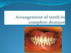
Arrangement of teeth in complete denture The four


- Slides: 66

Arrangement of teeth in complete denture

The four principal factors that govern the positions of the teeth for complete dentures are (1) the horizontal relations to the residual ridges, (2) the vertical positions of the occlusal surfaces and incisal edges between the residual ridges, (3) the esthetic requirements, and (4) the inclinations for occlusion 2

Guidelines for horizontal Placement of Anterior Teeth 3

Role of incisive papilla & mid palatal suture �Labial surface of maxillary incisors is approx. 8 to 10 mm anterior to incisive papilla. �A transverse line bisecting the middle of I. P. passes through the tip of canine. 4

Cuspid eminences When cuspid eminences are visible on cast, a line marking the distal of eminences co-incide with distal margin of cuspids. Relation to residual alveolar ridge Max. Anterior teeth are placed anterior to residual ridge, depending upon amount of resorption. 5

Arch Form And Shape �Square arch – C. I. in line with the canine �Tapering arch – C. I. at a greater distance forward than canine � Ovoid arch - in between 6

Esthetics �Vermilion border of upper lip. �Mento-Labial & Naso-Labial groove. �Everted upper lip. �Corner of mouth (no drooping appearance) 7

RELATION WITH THE UPPER LIP Incisal two-thirds of labial surface of teeth supports the lips. �If set too far posteriorly Lip looks unsupported. Vermilion border would not be visible. �If set too far anteriorly Lip would taut & stretch. Nasolabial fold may fill out. 8

MEDIO- LATERAL POSITION �Midline – midline of face passes between 2 upper & lower central incisors. �Ala of nose – line dropped from the Ala passes through tip of canine. 9

Guidelines for vertical Orientation of Anterior Teeth 10

Role of upper lip �Visibility of upper anterior teeth �Incisal edges are visible by 1 to 2 mm below the upper lip at rest. �Short or long or incompetent lip influences the amount of teeth visibility. �Some racial types have fuller lips, others have thinner. 11

Effect of aging �In young patients, incisal edges are visible by 1 to 2 mm below the upper lip at rest. �While smiling or during speech, incisal & middle 1/3 are visible in normal person. � With aging, tone of upper lip decreases, lesser amount of maxillary teeth visible and more of mandibular teeth become visible. 12

Relationship of lower lip to anterior teeth Lower canine & Ist premolar should be even with lower lip at the corner of mouth. If lower teeth are high -Anterior plane of occlusion may be too high -excessive VDO -Excessive vertical overlap reverse is true if mandibular teeth are below lower lip at corner of mouth. 13

Guides to position of posterior teeth 14

Retromolar pad The maximum extension posteriorly of any artificial tooth is anterior border of Retromolar pad. This is to avoid having a tooth over an incline which results in denture sliding. Sometimes space is available for only 3 mandibular posterior teeth, then drop Ist premolar. 15

Retromolar pad 16

Maxillary Tuberosity Teeth should not be set on the Tuberosity as it can lead to lever imbalance and might lead to cheek bite in posterior region. When space permits, 4 maxillary posterior teeth can be placed opposing 3 mandibular posterior teeth, to provide support to cheeks 17

OCCLUSAL PLANE Anterior occlusal plane parallel to interpupillary line & at the level of commissure. - Posterior occlusal plane should be at the level of 2/3 the height of retromolar pad 18

Stenson’s duct –it exits at Buccal mucosa in the region of 2 nd Molar. Occlusal plane is located of 1/8 of an inch below this. �With these anterio-posterior guidelines, occlusal plane is made parallel to lower mean foundation plane and Ala-Tragus plane. �Height of occlusal plane is also influenced by-length of lips -Ridge height -Amount of maxillomandibular space available 19

Relationship with tongue �Occlusal plane should be located in relation to lateral surface of tongue near demarcation zone b/w Dorsal keratinized mucosa & ventral nonkeratinized mucosa. 20

Buccal Limit �Teeth should not be set too far off the ridge. �Placing too far Buccally can cause: - Cheek Biting - Esthetic problems due to obliteration of Buccal corridor. - Denture instability due to lever imbalance & muscle function. 21

Lingual Limit �Lingual cusps of molars are in alignment with Mylohyoid ridge. �Placing too far lingually can cause � Crowding of tongue. � Tongue biting. � Imbalance due to tongue function. 22

Overjet & Overbite Class I – Normal , Class II – Retruded , Class III Protruded 23

Canine & Molar Relationship Mesial slope of cusp of upper canine opposes the distal slope of Lower canine cusp. OR Distal surface of lower canine is in line with tip of upper canine. M. B cusp of upper 1 st molar opposes the Buccal groove of lower 1 st molar. 24

Canine-retromolar Pad Reference Line �From tip of Canine to center of Retromolar pad. This designates centre of mandibular Ridge. �Central fossae of mandibular Posterior teeth should coincide with this line OR �This in turn corresponds to maxillary palatal cusps. 25

Individual orientation of maxillary teeth 26

ARRANGING TEETH FOR COMPLETE DENTURE OCCLUSION Maxillary Central Incisor: The long axis of the tooth is perpendicular to the horizontal (labiolingual inclination) Its long axis slopes towards the vertical axis ( mesiodistal inclination) Slopes labially about 15 degrees when viewed from the side. Incisal edge is in contact with the occlusal plane. 27

Teeth is set on the occlusal rim 28

Maxillary Lateral Incisor: Long axis slopes rather more towards the midline Inclined labially about 20 degrees when viewed from the side The neck is slightly depressed The incisal edge is about 1 mm short of the occlusal plane. 29

30

Maxillary Canine : Its long axis is parallel to the vertical axis when viewed from both the front and side or it may be slightly to the distal. The bulbous cervical half of the tooth provides its prominence. Its cusp is in contact with the horizontal plane. . The neck of the tooth must be prominent 31

32

Remaining maxillary teeth are arranged on the other side of the arch to complete the anterior set up. To maintain the set teeth in position, the wax supporting the teeth must be heated and sealed both to the teeth and to the record base. 33

34

35

First premolar: �Long axis is parallel to the vertical axis when viewed from the front or the side. �Its palatal cusp is about 1 mm short of, and its buccal cusp in contact with, the occlusal plane. 36

Second premolar: �Its long axis is parallel with the vertical axis when viewed from the front or the side. �Both buccal and palatal cusps are in contact with the occlusal plane. 37

First molar: �Long axis slopes buccally when viewed from the front, and distally when viewed from the side. �Only mesiopalatal cusp is in contact with the occlusal plane. 38

Second molar: �Long axis slopes buccally more steeply than the first molar when viewed from the front, and distally more steeply when viewed from the side. �All four cusps are clear of the occlusal plane, but the mesiopalatal cusp is nearest to it. 39

Maxillary teeth set checked on occlusal plane 40

41

Orientation and arrangement of mandibular teeth 42

Arranging the Mandibular Teeth Mandibular central incisor: �Long axis slopes slightly towards the vertical axis when viewed from the front. �Slopes labially when viewed from the side. �Incisal edge is about 2 mm above occlusal plane 43

Mandibular lateral incisor: �Long axis inclines to vertical axis when viewed from the front �Slopes labially when viewed from side but not so steeply as the central incisor. �Incisal edge is about 2 mm above occlusal plane 44

Mandibular canine: �Long axis leans very slightly towards the midline when viewed from the front. �Leans very slightly lingually when viewed from the side �Neck is slightly prominent and the tooth is tilted to the distal �Tip at same level as incisors. 45

46

47

Teeth arrangement checked in patient mouth 48

First molar: �Long axis leans lingually when viewed from the front and mesially when viewed from the side. �All cusps are at a higher level above the occlusal plane than those of the second premolar. �The buccal and distal cusps are higher than the mesial and lingual. �The mesiobuccal cusp occludes in the fossa between upper second premolar and first molar. 49

Mandibular 1 st Molar �Facial: Long axis leans mesially, when viewed from side. �Proximal : Long axis inclines Lingually, when viewed from front. �Occlusal: Buccal cusps are higher than Lingual cusps. Distal cusps are higher than Mesial cusps. 50

Second premolar: �Long axis is parallel to the vertical plane when viewed from both the front and the side. �Both cusps are about 2 mm above the occlusal plane. �The buccal cusp contacts the fossa between the two upper premolars. 51

Mandibular 2 nd Premolar �Facial & Proximal : Long axis is vertical from both views. �Occlusal : Both cusps are about 1 -2 mm above Occlusal plane 52

First premolar: �Long axis is parallel to the vertical plane when viewed from the front and the side. �Its lingual cusp is below the horizontal plane �Its buccal cusp about 2 mm above it as it contacts the mesial marginal ridge of the upper first premolar. 53

Mandibular Ist Premolar �Facial : Long axis is parallel to vertical plane. �Proximal : Long axis is parallel to vertical plane. �Occlusal : Buccal cusp is above the occlusal plane, whereas Lingual cusp is below occlusal plane. 54

55

Second molar: �Lingual and mesial inclination of the long axis is more pronounced than in the case of the first molar. �All the cusps are at a higher level above the occlusal plane than those of the first molar, the distal and buccal cusps more so than the mesial and lingual. �The mesiobuccal cusp contacts the fossa between the two upper molars. 56

Mandibular 2 nd Molar �Facial : Mesial inclination is more than 1 st molar. �Proximal : Lingual inclination is slightly more than 1 st molar. �Occlusal : Buccal cusps are higher than Lingual. Distal cusps are higher than Mesial. 57

58

59

Key of occlusion �Canine key of occlusion �The distal arm of the lower canine should align with the mesial arm of the upper canine. 60

�Molar key of occlusion �The mesiobuccal cusp of the maxillary permanent molars should coincide with the mesiobuccal groove of the mandibular permanent molar 61

Overjet & overbite �Overjet denotes the distance between the upper & lower incisor measured in horizontal plane - 2 mm �Overbite denotes the vertical overlap of the maxillary and mandibular anterior – 2 mm 62

Overjet overbite 63

64

65

66
 Vertical tooth
 Vertical
 Working side and balancing side
 Ivoclar blueline mould guide
 Every denture design
 Dangerous curves the zoo
 Apron complete denture
 Combination syndrome dental
 Steps of cd fabrication
 Classification of denture stomatitis
 Curve of wilson definition
 Quadrilateral clasp configuration
 Sequelae of denture wearing
 Nick and notch technique
 Balanced occlusion vs balanced articulation
 Genial tubercles
 Direct sequelae of wearing complete denture
 Mucostatic impression technique
 Diagnosis and treatment planning in complete denture
 Edentulous anatomy
 Ah line denture
 Atmospheric pressure in complete denture
 Complete the missing word to complete the three key words
 What are simple predicates
 Nguyên nhân của sự mỏi cơ sinh 8
 Một số thể thơ truyền thống
 Trời xanh đây là của chúng ta thể thơ
 Bảng số nguyên tố lớn hơn 1000
 Vẽ hình chiếu vuông góc của vật thể sau
 Các châu lục và đại dương trên thế giới
 Tư thế worm breton là gì
 Thế nào là hệ số cao nhất
 Hệ hô hấp
 Tư thế ngồi viết
 đặc điểm cơ thể của người tối cổ
 Cái miệng nó xinh thế
 Mật thư anh em như thể tay chân
 Bổ thể
 ưu thế lai là gì
 Tư thế ngồi viết
 Thẻ vin
 Thể thơ truyền thống
 Các châu lục và đại dương trên thế giới
 Từ ngữ thể hiện lòng nhân hậu
 Diễn thế sinh thái là
 Vẽ hình chiếu vuông góc của vật thể sau
 Thứ tự các dấu thăng giáng ở hóa biểu
 Phép trừ bù
 Bài hát chúa yêu trần thế alleluia
 Tỉ lệ cơ thể trẻ em
 Lời thề hippocrates
 Sự nuôi và dạy con của hổ
 đại từ thay thế
 Quá trình desamine hóa có thể tạo ra
 Công của trọng lực
 Thế nào là mạng điện lắp đặt kiểu nổi
 Hình ảnh bộ gõ cơ thể búng tay
 Dạng đột biến một nhiễm là
 Vẽ hình chiếu đứng bằng cạnh của vật thể
 độ dài liên kết
 Các môn thể thao bắt đầu bằng tiếng nhảy
 Voi kéo gỗ như thế nào
 Thiếu nhi thế giới liên hoan
 Sự nuôi và dạy con của hươu
 điện thế nghỉ
 4 straight sides
