ARRANGEMENT OF A MUSCLE Muscle Fascicle Muscle Fiber
 Myofibril Myofilaments (thick/thin)")
ARRANGEMENT OF A MUSCLE Muscle Fascicle Muscle Fiber (cell) Myofibril Myofilaments (thick/thin)
![MUSCLE FIBER [CELL] STRUCTURE up to. 5 meter long yet only. 1 mm in](http://slidetodoc.com/presentation_image_h2/8deabb2ff610687977a2dcfdbcfd2790/image-2.jpg "MUSCLE FIBER [CELL] STRUCTURE up to. 5 meter long yet only. 1 mm in")
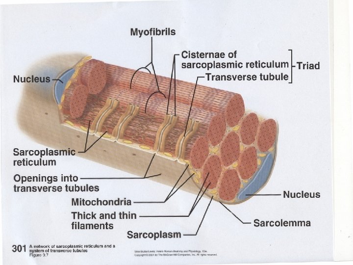
MUSCLE FIBER [CELL] STRUCTURE up to. 5 meter long yet only. 1 mm in thickness multinucleated (several nuclei) Sarcolemma – the membrane around a muscle fiber Sarcoplasm – the cytoplasm of a muscle cell Myofibril – parallel cords of protein, several make up a muscle fiber Sarcoplasmic Reticulum – Lattice-like network surrounding each myofibril [ Ca+ storage] Cisterns – larger tubules of the sarcoplasmic reticulum; 2 cisternae, along with a Transverse (T) Tubule form what is known as the triad T-Tubule – has openings that lead in from the sarcolemma surface

MYOFIBRIL Parallel cords of protein, several make up a muscle fiber

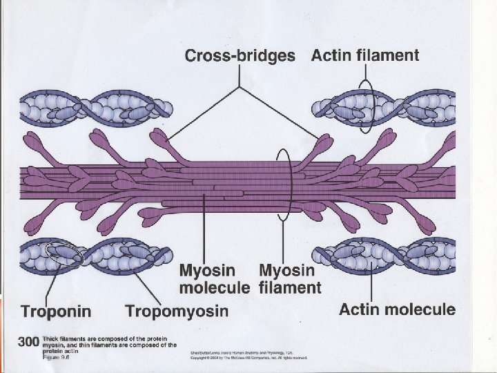
COMPOSITION OF A MYOFIBRIL 2 Types of Myofilaments: MYOSIN- thick filament protein ACTIN- thin filament /3 proteins [actin, troponin and tropomyosin]

SARCOMERE: structural and functional unit of the myofibril/ contractile unit of skeletal muscle Composed of Actin and Myosin Located between 2 Z Lines

PARTS OF THE SARCOMERE A-Band – where myosin and actin overlap I-Band – only actin filaments Z-line – perpendicular strands of protein fibers extend out from actin filaments H-Zone – center of A-Band, myosin filaments, w/ no overlapping actin filaments M-Line – center of H-Zone, proteins that attach to the center of the myosin filaments, holding them in place
- Slides: 8