Arches of the Foot DR SMITA SHIND ASSIST


. �Slings. �Tie beams.")
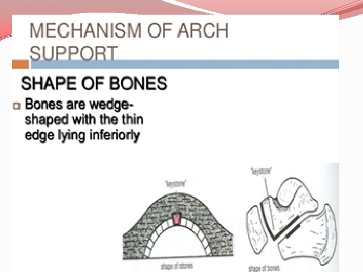


 2 Pillars (b) a summit (c) joints.")


 -which gives dynamic support to the head of talus")


 • Commonest of all foot problems. • Collapse")


: plantar flexedwalks on the toes-")


- Slides: 55
Arches of the Foot DR. SMITA SHIND ASSIST. PROF Dept. of anatomy
FUNCTIONS OF THE ARCHES �Arches of foot is distinctive feature of human �Helps in walking, running, jumping �Helps in weight bearing in upright posture �Foot prints are complete due to arches �Present since birth, masked in infant �The medial longitudinal arch gives a propulsive force during locomotion. �The lateral longitudinal arch functions as a support and weight transmission
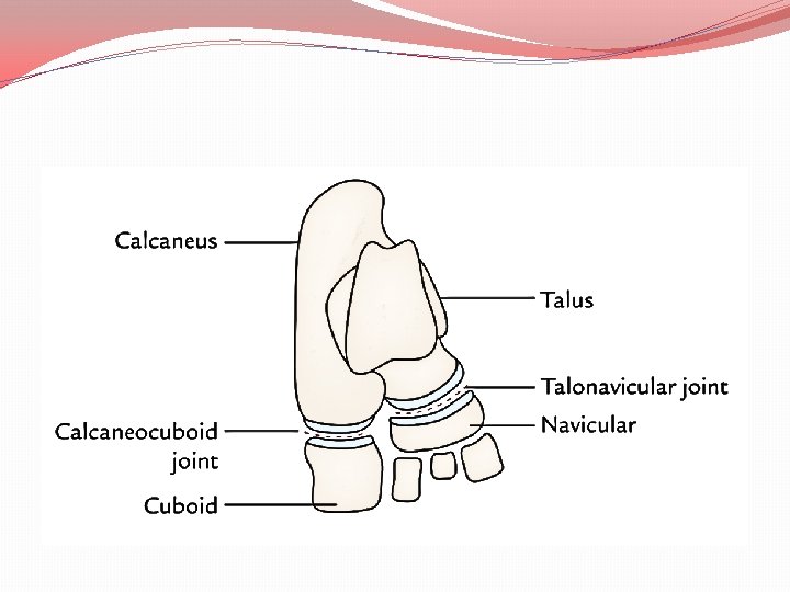
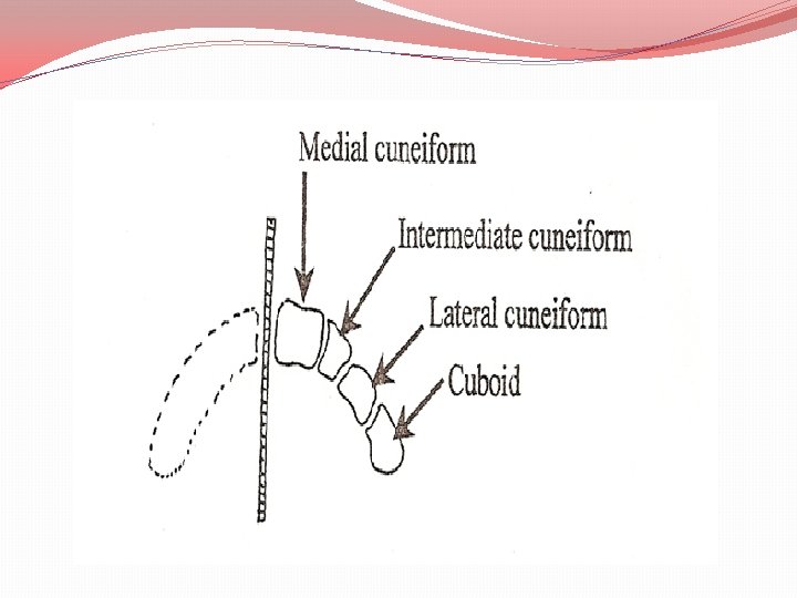
3 anatomical and functional divisions �The hindfoot -talus and calcaneus. �The midfoot navicular, cuboid, and cuneiforms. �The forefootmetatarsals and phalanges.
weight of the body is spread among 3 points �Posteroinferior tuberosity of the calcaneum (heel). �head of first metatarsal. �head of fifth metatarsal.
�Arches of foot is distinctive feature of human �Helps in walking, running, jumping �Helps in weight bearing in upright posture �Foot prints are complete due to arches �Present since birth, masked in infant
FACTOR MAINTAINING stone bridge are �Shape of stones. �Intersegmental ties (staples). �Slings. �Tie beams.
Intersegmental ties or ligament
Beams or bowstrings
SLING
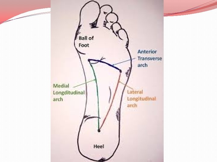
TYPES OF ARCHES
LONGITUDINAL ARCHES Every longitudinal arch has: (a) 2 Pillars (b) a summit (c) joints.
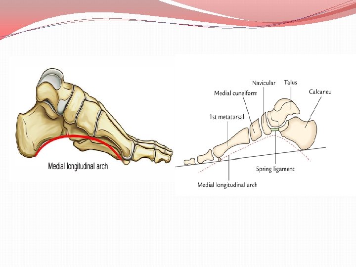
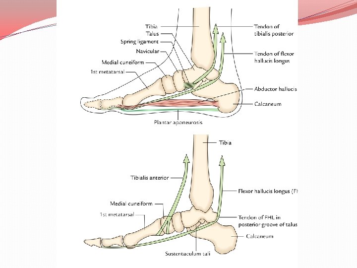
MEDIAL LONGITUDINAL ARCH �The medial longitudinal arch is formed by the calcaneum, talus, navicular, 3 cuneiforms, and medial 3 metatarsals.
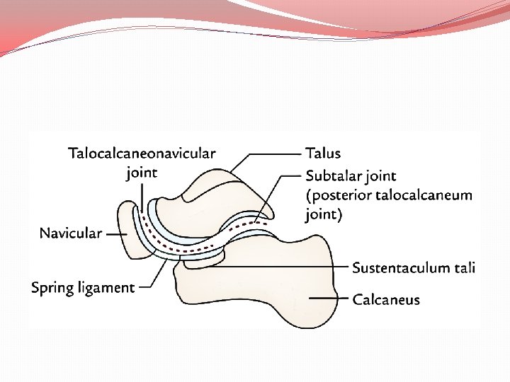
PILLARS �The medial half of the calcaneum forms the posterior pillar of the medial longitudinal arch. �The heads of the medial 3 metatarsals form the anterior pillar of the medial longitudinal arch. SUMMIT �The talus is located at the summit of the arch. For that reason, the talus is the keystone of the arch. JOINTS �The key joints of the medial longitudinal arch are talocalcaneonavicular and subtalar joints.
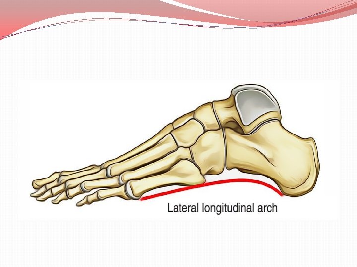
LATERAL LONGITUDINAL ARCH �The lateral longitudinal arch is composed by the calcaneum cuboid and lateral 2 metatarsals.
PILLARS �The posterior pillar of the lateral longitudinal arch is formed by the lateral tubercle of the calcaneum. �anterior pillar is formed by the heads of the lateral 2 metatarsals. SUMMIT �The summit of the lateral longitudinal arch is lies at the level of articular facets on the superior surface of calcaneum (i. e. , at the level of subtalar joint). JOINTS �Calcaneocuboid joint.
FACTOR MAINTAINING MEDIAL LONGITUDINAL ARCH BONES �The sustentaculum tali partially support the head of talus. �Shelf like projection
LIGAMENTS �plantar calcaneonavicular ligament(spring ligament) -which gives dynamic support to the head of talus �interosseous ligaments –connecting the adjacent bones �interosseous talocalcanean ligamentconnecting these bones. �These ligaments serve as intersegmental ties.
MUSCLES, TENDONS AND APONEUROSIS 1. Acting as slings- the tendon of tibialis posterior being located underneath the spring ligament supported by the tendons of flexor hallucis longus. �The flexor hallucis longus is the bulkiest and most powerful muscle 1. It strethetches arch like the string of a bow. 2. It supports the calcaneum by passing underneath the sustentaculum tali. 3. It supports the talus by passing along its posterior groove.
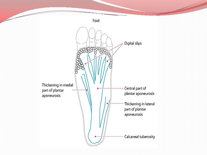
�The tendon of tibialis anterior also has a sling action. 2. Acting as tie beams ( structures which prevent separation of the Pillars). � The medial part of the plantar aponeurosis and abductor hallucis also by flexor hallucis brevis act as tie beam to maintain the height of the medial longitudinal arch.
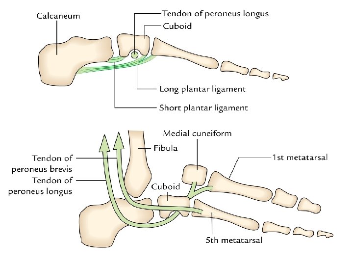
FACTORS MAINTAINING THE LATERAL LONGITUDINAL ARCH BONES �The appropriate formation of the distal end of calcaneus and proximal end of cuboid. �The cuboid is the keystone of longitudinal arch.
LIGAMENTS �Short plantar ligament: broad and thick. It is located deep to the long plantar ligament and supports the calcaneocuboid joint from below. �Long plantar ligament: long and supports the joints between the calcaneum, cuboid, and related metatarsals. �These ligaments serve as intersegmental ties.
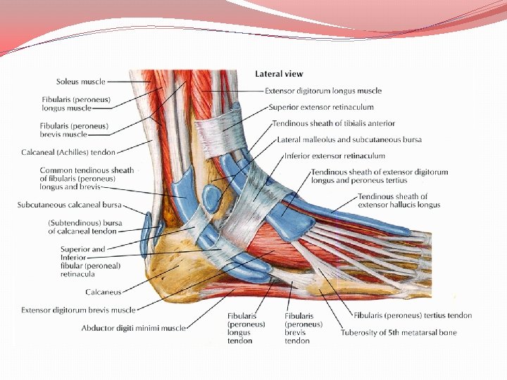
MUSCLES, TENDONS AND APONEUROSIS 1. working as tie beams: The lateral part of the plantar aponeurosis and the intrinsic muscles of the little toe (flexor digitorum brevis, abductor digiti minimi brevis, and flexor digiti minimi brevis) 2. Acting as slings: The tendons of peroneus brevis & peroneus tertius, �The tendon of peroneus longus-plantar aspect of cuboid then crosses attached to base of first metatarsal and adjoining part of medial cuneiform, supports the cuboid bone, it act like pulley
Medial longitudinal arch � Formed by more bones and more joints. �Characteristic feature is resiliency. �Higher and more mobile. � Involved in propulsion during locomotion Lateral longitudinal arch �Formed by less bones and less joints. �Characteristic feature is rigidity. � Lower and less mobile. � Involved in receiving and supporting the body weight
�Summit is formed by the talus. �Main joint is talocalcaneonavicular joint. �Summit is formed by the calcaneum. • Main joint is calcaneocuboid.
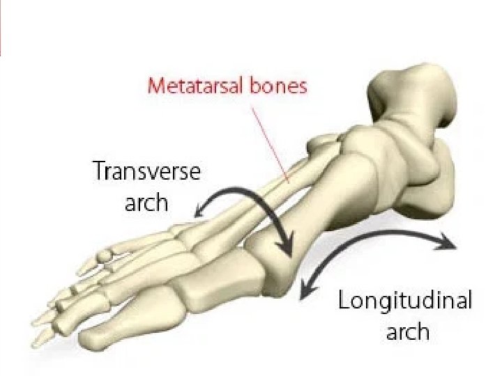
TRANSVERSE ARCHES ANTERIOR TRANSVERSE ARCH �The heads of the metatarsals create the anterior transverse arch. POSTERIOR TRANSVERSE ARCH �The posterior transverse arch is composed by greater parts of the tarsus and metatarsus. Incomplete arch & completed when entire dome is composed.
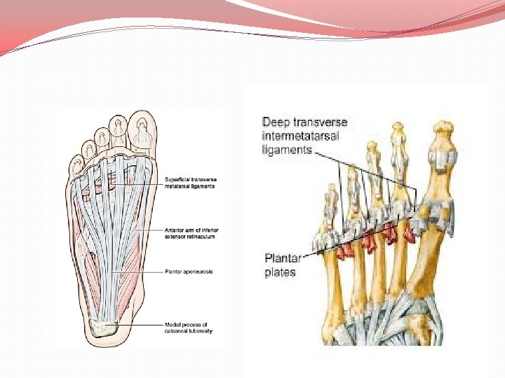
FACTORS MAINTAINING TRANSVERSE ARCHES BONES � The tarsal and metatarsal bones have larger dorsal and smaller plantar surfaces -wedge shaped leads to concavity LIGAMENTS �All ligaments, which bind together the cuneiform bones and metatarsals. �Superficial and deep transverse metatarsal ligaments at the heads of metatarsals function as intersegmental ties
MUSCLES AND TENDONS �Acting as tie beams: The tendons of peroneus longus and tibialis posterior support the transverse arch as tie beam. �Acting as slings: The peroneus tertius and peroneus brevis on the lateral side and tibialis anterior on the medial side support the transverse arch as slings. �Acting as intersegmental ties: The dorsal interossei serve as intersegmental ties.
CLINICAL ANATOMY FLAT FOOT (PES PLANUS) • Commonest of all foot problems. • Collapse of medial longitudinal arch. • During long periods of standing the plantar aponeurosis and spring ligament are overstretched. • the head of talus is lost and is push downward between the calcaneus and the navicular bones.
Flat foot �shuffling gait due to loss of spring in foot. �shock absorbing function lost �The compression of the nerves and vessels �Leads pain in the forefoot (metatarsalgia).
HIGH ARCHED FOOT OR PES CAVUS OR Claw foot �The exaggeration of the longitudinal arch of the foot causes pes cavus. �Planter flexed foot �Due to contracture at transverse tarsal joint
CLUB FOOT/TALIPES �Club foot points down and inwards, with the soles of the feet facing backwards. �Club foot isn’t painful for babies, but it can become painful as they get older and cause difficulties walking if it isn’t treated.
congenital or acquired. 5 types �Talipes equinus (horse like): plantar flexedwalks on the toes- with heel raised. �Talipes calcaneus: walks on the heelforefoot raised �Talipes varus: inverted- walks on the outer border of the foot.
�Talipes valgus: foot is everted- Walks on the inner border of his foot. �Talipes equinovarus: It’s the commonest deformity of the foot is inverted, adducted, and plantar flexed.
HALLUX VALGUS �Great toe adducted towards midline at metatarso phalangeal joint. �wearing of narrow pointed shoesprominence
HAMMER TOE �metatarsophalangeal and distal interphalangeal joints are hyperextended but the proximal interphalangeal joint is intensely bent. �This deformity generally changes the 2 nd and 3 rd toes.
March Foot �Neck of intermediate metatarsals undergo decalcification �Minor injury may lead to pathological fracture �Commonly observed in soldiers
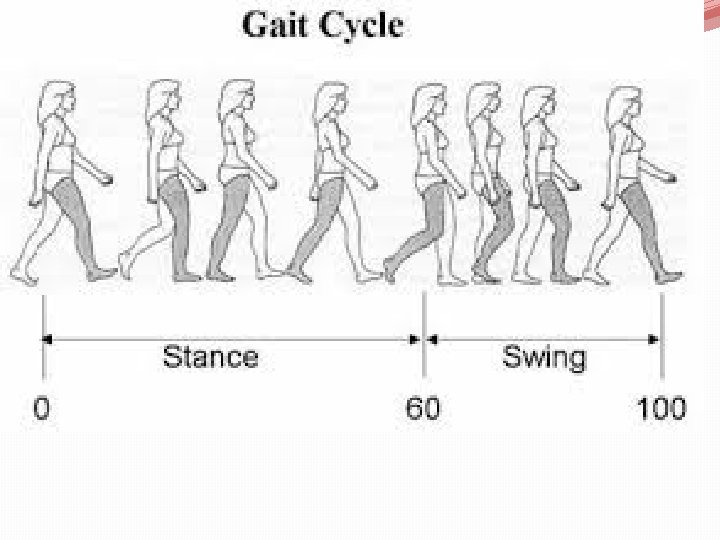
Gait cycle �It is defined as translatory progression of body as a whole produce by coordinated , rotatory movement of body segments. Phases of Gait �Stance �swing
STANCE 1. Heel strike 2. Flat foot 3. Mid stance 4. Heel off 5. Toe off
SWING 1. Acceleration 2. Mid –swing 3. Deceleration
Thank you