Arch of the foot Arches of the Foot


- Slides: 19
Arch of the foot
Arches of the Foot - The skeleton of the foot is built up in an arched form. ** Functions; 1 - Distribution of the body weight on the bones of the foot. 2 - Protection of the structures especially plantar nerve and vessels. 3 - Absorption of shock in falling and jumping. 4 - Act as lever as it propels the body forward in walking and running.
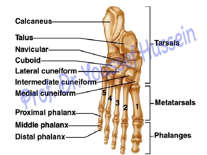
phalanges 3 cuneiform bones Navicular Talus Dorsal aspect Metatarsal bones cuboid Calcaneus
2 3 1 1 1 - Transverse Arch 4 2 34 C CU N T CAL 5 5 2 Longitudinal arch • The longitudin al arch is subdivided into : 1 - medial 2 - lateral
• Longitudinal arch: 1 - Medial longitudinal arch: is formed by - 3 bones: calcaneus ﺍﻟﻌﻘﺐ , talus & ﺍﻟﻘﻌﺐ navicular ﺍﻟﺰﻭﺭﻗﻰ. - 3 cuneiform ﺍﻻﺳﻔﻴﻨﻰ bones. - 3 medial metatarsal ﺍﻟﻤﺸﻄﻴﺎﺕ bones - 3 medial phalangeal bones. 2 - Lateral longitudinal arch: is formed of - 2 bones: calcaneus and cuboid ﺍﻟﻤﻜﻌﺐ. - 2 lateral metatarsal bones - 2 lateral phalangeal bones.
1 - Medial longitudinal arch: - It is higher than the lateral. - Posterior pillar ﺭﻛﻴﺰﺓ medial tubercle of the calcaneus. - Anterior pillar, head of the 1 st metatarsal bone. - Summit (highest point), talus. 2 - Lateral longitudinal arch: - Posterior pillar ﺭﻛﻴﺰﺓ , lateral tubercle of the calcaneus. - Anterior pillar, head of the 5 th metatarsal bone.
Transverse arches Anterior Bases of Metatarsal bones IC LC MC Posterior CU
v Deformities of the foot Flat foot Pes cavus • Flat Foot: loss of the arches of the foot • Pes Cavus: The arches of the foot is high • Hammer Toe: extension of metatarsophalangeal joint and flexion of proximal interphalangeal joint. • Hallux Valgus: lateral deviation of the big toe at the metatarsophalangeal joint. • Talipes Calcaneus, the heel rests on the ground and the toes pointed upwards (dorsiflexion) • Talipes Equinus, plantar flexion of sole of the foot and walking is done on toes without touching the heel to ground Hammer toe Hallux Valgus
** Factors supporting the arches of the foot; 1 - Shape of the bones. 2 - Muscles A. Muscles support the longitudinal arch: 1 - Flexor digitorum longus and flexor hallucis longus. 2 - Tendons of tibialis anterior and posterior. 3 - Short muscles of the sole of the toot • Muscles support the transverse arch 1 - Peroneus longus. 2 - Transverse head of adductor hallucis 3 - Short muscles of the sole of the toot A. 3 - Ligaments; a- Deltoid ligament. b- Spring ligament. c- Short and Long plantar ligaments. d- Superficial and deep transverse metatarsal ligaments 4 - Plantar aponeurosis
Factors support medial longitudinal arch of the foot Tibialis anterior Tibialis posterior Spring lig.
Abductor digiti minimi Abductor hallucis Short muscles of foot
flexor digitorum brevis Short muscles of foot
Transverse head adductor hallucis Short muscles of foot
Deltoid Ligament Neck of talus Tuberosity of Navicular bone Spring ligament Medial malleolus Body of talus Sustentaculum tali
Peroneus longus tendon Short planter ligament Long planter ligament Ligaments and long tendons
Planter aponeurosis It is a very dense thick layer of deep fascia that runs down the middle of the sole. It acts as tie beam maintaining the longitudinal arches of the foot by reinforcing the action of foot ligaments.
Th ank Qu you est ion s