ARBOVIRUSES Dr Mohit Bhatia Assistant Professor Department of

ARBOVIRUSES Dr. Mohit Bhatia Assistant Professor Department of Microbiology All India Institute of Medical Sciences, Rishikesh
 are diverse group of RNA viruses that are transmitted")
Definition: Arboviruses (arthropod-borne viruses) are diverse group of RNA viruses that are transmitted by bloodsucking arthropods (insect vectors) from one vertebrate host to another. Viruses must multiply inside the insects and establish a lifelong harmless infection in them. Viruses that are just mechanically transmitted by insects are not included in this group.

Togaviridae

Flaviviridae

Flaviviridae contd….

Bunyaviridae

Reoviridae

Rhabdoviridae

Arboviruses found in India Dengue, Chikungunya, and Japanese B encephalitis viruses are highly endemic in India

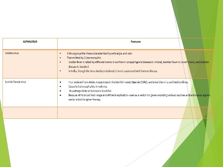
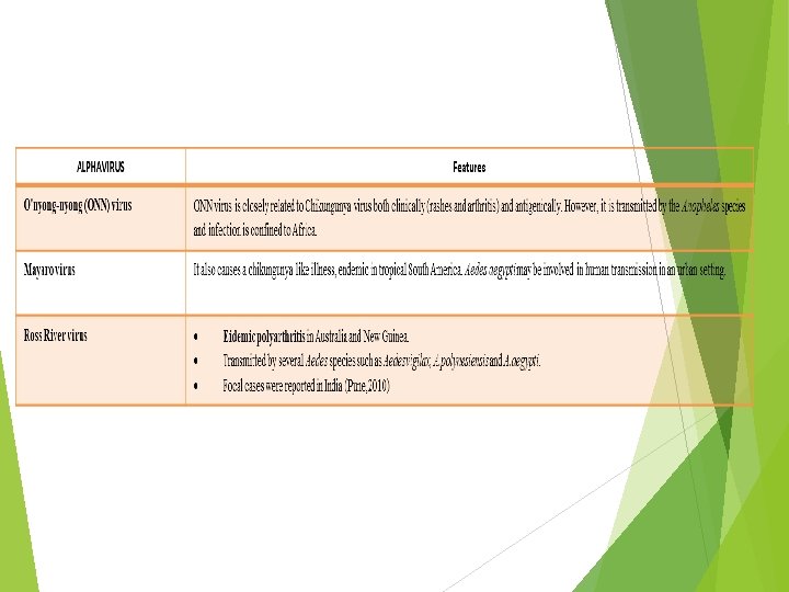
TOGAVIRIDAE- Classification Genus Alphavirus-Contains about 30 different mosquito borne viruses out of which about 13 are human pathogens. Genus Rubivirus -Contains rubella virus, which is not arthropod transmitted and is not an arbovirus.

Morphology Spherical, 50 -70 nm in diameter, Nucleocapsid Capsid contains 42 capsomeres Genome: positive-sense, ss. RNA Enveloped virus- Capsid is surrounded by a lipid envelope that contains two glycoproteins having hemagglutinating activity Replication: They replicate in the cytoplasm and release by budding through host cell membranes. All togaviruses are serologically related to each other.

Based on clinical manifestations, the pathogenic members of the genus Alpha virus can be categorized in to fever-arthritis group and encephalitic groups.

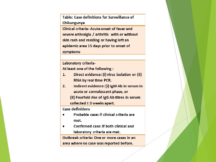
Chikungunya fever is a re-emerging disease characterized by acute fever with severe arthralgia. History-The name is derived from the Makonde word “kungunyala” meaning "that which bends up or gets folded" in reference to the stooped posture which develops as a result of the severe joint pain that occurs during the course of illness. Human Transmission- i) Aedes mosquito, primarily Aedes aegypti which bites during day time ii) Vertical transmission from mother to fetus. iii) Blood transfusion. Transmission cycle Urban transmission cycle- Human beings serve as reservoir during epidemic periods and the transmission occurs between humans and Aedes aegypti. Sylvan transmission cycle occurs usually in African forests involving the wild primates as reservoir (monkeys) and forest species of Aedes (e. g. A. furcifer, A. taylori) as vectors.
 Most common symptoms")
CLINICAL FEATURES Incubation period is about 5 days (3– 7 days) Most common symptoms are fever and severe joint pain (due to arthritis) Arthritis is polyarticular, migratory and edematous (joint swelling), predominantly affecting the small joints of wrists and ankles Other symptoms include headache, muscle pain, tenosynovitis or skin rashes Symptoms are often confusing with that of dengue. In general, chikungunya is less severe, less acute and hemorrhages are rare compared to dengue Most patients recover within a week, except for the joint pain (lasts for months) High risk group- includes newborns, older adults (≥ 65 years), and persons with underlying hypertension, diabetes, or heart disease.

Chikungunya vs Dengue
, was subsequently introduced into Asia")
Epidemiology Chikungunya virus was first reported in Africa(Tanzania, 1952), was subsequently introduced into Asia and had caused several outbreaks in various African and Southeast Asian countries (Bangkok and India) India- Several outbreaks were reported during 1963 -1973; e. g. Kolkata in 1963 and South India in 1964 (Pondicherry Chennai-Vellore) and Barsi in Maharashtra in 1973 Since then, it was clinically quiescent and no outbreaks were reported between 1973 -2005 from most parts of the world, except for the few sporadic cases, which occurred in various places in the world including India (Maharashtra) Re-emergence (Reunion Outbreak )- In 2005, Chikungunya re-emerged in Reunion Island of Indian Ocean and affected 2, 58, 000 people (almost one third of country’s population) Spread- Following this, it has been associated with several outbreaks in India and other Southeast Asian and African countries and has also spread to some areas of America and Europe India (at present)- Chikungunya is endemic in several states States: Karnataka, Tamil Nadu, Andhra Pradesh and West Bengal have reported higher number of cases In 2014, nearly 14, 452 cases were reported, much less than the previous years (95, 091 cases in 2008) Karnataka accounted for the maximum number of cases in the year 2013 & 2014

Genotypes Chikungunya virus has three genotypes- West African, East African and Asian genotypes Most Indian cases before 1973 were due to Asian genotypes However, Reunion outbreak was caused due to a mutated strain and is responsible for most of the current outbreaks in India as well as in other parts of the world The genotypes distribution is due to differences in their transmission cycles; for example, human cycle in Asia and forest cycle in Africa Reasons for re-emergence New mutation (E 1 -A 226 V)-Chikungunya virus underwent an important mutation. Alanine in the 226 position of E 1 glycoprotein gene is replaced by valine New vector (Aedes albopictus)-This mutation led to a shift of vector preference. Mutated virus was found to be 100 times more infective to A. albopictus than to A. aegypti.
and real time reverse transcriptase PCR are")
LABORATORY DIAGNOSIS Virus isolation (in mosquito cell lines)and real time reverse transcriptase PCR are best for early diagnosis (0 - 7 days) Serum antibody detection: Ig. M appears after 4 days of infection and lasts for 3 months. Ig. G appears late (after 2 weeks) and lasts for years. So, detection of Ig. M or a fourfold rise in. Ig. G titer is more significant. n MAC (Ig. M Antibody Capture) ELISA (using virus lysate) is the best format available showing excellent sensitivity (95%) and specificity (98%) with only little cross reactivity with other alphaviruses and dengue. In India, MAC ELISA kits are supplied by National Institute of Virology (NIV), Pune Several other rapid tests (e. g. ICT using envelope antigens) are also available Molecular method: Reverse-transcriptase PCR has been developed to detect specific gene (e. g. ns. P 1, ns. P 4) in blood Biological markers like IL-1β, IL-6 and are increased and RANTES(Regulated on Activation, Normal T Cell Expressed and Secreted) levels are decreased in chikungunya infection Hematological finding: Such as leukopenia with lymphocyte predominance, thrombocytopenia (rare), elevated ESR and C-reactive protein Treatment is by supportive measures, no specific antiviral drugs are available Vaccine- Recently, few vaccine trials are ongoing. In one of these trial, a live measles vaccine virus (Schwarz strain) is used as a vector; into the genome of which five structural genes from chikungunya virus are incorporated.

VEE
 Japanese B encephalitis is")
FLAVIVIRUSES MOSQUITO TRANSMITTED FLAVIVIRUSES ENCEPHALITIS VIRUSES Japanese B Encephalitis (JE) Japanese B encephalitis is the leading cause of viral encephalitis in Asia, including India It was so named as the disease was first seen in Japan (1871) as “Summer encephalitis epidemics” (however, it is now uncommon in Japan)and to distinguish it from encephalitis A (encephalitis lethargica /von Economo disease) which was endemic during that time Vector : Culex mosquito C. tritaeniorhynchusis the major vector worldwide including India C. vishnui is the next common vector found in India Transmission cycle- JE virus infects several extra human hosts, e. g. animals and birds. Two transmission cycles are predominant. Pigs→ Culex→ Pigs Ardeid birds→ Culex→ Ardeid birds

Animal hosts: Pigs have been incriminated as the major vertebrate host for JE. JE virus multiplies exponentially in pigs without causing any manifestation. Pigs are considered as the amplifier host for JE Cattle and buffaloes may also be infected with JE virus; although they are not the natural host. They may act as mosquito attractants Horses are probably the only animal to be symptomatic and show encephalitis Humans are considered as dead end; there is no man→ mosquito →man cycle (unlike in dengue) Bird hosts-Ardeid (wading) birds such as herons, cattle egrets, and ducks can also be involved in the natural cycle of JE virus Geographical distribution: Currently, JE is endemic in Southeast Asian region It is increasingly reported from India, Nepal, Pakistan, Thailand, Vietnam and Malaysia. Because of immunization, its incidence has been declining from Japan and Korea. It is estimated that nearly 50, 000 cases occur every year globally with 10, 000 deaths

In India: JE has been reported since 1955 JE is endemic in 15 states; Uttar Pradesh (Gorakhpur district) accounted for the largest burden Between 2010– 17; total 10, 710 number cases (average 1, 340 cases/year) and 1, 782 deaths (average 222 deaths/year) have been reported In 2017, nearly 2, 040 cases of JE were reported from India with 230 deaths. Maximum cases reported from UP followed by Assam, Manipur, West Bengal, Tamil Nadu, Tripura, Bihar, and Odisha.

Clinical Manifestations-JE is the most common cause of epidemic encephalitis Incubation period is not exactly known, probably varies from 5 -15 days Subclinical infection is common- JE typically shows iceberg phenomena. Cases are much less compared to subclinical/in-apparent infection with a ratio of 1: 300 -1000 Even during an epidemic the number of cases are just 1 -2 per village Clinical course of the disease can be divided into three stages Prodromal stage is a febrile illness; the onset of which may be either abrupt (1 -6 hours), acute (6 -24 hours) or more commonly subacute (2 -5 days) Acute encephalitis stage. Symptoms changes, meningeal signs or paralysis Late stage and sequelae- It is the convalescent stage in which the patient may be recovered fully or retain some neurological deficits permanently. Case fatality ratio is about 20 -40%. include convulsions, behavioral
 ELISA supplied by NIV, Pune has been")
Laboratory Diagnosis Ig. M Capture Antibody (MAC) ELISA supplied by NIV, Pune has been the recommended method for diagnosis of JE It is a two-step sandwich ELISA, uses JERA (JE recombinant antigen) to detect JE-specific Ig. M antibody in serum Reverse-transcriptase PCR has also been developed to detect JE virus specific envelope (E) gene in blood

Vaccine Prophylaxis Live attenuated SA 14 -14 -2 vaccine It is prepared from SA 14 -14 -2 strain It is cell line derived; primary hamster kidney cells are commonly used Single dose is given subcutaneously, followed by booster dose after 1 year It is manufactured in China, but now licensed in India Under Universal Immunization Programme, it is given to children (1 -15 years) targeting 83 endemic districts of four states –UP, Karnataka, West Bengal and Assam
: It is a mouse brain derived formalin inactivated")
Inactivated vaccine (Nakayama & Beijing strains): It is a mouse brain derived formalin inactivated vaccine It is prepared in Central Research Institute, Kasauli (India).
: It is a cell line derived vaccine Combined")
Inactivated vaccine (Beijing P 3 strain): It is a cell line derived vaccine Combined vaccine: A genetically engineered JE vaccine that combines the attenuated SA 14 -14 -2 strain and yellow fever vaccine strain 17 D (YF 17 D) virus as a vector for genes encoding the protective antigenic determinants, has been tested in several clinical trials.

Other Mosquito-borne Encephalitis Flaviviruses Other Flaviviruses Features West Nile virus • First described in West Nile region of Uganda (Africa). • Transmitted among wild birds by Culex mosquitoes in Africa, Middle East, Europe, Asia and recently in America. • Febrile illness (fever, myalgia with rashes on the trunk) • Occasionally it can also cause severe encephalitis. Murray Valley encephalitis viruses • Endemic in northern Australia and Papua New Guinea. • Major mosquito vector is Culex annulirostris

Other Mosquito-borne Encephalitis Flaviviruses Other Flaviviruses Features St. Louis encephalitis viruses • Transmitted by mosquito (Mansonia pseudotitillans). • Related to Japanese B encephalitis virus. • It has caused several outbreaks of encephalitis, mainly affecting the United States. Rocio encephalitis viruses It was first observed in São Paulo State, Brazil, in 1975. It had caused several epidemics of meningoencephalitis in coastal communities in southern São Paulo, Brazil, during 1975. Transmission is believed to be by Culex.

HEMORRHAGIC GROUP OF FLAVIVIRUSES Dengue viruses Dengue virus is the most common arbovirus found in India Serotypes-Dengue has four serotypes (DEN-1, to DEN-4). Recently, the fifth serotype (DEN-5) was discovered in 2013 from Bangkok Vector –Aedes aegypti is the principal vector followed by Aedes albopictus. They bite during the day time. A. aegypti is a nervous feeder (so, it bites repeatedly to more than one person to complete a blood meal) and resides in domestic places, hence is the most efficient vector Aedes albopictus is found in peripheral urban areas, it is an aggressive and concordant feeder i. e. can complete its blood meal in one go; hence is less efficient in transmission Aedes becomes infective only by feeding on viremic patients (generally from a day before to the end of the febrile period)

Extrinsic incubation period of 8 -10 days is needed before Aedes becomes infective Once infected, it remains infective for life Aedes can pass the dengue virus to it’s offspring by transovarial transmission Transmission cycle- Man and Aedes are the principal reservoirs. Transmission cycle does not involve other animals Pathogenesis Primary dengue infection occurs when a person is infected with dengue virus for the first time with any one serotype. Months to years later, a more severe form of dengue illness may appear (called secondary dengue infection)due to infection with another second serotype which is different from the first serotype causing primary infection.

Antibody response against dengue virus Infection with dengue virus induces the production of both neutralizing and non-neutralizing antibodies The neutralizing antibodies are protective in nature. Such antibodies are produced against the infective serotype (which last lifelong) as well as against other serotypes(which last for some time). Hence, protection to infective serotype stays lifelong but cross protection to other serotypes diminishes over few months The non-neutralizing antibodies are heterotypic in nature; i. e they are produced against other serotypes but not against the infective serotype Such antibodies produced following the first serotype infection, can bind to a second serotype; but instead of neutralizing the second serotype , it protects it from the host immune system by inhibiting the bystander B cell activation against the second serotype ADE-The above phenomena is called as antibody dependent enhancement (ADE) which explains the severity of secondary dengue infection Among all the serotypes combinations, ADE is remarkably observed when serotype 1 infection is followed by serotype 2, which also claims to be the most severe form of dengue infection.

Clinical Classification

Factors determining the outcome Infecting serotype- Type 2 is apparently more dangerous than serotypes Sequence of infection- Serotype 1 followed by serotype 2 seems to be more dangerous and can develop into DHF and DSS Age- Though all age groups are affected equally, children < 12 years are more prone to develop DHF and DSS

Global Scenario Dengue is endemic in >100 countries with 2. 5 billion people at risk Tropical countries of Southeast Asia and Western pacific are at highest risk About 50 million of dengue cases occur every year worldwide, out of which 5 lakh cases (mostly children) proceed to DHF

Situation in India Disease is prevalent throughout India in most of the urban cities/towns affecting almost 31 states/Union territories Between 2010– 17, >6 Lakh cases with >1560 deaths have been reported from India. Maximum cases have been reported (in descending order) from West Bengal, Tamil Nadu, Punjab, Kerala, Delhi, Karnataka, and Maharashtra In 2017, nearly 1, 57, 220 cases were reported; maximum of cases were reported from Tamil Nadu followed by West Bengal All four dengue serotypes have been isolated from India Serotype prevalence varies between seasons and places, but DEN-1 and DEN -2 are widespread. DEN-5 has not been reported yet

Laboratory diagnosis NS 1 antigen detection-ELISA and immunochromatography based rapid cards are available for detecting NS 1 antigen in serum. They gained recent popularity because of the early detection of the infection NS 1 antigen becomes detectable from day-1 of fever and remains positive up to 18 days Highly specific Antibody detection Primary infection- Antibody response is slow and of low titer. Ig. M appears first after 5 days of fever and disappears within 90 days. Ig. G is detectable at low titer in 14 -21 days of illness, and slowly increases Secondary infection- Ig. G antibody titers rise rapidly. Ig. G is often cross reactive with many flaviviruses and may give false positive result after recent infection or vaccination with yellow fever virus or JE. In contrast, Ig. M titer is significantly low and may be undetectable Past infection- Low levels of Ig. G remain detectable for over 60 years and, in the absence of symptoms, is a useful indicator of past infection.
: This is the recommended serological testing in")
MAC-ELISA (Ig. M antibody capture ELISA): This is the recommended serological testing in India. Kits are supplied by NIV, Pune Principle: It is a double sandwich ELISA; which captures human Ig. M antibodies on a microtiter plate using anti-human-Ig. M antibody followed by the addition of dengue virus four serotypes specific envelope protein antigens (this step makes the test specific). There is a signal enhancement due to use of avidin-biotin complex (ABC) which makes the test more sensitive Cross-reactivity with other flaviviruses is a limitation of this test.

Ig. G specific ELISA format is also available separately Rapid tests such as dipstick assays are also available
 CFT (Complement")
Other antibody detection assays used previously are: HAI (Hemagglutination inhibition test) CFT (Complement fixation test) Neutralization tests such as plaque reduction test, neutralization and micro neutralization tests
 for dengue Rapid diagnostic tests (e. g. ICT) for dengue")
Rapid diagnostic tests (RDT) for dengue Rapid diagnostic tests (e. g. ICT) for dengue Ig. M antibodies or NS 1 antigen are available, but have poor sensitivity and specificity. Government of India had passed an order in 2016, that a positive RDT for dengue NS 1 or Ig. M should be considered as probable diagnosis; must be confirmed by ELISA.

Virus isolation Dengue virus can be detected in blood from – 1 to +5 days of onset of symptoms. Virus isolation can be done by inoculation into mosquito cell line or in mouse.
 by real time")
Molecular Method Detection of specific genes of viral RNA (3’-UTR region) by real time RT-PCR: It is the most sensitive (80– 90%) and specific assay (95%), can be used for detection of serotypes and quantification of viral load in blood Viral RNA can be detected in blood from – 1 to +5 days of onset of symptoms Genotype detection: Each serotypes of dengue virus comprises of several genotypes which can be detected by molecular typing A total of 13 genotypes have been detected so far; three for DENV-1, two for DENV-2, four each for DENV-3 and 4 serotypes respectively Dengue virus keep undergoing genetic alterations leading to introduction of new genotypes and also a shift between the existing genotype lineages within a serotype; which may attribute to rapid increase in the clinical severity (DHF and DSS) in many parts of the world. Hence, there is need for close molecular monitoring of dengue virus.

Treatment There is no specific antiviral therapy. Treatment is symptomatic & supportive such as- Replacement of plasma losses Correction of electrolyte and metabolic disturbances Platelet transfusion if needed

Prevention Vaccine Mosquito control measures

Dengue vaccine After several trials, a dengue vaccine has been licensed for human use since 2015 It is a Chimeric Yellow Fever-Dengue, Live-Attenuated, Tetravalent Dengue Vaccine (CYD-TDV); commercially available as dengvaxia (developed by Sanofi Pasteur). It uses live attenuated yellow fever (YF) 17 D virus as vaccine vector in which the target genes of all four dengue serotypes are integrated by recombinant technique. Age: It is indicated for 9 -45 year of age. Schedule – 3 injections of 0. 5 m. L administered subcutaneously at 6 month intervals It is available as lyophilized form; reconstituted with normal saline. Contraindications: (i) allergic reactions to vaccine; (ii) immunodeficient Individuals (e. g. HIV) (iii) Pregnant and breast feeding women Efficacy against hospitalized dengue illness was found around 80 %. WHO recommends this vaccine to start in high burden countries (seroprevalence >70%). Currently, the vaccine is approved in Mexico, Philippines, Brazil, Indonesia, Thailand Singapore. In India, it is not available yet

Zika virus ss. RNA virus, belongs to family Flaviviridae Monkeys are the reservoirs Place of discovery (1947), Zika forest in Uganda Transmission: o Mosquito transmitted virus o Mosquito borne—Aedes aegypti, Aedes albopictus o Mother-to-child : Common in first trimester o Sexual transmission: Transmission has been observed from (i) asymptomatic males to their female partners. Longer shedding of Zika virus in semen has been reported, (ii) Symptomatic females to their male partners.

Epidemiology The first outbreak was reported in 2007 in Yap Islands (49 confirmed and 59 probable cases ) Aedes hensilli was the predominant Mosquito Recent Outbreak (2015 -2016): o It began in April 2015 in Brazil and then subsequently spread to other countries in South America, Central America, the Caribbean, Europe USA and Australia o In February 2016, the WHO declared the Zika virus outbreak a public health emergency of international concern Situation in India: 3 confirmed cases (Gujarat in 2017); first report from India. As the vector is prevalent, India has a higher risk of getting affected by ZIKV in near future.

Clinical Manifestations Incubation period - few days to 1 week Asymptomatic: symptomatic ratio is 5: 1 Zika fever- Symptomatic people develop minor illness such as fever and a rash, conjunctivitis Congenital transmission leads to development of fetal anomaly such as microcephaly Few cases of Guillain-Barré syndrome have been reported in patients with ZIKV disease.
 - investigation of choice. o o o Detect")
Lab Diagnosis Reverse transcriptase PCR (RT-PCR) - investigation of choice. o o o Detect ZIKV RNA in blood and urine up to 5 and 7 days of onset of symptoms respectively. Multiplex real time RT-PCR – available Targets - non-structural 5 (NS 5) region of ZIKV, non-structural protein 4 (ns. P 4) from CHIKV and 3′ untranslated region (3′UTR) of DENV 1– 4 Ig. M antibody detection (ELISA): It appears in blood after 1 week of symptoms and remain positive up to several months. It cross reacts with dengue antibodies Plaque-reduction neutralization test is more specific serological (antibody detection) test; but it is cumbersome, not widely used.

Treatment Zika virus No effective treatment Vaccine Only – not available symptomatic treatment available such as fluid replacement and analgesic such as acetaminophen.
: o Most advanced trial is the one")
ZIKV Vaccine ZIKV DNA vaccine (VRC 705): o Most advanced trial is the one conducted by Vaccine Research centre (VRC) under NIAID, USA o Genetically engineered plasmid encoding Zika virus protein o Entered phase-2 vaccine trail in 2017 and is expected to get completed by 2019 Killed ZIKV vaccine: Another trial (phase-I) is been evaluated by Bharat Biotech’s Hyderabad, India. It uses an African strain of ZIKV.

General preventive measures Mosquito control measures Infected patients should prevent mosquito bites for the first week of illness Sex/ pregnancy restriction: CDC has recommended for considering condom use or abstinence for at least 6 months for males and 8 weeks for females after travel to endemic area or developing symptoms or ZIKV diagnosis During outbreak time (2015 -16): o Affected countries such as Brazil, and others have advised women to postpone getting pregnant until more was known about the risks. o Travel of pregnant women from other countries (including India) to ZIKV affected countries have been restricted.

Yellow fever virus Acute, febrile illness Geographical distribution- Endemic in West Africa and Central South America. Not found in the rest of the World including India. Typing- Seven genotypes and one serotype. Vector- Aedes aegypti or the tiger mosquito. Transmission cycle: o Jungle cycle - Between monkeys and forest mosquitoes. Humans can only get infection occasionally during their forest visits. o Urban cycle - Occurs between humans and urban mosquitoes (Aedes aegypti)

Yellow fever virus India- Not invaded, but potential of developing because the vector, A. aegypti, is widely distributed here, and India has the tropical climatic condition similar to Africa. Various reasons have been hypothesized to explain the absence of yellow fever in India: Measures in airports: Govt. of India has laid down strict guidelines for vigilance and quarantine of the travelers in the international airports Unprotected (i. e. unvaccinated) travelers coming from endemic zone to India will be kept in quarantine for 6 days Breteau index or the Aedes aegypti index should be less than one, surrounding 400 mt of an airport (Breteau index - No. of containers showing breeding of Aedes aegypti larvae / No. of houses surveyed × 1000)

Yellow fever virus India has all the potential of developing yellow fever in future because the vector A. aegypti, is widely distributed here, and India has the tropical climatic condition similar to Africa. Various reasons have been hypothesized to explain the absence of yellow fever in India: o Measures in airport: Govt. of India has laid down strict guidelines for vigilance and quarantine of the travelers in the international airports. o Cross reacting dengue antibody provides protection against yellow fever. However, yellow fever immunization does not protect from dengue Period of communicability: o Man- Patients are infective to mosquito during the first 3 -4 days of illness o Aedes- After an extrinsic incubation period of 8 -10 day, the mosquito becomes infective and once infected, remains infective for life.

Clinical manifestations Incubation period is about- 3 -6 days Febrile illness Severe cases – o Hemorrhagic manifestations o Platelet dysfunction, o Features of liver involvement (hepatitis) are- o Mid-zonal necrosis and presence of councilman bodies o Intranuclear inclusions may be seen inside the hepatocytes called as Torres bodies. o Appearance of jaundice o Renal dysfunction o Encephalitis occurs very rarely. o Mortality rate is high (>20%), especially among children & elderly.

Epidemiology It is estimated that about 200, 000 cases of yellow fever with 30, 000 deaths occur annually and majority of outbreaks (~90%) occur in Africa Epidemics usually occur in humid and semi-humid savanna area adjoining a rain forest Infection: case ratio during epidemic ranges from 20: 1 to 2: 1 All age groups are susceptible

Laboratory diagnosis Serology: Ig. M ELISA can be done after 3 days of onset of symptoms Molecular method: RT-PCR detecting specific viral RNA (NS 5 region) in blood is more confirmatory than serology

Yellow fever 17 D vaccine Live attenuated vaccine Prepared from allantoic cavity of chick embryo In India- It is prepared in Central Research Institute(CRI), Kasauli Dosage- Single dose, given subcutaneously Vaccine is effective within 7 days of administration, which lasts for 35 years Validity of yellow fever vaccine certificate: Certificate is issued after 10 days of vaccination and renewed (i. e. re-immunization) every 10 years

Yellow fever 17 D vaccine Contraindications of yellow fever vaccine includeo Children <9 m, (<6 m- during epidemic), o Pregnancy(except during outbreak), o HIV infected people o People with allergy to egg
 Viruses Powassan Encephalitis Viruses Louping-ill Virus")
ENCEPHALITIS GROUP Tick-borne Encephalitis (TBE) Viruses Powassan Encephalitis Viruses Louping-ill Virus
 Virus Vector: Hard ticks (Haemaphysalis spinigera) Hosts:")
HEMORRHAGIC FEVER GROUP Kyasanur Forest Disease (KFD) Virus Vector: Hard ticks (Haemaphysalis spinigera) Hosts: Monkeys, rodents and squirrels are common hosts Seasonality: KFD is increasingly reported in dry months (January-June) which coincides with human activity in forest

Clinical Manifestation in Humans Incubation period varies from 3– 8 days o First stage (hemorrhagic fever) o Second stage in the form of meningoencephalitis

Laboratory Diagnosis is made by virus isolation from blood or by Ig. M antibody detection by ELISA Recently, nested RT-PCR and real time RT-PCR have been developed detecting viral RNA (NS-5 non-coding region) in serum samples and can provide early, rapid and accurate diagnosis of the infection

VACCINE Killed KFD Vaccine - formalin-inactivated chick embryo vaccine - developed for KFD in the Haffkine institute, Mumbai Schedule: Two-doses at interval of 2 months, followed by booster doses at 6– 9 months and then every 5 years Target area: KFD vaccine is recommended in endemic areas of Karnataka (villages within 5 km of endemic foci).

Thank you
- Slides: 70