April 26 2018 Scottsdale AZ Flexible Rigid Endoscopy

April 26, 2018 Scottsdale, AZ Flexible & Rigid Endoscopy Workshop: Advanced Course Marti Evans, PA-C Eighth Annual ENT for the PA-C | April 25 – 29, 2018| Scottsdale, AZ

Flexible & Rigid Endoscopy Workshop: Advanced Course Basic Instruction Demonstration Hands-On Practice Learn by doing Identify abnormal pathology Perform flexible endoscopy adult Perform flexible endoscopy child/infant Perform rigid endoscopy

Introduction There are multiple methods and techniques available to successfully complete all the topics presented in this workshop. Some are based on patient request, available equipment or supervising physician’s preference. The goal of this workshop is to correctly demonstrate the most common methods and give participants time for hands-on training. Eighth Annual ENT for the PA-C | April 25 – 29, 2018| Scottsdale, AZ

Objectives Learning Objectives • Identify normal anatomy, normal variants and abnormal findings visible via flexible nasopharyngoscopy. • Understand indications and perform flexible and rigid scope examination - adult. • Understand indications and perform flexible scope examination - child/infant. • Perform exam, intranasal culture and sinus debridement using rigid scope adult. Additional opportunities: Practice packing on epistaxis trainer Practice placing drug-eluting implant

Flexible Endoscopy See workstations for demonstration and practice of flex endoscopy Eighth Annual ENT for the PA-C | April 25 – 29, 2018| Scottsdale, AZ

Indications for Flexible Endoscopy • Strong gag reflex* • Failed mirror exam* • Nasal obstruction – – – Foreign Body Septal deviation Adenoid hypertrophy Nasal mass Unilateral otitis media Polyps • Sinusitis • Chronic throat pain • Chronic cough • Dysphonia – – – Presbylarynx VC paralysis VC Nodules LPR Neoplasms • Dysphagia – Candidiasis • Odynophagia • Symptoms of aspiration – Laryngomalacia – Angioedema *Documentation of a strong gag reflex and/or failed mirror exam should be included in notes. It may justify procedure for billing purposes.
 • Relative: – Coagulopathy – Craniofacial trauma Eighth Annual")
Contraindications • Epiglottitis (by inexperienced) • Relative: – Coagulopathy – Craniofacial trauma Eighth Annual ENT for the PA-C | April 25 – 29, 2018| Scottsdale, AZ

Know Your Anatomy
 *With Mirror Laryngoscopy, image is inverted. 1. True vocal cords 2.")
Laryngeal Anatomy (Mirror*) *With Mirror Laryngoscopy, image is inverted. 1. True vocal cords 2. False cords 3. Epiglottis 4. Aryepiglottic folds 5. Arytenoids 6. Pyriform sinuses 7. Base of Tongue

Laryngeal Anatomy, Endoscopic view* True Vocal Cords abducted True Vocal Cords adducted *With flexible endoscopy, images are true.
")
Review Tips For Starting the Exam • Patient informed of the procedure (obtain consent) • Proper positioning – Sniffing, head supported, use non-dominant hand to steady the patient’s head – Choose the more patent of the nares • Appropriate equipment – Adult vs. Pedi – Decongestant/anesthetic – Gloves – Chair – Photographic/video accessories – Biopsy materials if needed – Lubricant +/-

Preparation of the Patient for Nasal Endoscopy • Ask the patient to blow the nose. • Assess which is more patent of the nares. • Apply topical decongestant • Apply topical anesthetic • OR apply combination anesthetic/decongestant spray Photo Courtesy Bernadine Sonnier 2011

Flexible Nasal Endoscopy: Technique • • • Topical decongestant/anesthetic Two hands required to drive scope Thumb drive or index finger drive May add lubricant to leading edge of scope Improved image quality with digital flexible endoscopes Generally smaller diameter and well tolerated even in tight areas Flexible end allows wide range of visualization Can easily pass into NP and visualize pharynx and larynx Flex scope is a poor tool for obtaining culture or biopsy

Flexible Nasal Endoscopy Eighth Annual ENT for the PA-C | April 25 – 29, 2018| Scottsdale, AZ

Causes of Nasal Obstruction Nasal Foreign Body Septal deviation Synechiae Turbinate hypertrophy Sinusitis Adenoid hypertrophy Nasopharyngeal masses Nasal polyps Eighth Annual ENT for the PA-C | April 25 – 29, 2018| Scottsdale, AZ

Septal Deviation/ Turbinate Hypertrophy

Synechiae • Fused intranasal tissues are called adhesions or synechiae. • Adhesions are a common, usually minor complication of nasal or sinus surgery and nasal packing. • They also may develop because of trauma (nosepicking or cocaine use) and in such conditions as syphilis, tuberculosis, lupus, or sarcoidosis.

Nasal Polyps • Nasal polyps are common, noncancerous, teardrop-shaped growths that form in the nose or sinuses, usually around the area where the sinuses open into the nasal cavity. • Mature nasal polyps look like seedless, peeled grapes Top image polyps viewed via nasal speculum. Bottom image polyps viewed via flex scope intranasal exam. Mercado 2011 ©

Sinusitis Mercado 2013 ©

Sinusitis • Sinusitis is diagnoses by history and physical examination. • Fiberoptic endoscopy (flexible or rigid) is ideal for evaluating the osteomeatal complex Image of fungal sinusitis. Note thick debris vs purulent secretions in previous images.

Adenoid Hypertrophy Adenoids are described based on % of obstruction – No obstruction – Partial (percentage) obstruction – Complete obstruction • Endoscopic view of the nasopharynx showing adenoidal obstruction of the choana. Mercado 2011 © Mercado 2013 © http: //icarus. med. utoronto. ca/carr/atlasoutline. htm

Juvenile Angiofibroma Juvenile angiofibroma – benign, highly vascular invasive mass that occurs in the posterior nasal cavity in less than 0. 5% of head and neck tumors. Almost always found in adolescent boys. Presents with epistaxis and nasal congestion or both. CT Scan w/contrast confirmed nasopharyngeal mass measuring 4. 0 cm x 3. 8 cm consistent with a juvenile angiofibroma.

Tornwaldt Cyst • A Tornwaldt cyst is a midline pouch within the posterior roof of the nasopharnx which is caused by an adherence of the notochord to the pharyngeal ectoderm during development, resulting in the outpouching of ectoderm into the pharyngobasilar fascia. • They are typically asymptomatic but may present with middle ear symptoms (eustachian tube obstruction), halitosis (when associated with a leaking sinus tract), foul taste in mouth, and, rarely, occipital headaches. • These lesions will demonstrate low to increased signal on T 1 -weighted images depending on the protein content of the mucus. They will be high signal on T 2 weighted images and will not enhance.

Unilateral Otitis Media Mercado 2011 © Unilateral Otitis Media in adults should raise suspicion of Nasopharyngeal mass blocking Eustachian Tube orifice which can cause unilateral middle ear effusion. • 1. 5: 100, 000 patients, more common in Asian and Alaskan people 20: 100, 000 • Top image normal Torus Tubarius • Bottom image opening covered with mucosal tissue (squamous cell carcinoma).

Voice Change Voice overuse/misuse Vocal cord nodules/polyps Laryngopharyngeal Reflux Vocal Cord Paralysis Presbylarynx Laryngeal neoplasms Eighth Annual ENT for the PA-C | April 25 – 29, 2018| Scottsdale, AZ

Mercado 2014 © • Cervical osteophytes are bone spurs that grow on any of the seven vertebrae in the cervical spine (C 1 - C 7 vertebrae). • More than half of people over the age of 60 have osteophytes somewhere in their bodies. Osteophytes in the spine are a normal sign of aging and usually do not cause symptoms. However, neurological symptoms or pain may occur if the osteophytes encroach upon the individual spinal nerves, the spinal cord itself, the vertebral discs, or the blood vessels in the region of the cervical vertebral column. • The inflamed or damaged tissue that stimulates cervical osteophyte growth is often caused by cervical osteoarthritis, a degradation in the neck joints that occurs in many older people. These joints include the disc spaces themselves (a modified joint) and the facet joints, and this condition of cervical osteophyte formation is referred to as cervical spondylosis.

Adult Mannequin Mercado 2013 © Advanced airway and custom laryngeal mannequins available to practice flexible endoscopy technique.

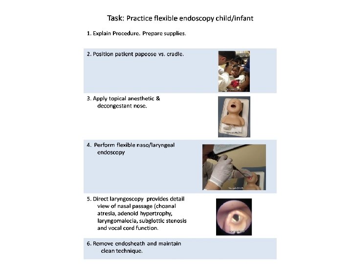
Task: Practice flexible endoscopy 1. Explain Procedure. Prepare supplies. 2. Position patient. 3. Apply topical anesthetic & decongestant nose. Mercado 2011 © 4. Perform flexible naso/laryngeal endoscopy. Mercado 2011 © 5. Direct laryngoscopy provides detail view of nasal passage and vocal cord function. 6. Remove endosheath and maintain clean technique. Mercado 2011 ©

Respiratory Complications Children • Must rule out foreign body aspiration • Choanal Atresia • Laryngomalacia • Subglottic stenosis Adults • Angioedema Eighth Annual ENT for the PA-C | April 25 – 29, 2018| Scottsdale, AZ

Angioedema Vascular leakage beneath the dermis and subcutanis. Response is mediated by vasoactive mediators, i. e. , histamine, serotonin, and kinins (eg, bradykinins), which cause the arterioles to dilate while inducing a brief episode of vascular leakage in the venules, where the junction between the endothelial cells appears looser than in the capillaries and arterioles. Angioedema with or without urticaria, is classified as allergic, hereditary, or idiopathic. Complications range from dysphonia or dysphagia to respiratory distress, complete airway obstruction, and death. Symptoms - Severe facial/oral edema, Urticaria – (hives) food allergy, erythema. Mercado 2011 © Most common cause - ACE Inhibitor sensitivity, Food allergies such as fresh berries, shellfish, nuts, tomatoes, eggs, milk, chocolate, food additives, and preservatives Treatment - H 1 (antihistamine), H 2 (antacid), Steroids. Fresh Frozen Plasma, Protect airway. Mercado 2011 ©

Angioedema Mercado 2011 © Edema floor of mouth Edema epiglottis Edema uvula Edema arytenoid

Flexible scope exam on children. • Generally well tolerated by children. • Explain procedure in detail. • Secure patient (papoose vs. cradle). • Anesthesia vs. decongestant? • Give adequate time for anesthesia/decongestant. • Provides better visualization Mercado 2013©©

Flexible scope exam on infants. • Generally well tolerated by infants. • Explain procedure in detail. • Secure patient (papoose vs. cradle) • Anesthesia vs. decongestant? • Give adequate time for anesthesia/decongestant. • Provides better visualization Mercado 2013©©

Flexible scope exam on infants and children Evaluating pediatric airway via flexible scope • Assess nares/choanae (choanal atresia) • Assess adenoid and lingual tonsil (hypertrophy) • Assess Airway – Epiglottis (laryngomalacia) – TVC mobility (paralyzed vocal cords) – Assess laryngeal structures (stenosis) – Foreign bodies

Flexible scope exam on infants and children • A child's airway differs from that of an adult in that the child's tongue is proportionately larger in the oropharynx compared to that of an adult. • A child's airway is smaller, softer and more prone to foreign body obstruction. • The trachea is usually about the diameter of a pencil.

Flexible fiberoptic exam on infants and children Difference Pediatric vs Adult Airway Relatively larger tongue Angled vocal cords Obstructs airway Infant’s vocal cords have more angled Obligate nasal breathers attachment to trachea, whereas adult vocal Difficult to visualize larynx cords are more perpendicular Image from: http: //www. utmb. edu/otoref/Grnds/Pedi-airway-2001 -01 -slides. pdf

Laryngomalacia • Most common congenital abnormality of the larynx. • Most prominent symptom: inspiratory stridor. • Immature development results in soft laryngeal walls that close airway. • Child outgrows. Usually, no treatment necessary. • Omega-Shaped epiglottis Mercado 2011 ©

http: //www. drrahmatorlummc. com/congenitaldevelopmental. htm Class III Narrowing of subglottis can be congenital or acquired in etiology. Its nature can be membranous, cartilaginous, or mixed, with or without combination of glottic or upper tracheal stenosis. The lower limit of normal subglottis dimension in full term infant is 4. 0 mm and in premature infant 3. 5 mm. Circumferential edema of 1 mm reduces its crosssectional area by 60%. Myer-Cotton grading system is a useful classification for mature circumferential subglottic stenosis. It is divided into 4 grade as below: : Grade I - Obstruction of 0 -50% of the lumen obstruction Grade II - Obstruction of 51 -70% of the lumen Grade III - Obstruction of 71 -99% of the lumen Grade IV - Obstruction of 100% of the lumen (ie, no detectable lumen) Class I Subglottic Stenosis

Infant/Child Mannequin Mercado 2013 © Practice mannequins available to practice flexible fiberoptic endoscopy technique.

Rigid Endoscopy See workstations for demonstration and practice of rigid endoscopy Eighth Annual ENT for the PA-C | April 25 – 29, 2018| Scottsdale, AZ

Rigid Scope

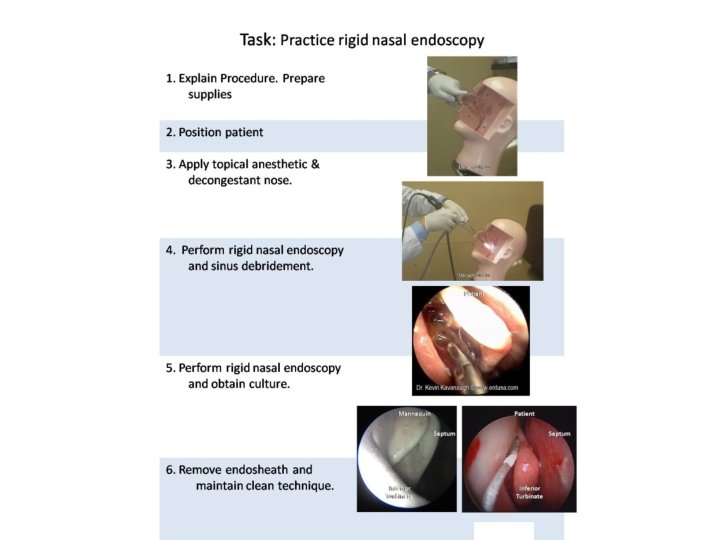
Rigid Scope The rigid endoscope provides superior image clarity, facilitates culture and tissue sampling, controls epistaxis, and affords the endoscopist the ability to perform surgery. Rigid endoscopes for the nose come in diameters of 2. 7 -4 mm and have tips of different angles (generally 0 -70º), allowing the clinician to visualize various sinuses and areas within the nasal cavity. This facilitates culture of the sinuses and debridement postoperatively.

Rigid Nasal Endoscopy One hand to drive scope • Second hand available for: 1. culture 2. debridement 3. epistaxis 4. biopsy • • • Superior illumination & image quality Identify pathology in 40% of patients with normal anterior rhinoscopy Generally 0, 30, 45, 70 degree angles 2. 7 mm and 4. 0 mm Uncomfortable if contacts septum, middle turbinate and sphenoethmoidal recess

Nasal Endoscopy 3 Passes of Endoscope • Low - Nasal floor to Nasopharynx • Mid - Middle turbinate/M. Meatus to SER • High - Cribriform fossa

Complications • • • Tearing Epistaxis Coughing Laryngospasm – rare Bleeding Advise patient not to eat or drink anything 1 hour after procedure.

Rigid Scope for cultures Nasal Septum Mercado 2011 © Inferior Turbinate Nasal endoscopic examination and culture for definitive diagnosis best accomplished with rigid scope. Small culturette used to obtain mucous & pus sample from hiatus semilunaris. Be sure not to touch other tissue as this may contaminate specimen.

Nasal Cultures • Nasal cultures are not routinely indicated in firstline management of Acute Rhinosinusitis • Endoscopically guided microswab or suction aspiration culture of a draining sinus ostium are a strong consideration in Chronic Rhinosinusitis, especially when poorly responsive to prior antibiotics. Middle Turbinate Microswab

Rigid Endoscopy After FESS Mercado 2011 © Dr. Kevin Kavanaugh © www. entusa. com Post operative debridement following FESS is best accomplished with rigid scope. Rigid scope allows clinician to visualize area and suction or use forceps.

Steroid Eluting Sinus Implants a. k. a. “Stents” Eighth Annual ENT for the PA-C | April 25 – 29, 2018| Scottsdale, AZ

Steroid-Releasing Implants • FDA-approved localized, controlled drug delivery technology for sinus surgery patients, placed intraoperatively – delivery of Mometasone Furoate over 30 days – provide middle turbinate and/or ostial support – bioreabsorbs after 4 -6 weeks • Intended to maintain surgical result by preventing inflammation & scarring

Intraoperative Placement of Steroid Eluting Implants Placement in Ethmoid Sinus Anterior edge of MT Plunger Achieve Hemostasis Advance Delivery System with 10° tip angled superiorly Align distal tip of green plunger w/ Anterior Edge Middle Turbinate Placement in Frontal Sinus placement after Frontal Sinus Surgery Slow controlled deployment to ensure uniform apposition

Steroid-eluting Sinus Implant • Newly-approved for treatment of nasal polyps in adults who have had previous ethmoid sinus surgery, placed in the office setting. Eighth Annual ENT for the PA-C | April 25 – 29, 2018| Scottsdale, AZ

Placement of Implant STEP 1 STEP 2 Orient 10 curve of distal Advance Delivery System tip superiorly with shaft parallel to roof of ethmoid sinus STEP 3 STEP 4 Release Implant by pressing down on Thumb Rest while pulling back on Finger Rests Position Implant in nasal polyps with cap pointing posteriorly, and as superiorly as possible Note: Implant should not be re-crimped / re-implanted more than twice to maintain radial strength.

Rigid Scope Practice Mercado 2013 © Demonstration of Rigid Endoscopy technique. Custom-made models available to practice Post-FESS Debridement and collecting culture specimen of sinus

Rigid Scope EXAM: Practice on Phacon Sinus model Post-FESS Polyp *PLEASE DO NOT REMOVE!

Care of Scope Eighth Annual ENT for the PA-C | April 25 – 29, 2018| Scottsdale, AZ

Endosheaths Loosen endosheath Mercado 2011 © Pull endosheath from distal end Mercado 2011 ©

Review of Scope Care Avoid bending scope in tight angles. Clean lens with lens cleaner/paper. Pre-clean with enzymatic cleaner. Soak only for required period depending on brand manufacture. • Store in dry safe place. • Perform regular leak testing to avoid damage. • • Eighth Annual ENT for the PA-C | April 25 – 29, 2018| Scottsdale, AZ

Cold Sterilization • Flexible scopes are nonautoclavable. • Rigid scopes can be autoclaved. • Clean length of scope with an enzymatic detergent solution like ENZOL® to remove debris and reduce bacterial burden before instruments are disinfected or sterilized. DO NOT ALLOW debris to dry. • Soak scope in a glutaraldehyde solution like Cidex ® which provides quick high-level disinfection. • Noncorrosive solution reduces instrument damage and associated repair costs. • Soaking times vary by product. Mercado 2011 ©

Leak Testing 1. Routine leak testing in accordance with specific manufacture depending on volume of use. 2. Introduce air pressure via attached bulb (DO NOT overinflate) and submerge looking for leaks. 3. Leaks can slowly damage internal parts causing expensive yet preventable damage. Mercado 2011 ©

Flexible & Rigid Scopes: Advanced Screen D o o r Station 5 Video Tower Rigid Phacon model Practice exam BALLROOM D 35 x 72 24 learners per session 33 chairs, 10 6 ft tables, AV setup Station 6 Video Tower Rigid Phacon model Practice stents Proctors Station 2 Station 3 Projector Speaker Station 4 Collect culture Rigid and suction cutaway models POSSIBLE Station 7 ADVANCED EPISTAXIS TRAINER Tower or tabletop system D o u b l e Station 3 DScope system Adult exam Station 2 DScope system Adult exam D o o r Station 1 Pediatric Station 1 Towers pull 10 amps each

Reminder: complete your workshop cards and turn them in at the end of this session. • Score cards will be used for admission to workshops and attendance. • Credit will only be awarded for completed score cards. • Rotate and complete each station. • Completion of workshop is NOT contingent on pass/fail Eighth Annual ENT for the PA-C | April 25 – 29, 2018| Scottsdale, AZ

Flex and Rigid Scopes: Advanced Session Evaluation Score cards will be used for admission to workshops and attendance. Credit will only be awarded for completed score cards. Name Scale: 1=NO or LOW, to 5=YES or most likely/most positive Session 1 2 3 Scale 1 -5 1. Were learning objectives met? 2. Was instruction free of commercial bias? 3. Was there adequate instruction before practice? 4. Was there adequate supervision during practice? 5. Were training aids useful/realistic in learning skill? 6. How likely are you to perform these skills in future 7. Did this training improve your skills? Comments: ATTENDEE NAME (print) _______________ ATTENDEE SIGNATURE:

Flex & Rigid Scopes: Advanced Score Card Rotate and complete each station. “Go/No Go” for internal use only. Completion of workshop is NOT contingent on pass/fail. Task 1. Perform flexible endoscopy child/infant mannequin. 2. Perform flexible endoscopy adult mannequin. 3, Perform flexible endoscopy on simulated patient. 4. Control epistaxis on trainer model. 5. Perform rigid endoscopy culture &/or stent. 6. Perform rigid endoscopy debridement. Comments Proctor Name and Signature: Go No Go

Resources On-Line Sinus Videos by Dr. Ralph Metson of MEEI Boston https: //www. youtube. com/user/sinusvideos/videos New England Journal of Medicine Video http: //www. youtube. com/watch? v=3 tbu. F 7 Qwmps Excellent pictures and videos by Dr. Kevin Kavenaugh http: //www. entusa. com/larynx_photo. htm Dr Rahmat Omar http: //www. drrahmatorlummc. com/ Direct Laryngoscopy video http: //www. airwaycam. com/video-library. html Functional Voice Disorders Neil N Chheda, MD http: //emedicine. medscape. com/article/865191 -overview Eighth Annual ENT for the PA-C | April 25 – 29, 2018| Scottsdale, AZ

Recommend Reading Examination of the Larynx and Pharynx n engl j med 358; 3 www. nejm. org january 17, 2008 Laryngeal Evaluation by Kendall & Leonard Publication Date: August 2010 324 pp, 309 illustrations hardcover & video ISBN (Americas): 9781604062724 ISBN (EUR, Asia, Africa, AUS): 9781604062724 Eighth Annual ENT for the PA-C | April 25 – 29, 2018| Scottsdale, AZ
- Slides: 68