April 21 2017 Chicago Videostroboscopy Workshop Jeffrey Lehman

April 21, 2017 Chicago Videostroboscopy Workshop Jeffrey Lehman, MD Updated 2/21/2017 Seventh Annual ENT for the PA-C | April 21 -23, 2017| Chicago, IL

LARYNGEAL STROBOSCOPY Attribution and thanks to Author RALPH IANNUZZI, MD Seventh Annual ENT for the PA-C | April 21 -23, 2017| Chicago, IL

Learning Objectives • Recognize the usage and care of videostroboscopy equipment. • Discuss indications for videostroboscopy examination of the adult. • Identify normal anatomy, normal variants and abnormal findings visible via videostroboscopy • Practice performing and recording a videostroboscopy exam on a simulated adult patient. Seventh Annual ENT for the PA-C | April 21 -23, 2017| Chicago, IL

Intro to Stroboscopy • • Identify System Components Understanding How a Strobe Works System Operations Introduction to the Clinical Exam Seventh Annual ENT for the PA-C | April 21 -23, 2017| Chicago, IL
 • Camera")
Identifying System Components • Monitor • Strobe Light Source ( foot pedals) • Camera and Lens • Endoscope (flexible or Rigid) • Microphones • Recording System (computer, DVD or VCR) • Printer • Uninterrupted Power Supply (UPS) OPTIONAL ITEMS • EPK Processor • Electroglottograph • Cart Seventh Annual ENT for the PA-C | April 21 -23, 2017| Chicago, IL

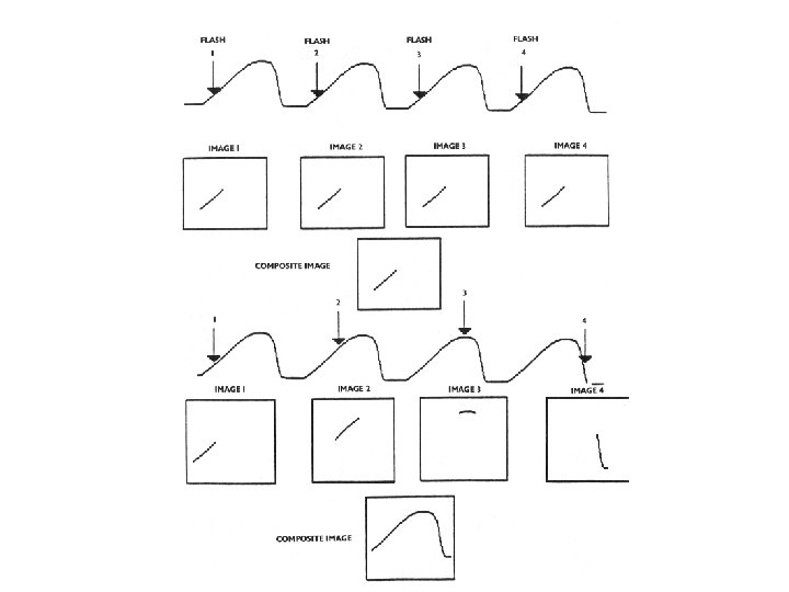
Understanding How A Strobe Works • Strobe box has two types of light, a flashing Xenon and a constant Halogen for constant viewing. • A microphone is used to detect the Fundamental Frequency of the vocal folds. • The flash is synchronized (video rate 30 times a second) or Pitch rate. Seventh Annual ENT for the PA-C | April 21 -23, 2017| Chicago, IL

Understanding How A Strobe Works • The flashing Strobe light creates an illusion that the cords are moving in slow motion, but it is actually a composite image, sampled from a variety of glottal cycles. Seventh Annual ENT for the PA-C | April 21 -23, 2017| Chicago, IL
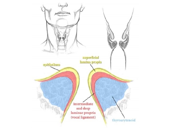

Muscle Vibratory layer Any change that affects this layer – stiffness of vocal fold layers, weakness or failure of closure, imbalance between R and L vocal folds from a lesion on one vocal fold – causes voice problems. Seventh Annual ENT for the PA-C | April 21 -23, 2017| Chicago, IL

Diagnostic Value of Stroboscopic Examination in Hoarse Patients • “Videostroboscopy contributed significant diagnostic information in 27. 2% of the cases” (versus non-stroboscopic means). • “It was instrumental in changing the diagnosis in 10% of the cases. ” • Case no. 4: original diagnosis was “recurrent laryngeal cancer”; changed to “excessive mucous” • Conclusion: “Stroboscopy should be performed in selected patients suspected of vocal fold pathology. It is especially useful in serial examinations to help assess treatment progress. ” – “Diagnostic Value of Stroboscopic Examination in Hoarse Patients”, Woo, Peak, et. al. ; 1991: Journal of Voice, Vol. 5; No. 3 Seventh Annual ENT for the PA-C | April 21 -23, 2017| Chicago, IL

More Pronounced Results In Study Done By Sataloff, et. al • Diagnoses were noted before and after employing use of videostroboscopy in 377 patients • Results: – 29% of original diagnoses had additional diagnosis added – 18% of original diagnoses were incorrect – 47% of original diagnoses were modified • Sataloff, et. al. ; “Strobovideolaryngoscopy: Results and Clinical Value; Annals of Oto, Rhino, Laryngo, September 1991 Seventh Annual ENT for the PA-C | April 21 -23, 2017| Chicago, IL

Database Seventh Annual ENT for the PA-C | April 21 -23, 2017| Chicago, IL

• Assessment form and selections based on Bless and Hirano Template. • Data and still images from database are imported into a Microsoft Word template customized to your institution.

REVIEW OF STROBES RECORDINGS -See CD Seventh Annual ENT for the PA-C | April 21 -23, 2017| Chicago, IL

Videostroboscopy Workshop: Evaluation Score cards will be used for admission to workshops and attendance. Credit will only be awarded for completed score cards. Name Session 1 On scale of 1 through 5 with 5 being most likely 1. Were learning objectives met? 2. Was instruction free of commercial bias? 3. Was there adequate instruction before practice? 4. Was there adequate supervision during practice? 5. Were training aids useful/realistic in learning skill? 6. How likely are you to perform these skills in future 7. Did this training improve your skills? Comments: Scale 1 -5 2

Videostroboscopy Workshop: Score Card Rotate and complete each station. “Go/No Go” for internal use only. Completion of workshop is NOT contingent on pass/fail. Task Go Understand indications & contraindications to exam. Properly explain procedure. Apply topical anesthetic & decongestant. Perform supervised videostroboscopy on simulated patient. Identify normal anatomy. Demonstrate proper equipment handling technique. Comments Proctor Name Proctor Signature No Go

Videostroboscopy: Room Set Up Screen Station 2 Video Tower Station 1 Video Tower Station 4 Projector Speaker Equipment Table Proctors
- Slides: 18