Approach to upper GI bleeding RAHAF ALNAJJAR 1635099

Approach to upper GI bleeding RAHAF ALNAJJAR -1635099

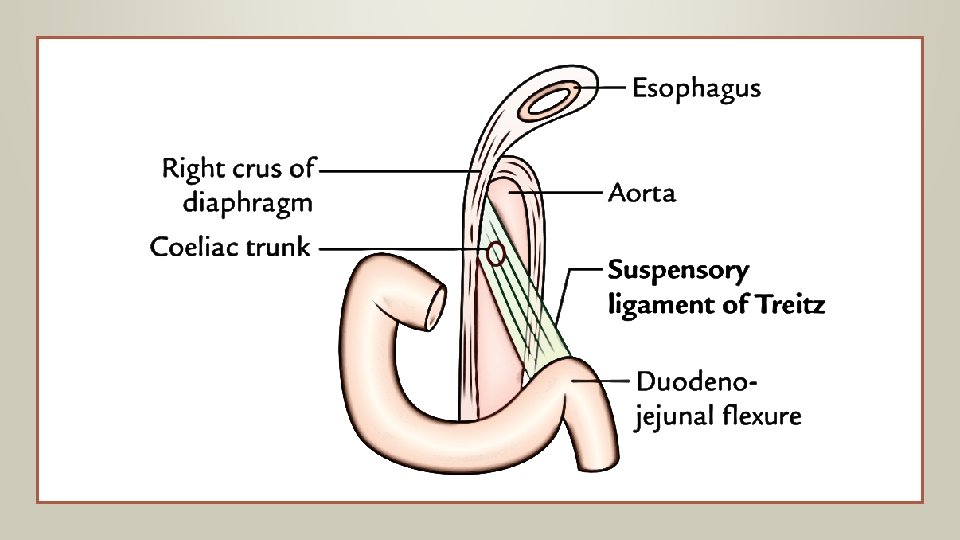
introduction Acute gastrointestinal bleeding is a potentially life-threatening abdominal emergency that remains a common cause of hospitalization. Upper GI bleeding is a bleeding in the GI tract which is proximally to the ligament of Treitz. The ligament of Treitz, also known as the suspensory ligament of the duodenum, is a double fold of peritoneum suspending the duodenojejunal flexure from the retroperitoneum. UGIB is 4 times as common as LGIB, with a higher incidence in males.
 bleeding commonly present with hematemesis (vomiting of")
introduction Patients with acute upper gastrointestinal (GI) bleeding commonly present with hematemesis (vomiting of blood or coffee-ground-like material) and/or melena (black, tarry stools). The initial evaluation of patients with acute upper GI bleeding involves an assessment of hemodynamic stability and resuscitation if necessary. Diagnostic studies (usually endoscopy) follow, with the goals of diagnosis, and when possible, treatment of the specific disorder.

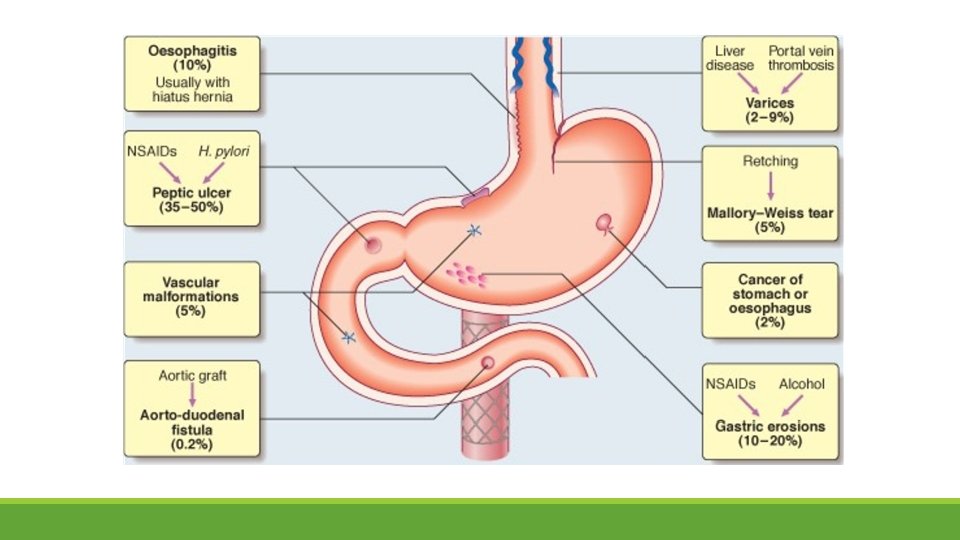
Causes Esophageal causes: ◦ ◦ ◦ Esophageal varices Mallory-Weiss tear Esophagitis Esophageal cancer Esophageal ulcers Gastric causes: ◦ ◦ ◦ Gastric ulcer Gastric cancer Gastritis Gastric varices Dieulafoy's lesions • Duodenal causes: • Duodenal ulcer • Vascular malformation including aorto-enteric fistulae • Hematobilia, or bleeding from the biliary tree • Bleeding from the pancreatic duct • Severe superior mesenteric artery syndrome

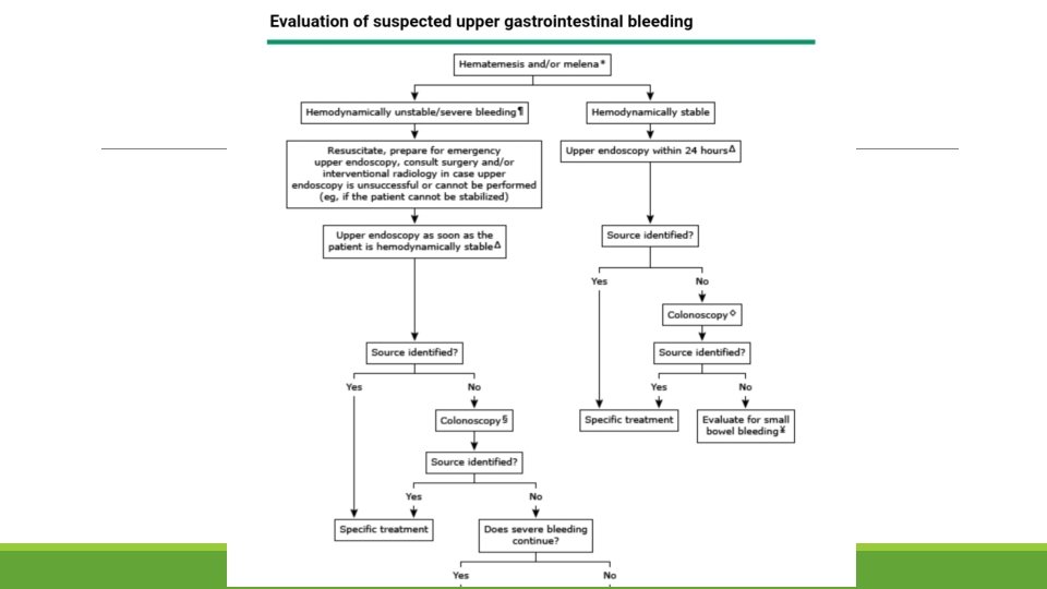
evaluation The initial evaluation of a patient with a suspected clinically significant acute upper GI bleed includes a history, physical examination, laboratory tests, and in some cases, nasogastric lavage. The goal of the evaluation is to assess: 1. the severity of the bleed 2. identify potential sources of the bleed 3. determine if there are conditions present that may affect subsequent management
 suggests bleeding proximal to the ligament")
History Hematemesis (either red blood or coffee-ground emesis) suggests bleeding proximal to the ligament of Treitz. ◦ The presence of frankly bloody emesis suggests moderate to severe bleeding that may be ongoing ◦ Coffee-ground emesis suggests more limited bleeding. Melena: the majority of melena (black, tarry stool) originates proximal to the ligament of Treitz ◦ Melena may be seen with variable degrees of blood loss, being seen with as little as 50 m. L of blood Hematochezia (red or maroon blood in the stool) is usually due to lower GI bleeding. However, it can occur with massive upper GI bleeding, which is typically associated with orthostatic hypotension.

History Patients should be asked about symptoms as part of the assessment of the severity of the bleed and as a part of the evaluation for potential bleeding sources. Symptoms that suggest the bleeding is severe include 1. 2. 3. 4. 5. Orthostatic dizziness Confusion Angina Severe palpitations Cold/clammy extremities.

history Specific causes of upper GI bleeding may be suggested by the patient's symptoms: 1. Peptic ulcer: Upper abdominal pain 2. Esophageal ulcer: Odynophagia, gastroesophageal reflux, dysphagia 3. Mallory-Weiss tear: Emesis, retching, or coughing prior to hematemesis 4. Variceal hemorrhage or portal hypertensive gastropathy: Jaundice, abdominal distention (ascites) 5. Malignancy: Dysphagia, early satiety, involuntary weight loss, cachexia

History Past Medical History: Patients should be asked about prior episodes of upper GI bleeding, since up to 60 percent of patients with a history of an upper GI bleed are bleeding from the same lesion. Potential bleeding sources suggested by a patient's past medical history include: 1. Varices or portal hypertensive gastropathy in a patient with a history of liver disease or alcohol abuse. 2. Aorto-enteric fistula in a patient with a history of an abdominal aortic aneurysm or an aortic graft.

Past medical history 3. Angiodysplasia in a patient with renal disease, aortic stenosis, or hereditary hemorrhagic telangiectasia. 4. Peptic ulcer disease in a patient with a history of Helicobacter pylori infection, nonsteroidal anti-inflammatory drug (NSAIDs) use, or smoking. 5. Malignancy in a patient with a history of smoking, alcohol abuse, or H. pylori infection

Past medical history Comorbid illnesses may influence patient management in the setting of an acute upper GI bleed. Comorbid illnesses may: 1. Make patients more susceptible to adverse effects of anemia (eg, coronary artery disease, pulmonary disease). 2. Predispose patients to volume overload in the setting of vigorous fluid resuscitation or blood transfusions(eg, renal disease, heart failure). 3. Result in bleeding that is more difficult to control (eg, coagulopathies, thrombocytopenia, significant hepatic dysfunction). 4. Predispose to aspiration of GI contents into the lungs (eg, dementia, hepatic encephalopathy).

Medication history Particular attention should be paid to drugs that: 1. redispose to peptic ulcer formation, such as aspirin and other NSAIDs, including COX-2 inhibitors 2. Are associated with pill esophagitis 3. Promote bleeding, such as antiplatelet agents (eg, clopidogrel) and anticoagulants (including the direct oral anticoagulants) 4. Have been associated with GI bleeding, including selective serotonin reuptake inhibitors (SSRI), calcium channel blockers, and aldosterone antagonists 5. May alter the clinical presentation, such as bismuth and iron, which can turn the stool black

Physical examination The physical examination is a key component of the assessment of hemodynamic stability. Signs of hypovolemia include: 1. 2. 3. Resting tachycardia. (less than 15% of blood volume lost) Orthostatic hypotension. (at least 15% of blood volume lost) Supine hypotension. (at least 40% of blood volume lost) The presence of abdominal pain, especially if severe and associated with rebound tenderness or involuntary guarding, raises concern for perforation.

Laboratory data Laboratory tests that should be obtained in patients with acute upper gastrointestinal bleeding include: 1. Complete blood count: chronic or subacute bleeding leads to anemia but the hemoglobin concentration may be normal after sudden, major bleeding until hemodilution occurs 2. Serum chemistries and KFTs 3. LFTs: these include PT, INR and serum albumin concentration. They may show evidence of chronic liver disease or anti-coagulated patients 4. Cross-matching: at least two units of blood should be cross-matched.

Nasogastric lavage Whether all patients with suspected acute upper GI bleeding require nasogastric tube (NGT) placement is controversial. More often, NGT lavage is used when it is unclear if a patient has ongoing bleeding and thus might benefit from an early endoscopy. In addition, NGT lavage can be used to remove particulate matter, fresh blood, and clots from the stomach to facilitate endoscopy. The presence of red blood or coffee ground material in the nasogastric aspirate also confirms an upper GI source of bleeding
This should be")
Initial management Hemodynamically unstable patients : ◦ ABC : (give Oxygen: )This should be given to all patients in shock. Intravenous access : peripheral access should be attained with either two 18 gauge or larger intravenous catheters. Fluid resuscitation: Crystalloid fluids should be given to raise the blood pressure Transfusion — Patients with active bleeding and hypovolemia may require red blood cell transfusion despite an apparently normal hemoglobin, particularly if the patient remains hemodynamically unstable despite appropriate fluid resuscitation. Patients without active bleeding who become hemodynamically stable with fluid resuscitation should receive a blood transfusion if the hemoglobin is <9 g/d. L (90 g/L) for high-risk patients and if it is <7 g/d. L (70 g/L) in low-risk patients

Initial management Platelets : Patients with active bleeding and a low platelet count (<50, 000/micro. L) FFP — Patients with a coagulopathy that is not due to cirrhosis and with a prolonged prothrombin time with INR >2. 0 should generally be transfused with FFP. We will perform upper endoscopy once the INR is <2. 5
 Prokinetics — Both erythromycin and metoclopramide have")
Initial management Medications: Acid suppression — (PPI) Prokinetics — Both erythromycin and metoclopramide have been studied in patients with acute upper GI bleeding. The goal of using a prokinetic agent is to improve gastric visualization at the time of endoscopy by clearing the stomach of blood, clots, and food residue. Antibiotics for patients with cirrhosis. Vasoactive medications — Somatostatin, its analog octreotide, and terlipressin are used in the treatment of variceal bleeding

Diagnostic studies Upper endoscopy — Upper endoscopy is the diagnostic modality of choice for acute upper GI bleeding. Endoscopy has a high sensitivity and specificity for locating and identifying bleeding lesions in the upper GI tract. In addition, once a bleeding lesion has been identified, therapeutic endoscopy can achieve acute hemostasis and prevent recurrent bleeding in most patients Risks of endoscopy — Risks of upper endoscopy include pulmonary aspiration, adverse reactions to medications used to achieve conscious sedation, GI perforation, and increasing bleeding while attempting therapeutic intervention.

Evidence Supports Performing Early Endoscopy Early endoscopy within first 24 hrs of acute GIB is associated with: 1. Decreased hospital stay 2. Decreased rate recurrent bleeding 3. Decreased need for surgery 4. 50% reduction in mortality rates

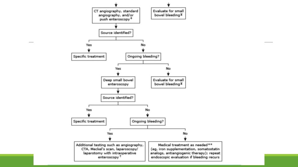
Diagnostic studies Other diagnostic tests — Other diagnostic tests for acute upper GI bleeding include angiography, which can detect active bleeding, deep small bowel enteroscopy, and rarely, intraoperative enteroscopy. Upper GI barium studies are contraindicated in the setting of acute upper GI bleeding because they will interfere with subsequent endoscopy, angiography, or surgery

Triage 1. All patients with hemodynamic instability or active bleeding (manifested by hematemesis, bright red blood per nasogastric tube, or hematochezia) should be admitted to an intensive care unit for resuscitation and close observation with automated blood pressure monitoring, electrocardiographic monitoring, and pulse oximetry. 2. Other patients can be admitted to a regular medical ward, though we suggest that all admitted patients with the exception of low-risk patients receive electrocardiographic monitoring. 3. Outpatient management may be appropriate for some low-risk patients. Determining the appropriate site of care for a patient can be facilitated using risk stratification scores, such as the Glasgow-Blatchford score. Use of these scores is recommended in the International Consensus Group guideline

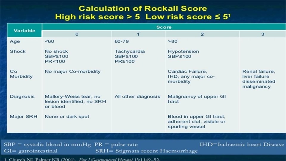
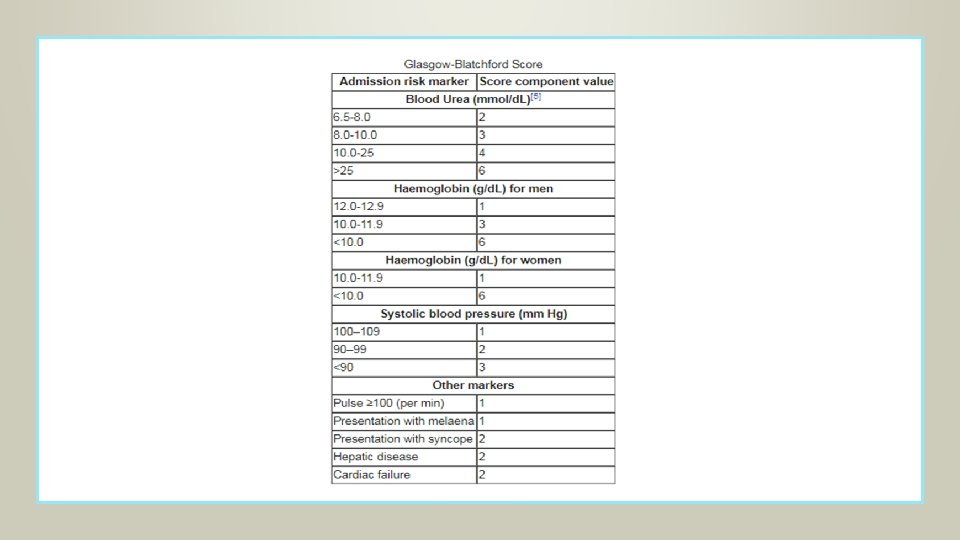
Risk stratification Two commonly cited scoring systems are the Rockall score and the Blatchford score The Rockall score is based upon age, the presence of shock, comorbidity, diagnosis, and endoscopic stigmata of recent hemorrhage The Blatchford score (also known as the Glasgow Blatchford score), unlike the Rockall score, does not take endoscopic data into account and thus can be used when the patient first presents

Blatchford score The score is based upon the blood urea nitrogen, hemoglobin, systolic blood pressure, pulse, and the presence of melena, syncope, hepatic disease, and/or cardiac failure. The score ranges from zero to 23 and the risk of requiring endoscopic intervention increases with increasing score. A simpler version of the score, known as the modified Glasgow Blatchford score, is calculated using only the blood urea nitrogen, hemoglobin, systolic blood pressure, and pulse.
 Indications for surgery : Only about 10% of patients will")
Treatment(ACCORDING TO THE CAUSE) Indications for surgery : Only about 10% of patients will require surgery. 1. Exsanguinating hemorrhage. A patient with uncontrollable hemorrhage who is losing blood faster than it can be replaced must be sent to the operating room immediately for control of the site of bleeding. 2. Profuse bleeding, especially in association with hypotension. Patients should be treated surgically: ◦ If more than 4 Units of blood are required for initial resuscitation. ◦ If bleeding continues at a rate of more than 1 Unit every 8 hours. ◦ If a brief hypotensive episode could have catastrophic results, as in patients with coronary artery disease or cerebrovascular disease or in patients older than 60 years of age.

treatment 3. Recurrent bleeding after its initial cessation. 4. Pathologic features of the bleeding site that increase the risk of recurrent bleeding include: ◦ A posterior duodenal ulcer with the gastroduodenal artery visible in its base ◦ A giant gastric ulcer

Chronic gi bleeding Acute UGIB is more recognizable than chronic GI bleeding, it happens suddenly and can have very serious consequences. Chronic UGIB is a slow, long term bleeding or occurs at an on/off pattern. It is more difficult to diagnose because the symptoms are not easily detected.

Management of chronic gi bleeding Chronic UGIB usually does not present as an emergency. It is usually discovered due to signs and symptoms of anemia. Management: includes resuscitation and identifying the underlying cause, and hence treatment depends on the cause itself.

references 1. Up. To. Date 2. Past seminars

Lower Gastrointestinal Bleeding Eman Humaidat
: Refers to blood loss of recent onset originating from")
Definition Lower gastrointestinal bleeding (LGIB): Refers to blood loss of recent onset originating from a site distal to the ligament of Treitz. Note: The ligament of Treitz, also known as the suspensory ligament of the duodenum, is a double fold of peritoneum suspending the duodenojejunal flexure from the retroperitoneum.
 bleeding typically reports haematochezia (passage of")
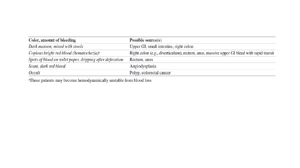
Presentation • A patient with lower gastrointestinal (GI) bleeding typically reports haematochezia (passage of maroon or bright red blood or blood clots per rectum). • Bleeding from the left colon >> usually bright red in colour • Bleeding from the right >> usually dark or maroon coloured, may be mixed with stool. Rarely, may present with melena. • Symptoms range from: Occult bleeding Small amounts of blood on toilet paper Bloody stools Symptoms and signs of anaemia Signs of volume depletion (presents as pallor, dizziness, weakness, syncope, and signs of shock may be present). • Nonspecific complaints: anorexia, dyspnoea, abdominal pain, chest pain, and fatigue. • • •

Initial Evaluation History Physical Examination Age Onset and duration Colour and amount Exacerbating and relieving factors (Antibiotics, stress, steroids, UC…) • Associated symptoms • PMH: • Vital signs, signs of anaemia, pallor, ecchymoses • Assess haemodynamic stability • Extraintestinal manifestation for IBD (eye skin joint) • Abdominal pain suggests the presence of an inflammatory bleeding source such as ischemic or infectious colitis or a perforation (e. g. a perforated peptic ulcer in a patient with severe upper GI bleeding) • DRE… • • • Atrial fibrillation in children or atherosclerosis in elderly • Prolonged hypotension: non occlusive ischemia • Family history • Social history • Medications
 >> may indicate an infectious or inflammatory")
Cont. • Systemic symptoms (fever, bloody diarrhoea…) >> may indicate an infectious or inflammatory cause. • Weight loss >> should raise suspicion for malignancy, especially in older patients who have changes in bowel habits and/or iron- deficiency anaemia. • Iron-deficiency anaemia in a man or a postmenopausal woman should raise suspicion for malignancy (especially colorectal cancer, particularly if the patient has never had screening). • Bleeding that follows straining at stool >> suggests an anorectal cause. • Tenesmus, a sense of incomplete evacuation of stool >> most often seen with ulcerative colitis and infectious aetiologies. • Bleeding from diverticulosis and angiodysplasia tends to be painless. • Abdominal pain (especially in elderly patients) should raise suspicion for ischaemic colitis.

Initial Evaluation & Management • The first step is to place two large-bore IVs and order lab tests: CBC, chemistry, LFT and INR/ PTT. Send blood for type & cross-matching. Insert foley’s catheter • If the patient demonstrates evidence of significant blood loss, resuscitation should be instituted with crystalloid (normal saline or lactated Ringer’s) followed by packed red blood cells as needed. • General supportive measures: Patients should receive supplemental oxygen by nasal cannula and initially should receive nothing per mouth in the event urgent upper endoscopy is needed.

Consider an upper-GI source Findings that are suggestive of UGIB Findings that decrease the likelihood of UGIB • Haemodynamic instability • Orthostatic hypotension • Elevated BUN-to-creatinine or urea-to-creatinine ratio (>20 to 30: 1 or >100: 1, respectively) • Blood clots in the stool The first thing to do is to insert an NGT.

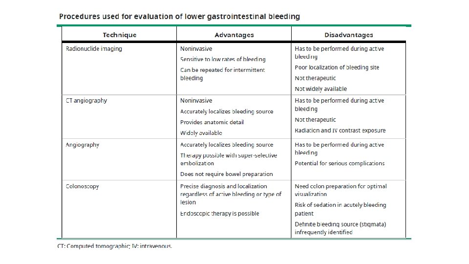
NGT Blood Bile, no blood Clear: no bile, no blood EGD Anoscopy / proctoscopy EGD Rx Rx Depends on bleeding rate > 1 m. L/min 0. 5 -1 m. L/min < 0. 5 m. L/min Tagged RBCs Wait for bleeding to stop, then colonoscopy Angiogram Rx
")
Aetiology of LGIB The causes of LGIB may be grouped into: • Anatomic (diverticulosis) • Vascular (angiodysplasia, ischemic, radiation-induced) • Inflammatory (inflammatory bowel disease, infectious) • Neoplastic The most common causes of massive lower GI bleeding? 1. Diverticulosis (usually right-sided is severe bleeding) 2. Angiodysplasia

Diverticulosis • Condition in which diverticula can be found within the colon, (90% in sigmoid colon) • These are actually false diverticula in that only mucosa and submucosa herniate through the bowel musculature • Prevalence of diverticular disease is age-dependent, increasing from less than 20 percent at age 40 to 60 percent by age 60. • People with low-fibre diets, chronic constipation, and a positive family history have a higher risk • The high prevalence of diverticulosis explains why it is the most common cause of LGIB, even though bleeding is a rare complication of this common disease.

• Diverticular bleeding may be massive and life-threatening since diverticula often form at the site of arterial vascular penetration. • The bleeding is usually painless except for mild abdominal discomfort and cramping due to colonic spasm from intraluminal blood. • Diverticular bleeding is self-limited in 70 -80% of cases. • Risk factors for diverticular bleeding include the use of aspirin and NSAID

Angiodysplasia • Refers to dilated, tortuous submucosal vessels. It is the most common vascular abnormality of the GI tract. • Angiodysplasia may occur throughout the colon, although bleeding most often originates from the caecum or ascending colon. • Bleeding from angiodysplasia is venous in origin (in contrast to arterial bleeding with diverticula) and therefore tends to be less massive than diverticular bleeding.

Colitis Infectious and ischemic colitis and inflammatory bowel disease can all present initially with haematochezia. Mucosal inflammation (colitis) is the common response to acute injury, resulting in activation of the immune system and inflammatory cascade. • Infectious colitis — There are many infectious causes of colitis. A routine stool culture will identify Salmonella, Campylobacter, and Shigella.

• Ischaemic colitis —The pathophysiology is decreased blood flow to the colon, causing non-occlusive ischemic colitis. • The mucosa is affected first , and depending on the severity progression may occur through all layers of the colon. • The most common areas to be affected are “watershed” areas which have relatively poor perfusion as they are in between two areas of the colonic blood supply (splenic flexure rectosigmoid junction) • CT imaging in IC can show oedema and air (pneumatosis} in the bowel wall. • Colonoscopy shows segments of cyanotic mucosa and haemorrhagic ulcerations. with a sharp transition from affected to unaffected mucosa.

Modalities of Treatment 1 - Endoscopy therapy Thermal, sclerosing agent, mechanical 2 - Angiography therapy Vasopressin, embolization 3 - Surgery • Persistent hemodynamic instability with active bleeding • Transfusion of more than 4 units packed red bloods cells in a 24 -hour period, with active or recurrent bleeding.
- Slides: 52