Approach to thyroid nodules Mohsen Eledrisi MD FACP


 FNA not needed Very low suspicion FNA")

- Slides: 16
ﺑﺴﻢ ﺍﻟﻠﻪ ﺍﻟﺮﺣﻤﻦ ﺍﻟﺮﺣﻴﻢ Approach to thyroid nodules Mohsen Eledrisi, MD, FACP, FACE Department of Medicine Hamad Medical Corporation Doha, Qatar eledrisi@yahoo. com
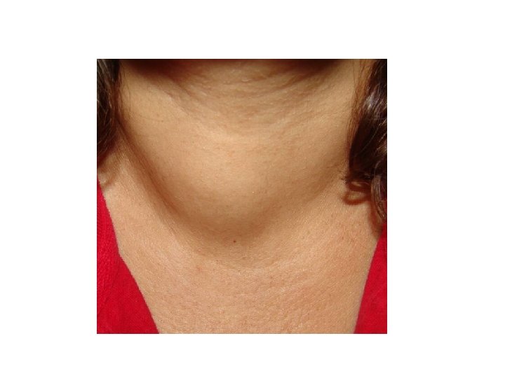
CASE presentation • A 38 -year-old woman with neck swelling noted by her husband about 2 months • She has no complaints • Past history: none • Exam showed a mildly enlarged thyroid with about 3 × 2 cm mass in the lower part of the left thyroid lobe • How to approach?
Thyroid nodule: history • Shortness of breath, dysphagia, dysphonia, choking with position • Rate of growth • Symptoms of hyperthyroidism or hypothyroidism • History of head/neck irradiation • Family history of thyroid cancer
Labs for thyroid nodules: choose best answer • TSH, FT 4, FT 3 • TSH, TG Ab • TSH, TPO Ab American Association of Clinical Endocrinologist. Endocr Pract 2010; 1: 63
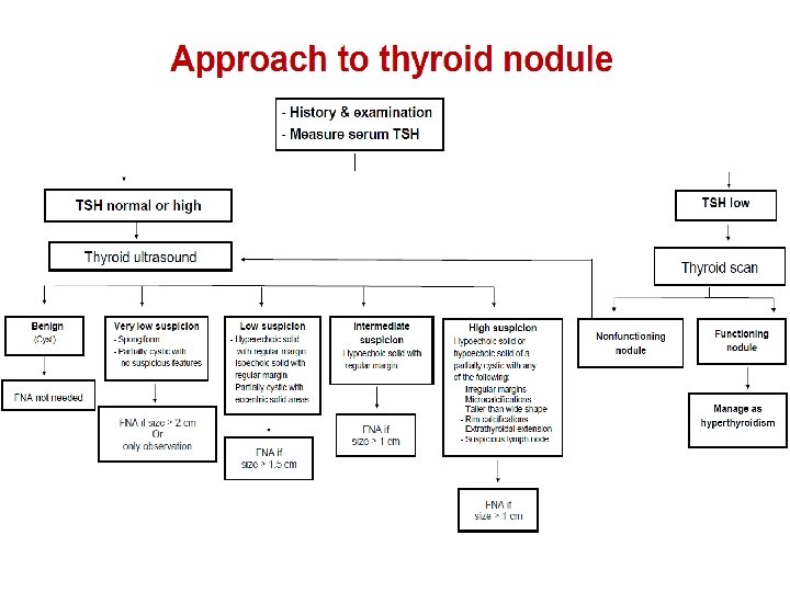
Evaluation of thyroid nodule - History & Physical - Check TSH Low TSH Normal Thyroid scan Hyperthyroid Manage accordingly Non-functional or Ultrasound Go to FNA algorithm
Thyroid ultrasound • Confirms the presence of nodules • Detects non-palpable nodules • Size and nature of nodules and risk of malignancy • Follow up of nodules • FNA-biopsy (U/S-guided)
When to be concerned about possible malignancy? • • • History of head and neck irradiation Age < 14 or > 70 years Male Family history of thyroid carcinoma, MEN type 2 Growing nodule Firm or hard consistency Cervical lymphadenopathy Fixed nodule Persistent dysphonia, dysphagia, or dyspnea American Association of Clinical Endocrinologist. Endocr Pract 2010; 1: 63
High suspicious thyroid nodule features on ultrasound - Solid hypoechoic or solid hypoechoic component of a partially cystic nodule with any of the following: - Irregular margins - Microcalcifications - Rim calcifications - Taller than wide shape - Extrathyroidal extension - Suspicious lymph node
Intermediate suspicious thyroid nodule features on ultrasound Hypoechoic solid with regular margin
Low suspicious thyroid nodule features on U/S • Hyperechoic solid with regular margin • Isoechoic solid with regular margin • Partially cystic nodule with solid areas
Very low suspicious features on ultrasound • Spongiform • Partially cystic nodule Cyst Benign
When to do thyroid FNA? Benign (Cyst) FNA not needed Very low suspicion FNA if size ≥ 2 cm Or only observation American Thyroid Association. Thyroid 2016; 26; 1. Low suspicion FNA if size ≥ 1. 5 cm Intermediate & high suspicion FNA if size ≥ 1 cm
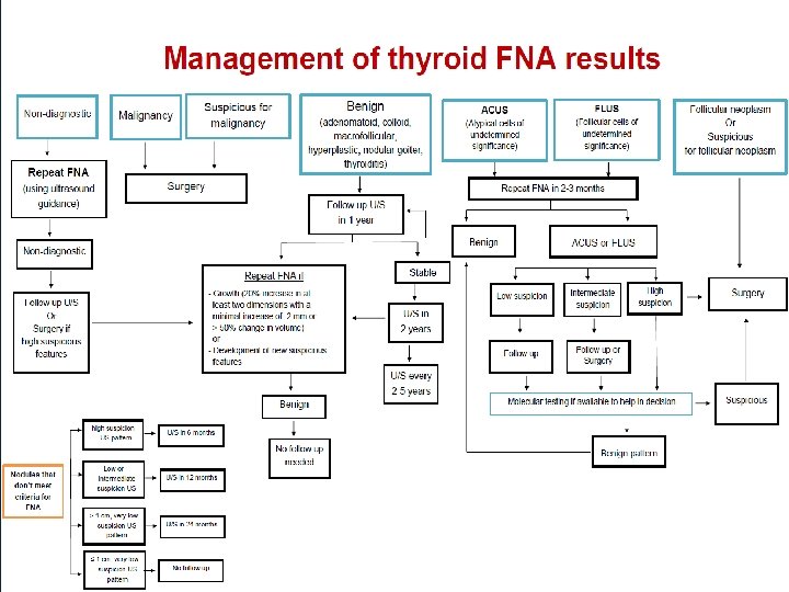
Thyroid nodules: when to do surgery ? • Malignancy • Suspicious for malignancy • Indeterminate cytology + high risk • Repeat non-diagnostic + high risk • Follicular neoplasm (FN) or suspicious for FN