Approach to the patient with chronic kidney disease


 • CKD is defined as abnormalities of kidney structure or")


")
α X max(Scr/κ, 1)-1. 209 X 0. 993")


")


 140 -age)x Wt 72 x p. Cr For")


")


 Stage 5")


- Slides: 81
Approach to the patient with chronic kidney disease Gülçin Kantarcı, M. D. Nephrology Department
Learning objectives and training goals of this lecture • Define chronic kidney disease. • Explain the pathophysiology of chronic kidney disease. • Describe the clinical findings of chronic kidney disease. • Take preventive measures against the development of chronic kidney disease. • List the possible etiology of chronic kidney disease and make a differential diagnosis. • Arrange the initial treatments and refer to a specialist.
REFERENCE &SUGGESTED READING • Current Medical Diagnosis and Treatment, Maxine A. Papadakis, Stephen J. Mc. Phee, Eds. Michael W. Rabow, Associate Ed. http: //accessmedicine. com Chapter 22. Kidney Disease • http: //www. uptodate. com. (Definition and staging of chronic kidney disease in adults, Screening for chronic kidney disease, Epidemiology of chronic kidney disease)
chronic renal diseases (CKD) • CKD is defined as abnormalities of kidney structure or function, present for ≥ 3 months, with implications for health
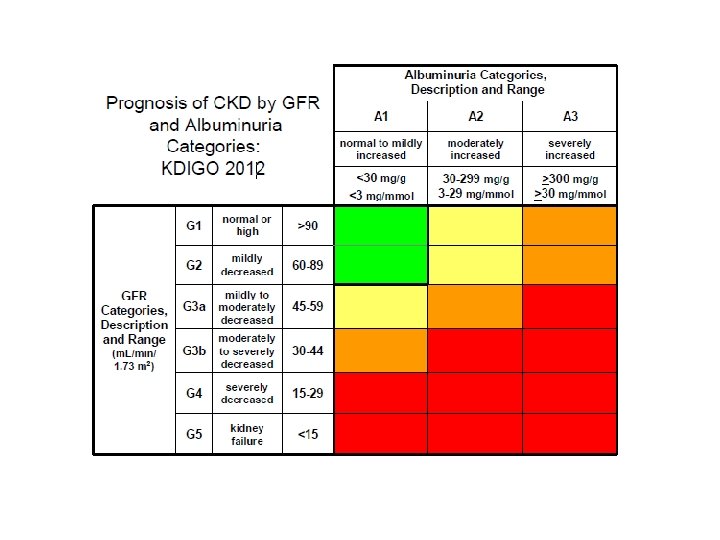
Criteria for CKD KDOQI Clinical Practice Guidelines for Chronic Kidney Disease: Evaluation, Classification, and Stratification 2012
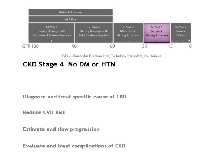
Staging of CKD KDOQI Clinical Practice Guidelines for Chronic Kidney Disease: Evaluation, Classification, and Stratification 2012
DIAGNOSIS OF CKD GFR ü Serum creatinine (muscle mass, dietary meat intake, simetidin, trimetoprim) ü Creatinine Clearance = Ucr x V 1440 P cr x Body weight ü Cockcroft formula= 140 -age (women x 0. 85) 72 x P cr ü Estimated Cr. Cl (MDRD Study)
CKD-EPI GFR = 141 X min(Scr/κ, 1)α X max(Scr/κ, 1)-1. 209 X 0. 993 Age X 1. 018 [if female] X 1. 159 (if black) • The CKD-EPI (Chronic Kidney Disease Epidemiology Collaboration) equation was developed in an effort to create a formula more precise than the MDRD formula, especially when actual GFR is > 60 m. L/min per 1. 73 m 2. Researchers pooled data from multiple studies to develop and validate this new equation. • The CKD-EPI equation performed better than the MDRD (Modification of Diet in Renal Disease Study) equation, especially at higher GFR, with less bias and greater accuracy.
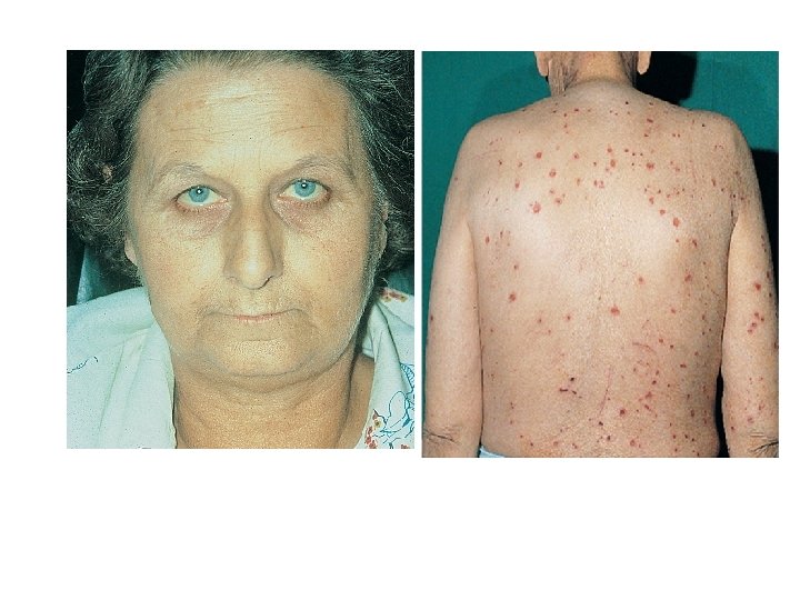
CKD Symptoms • In the early stages, CKD is asymptomatic. Symptoms develop slowly with the progressive decline in GFR, are nonspecific, and do not manifest until kidney disease is far advanced (GFR < 10– 15 m. L/min/1. 73 m 2). • General symptoms of uremia may include fatigue and weakness; anorexia, nausea, vomiting, and a metallic taste in the mouth are also common. Patients or family members may report irritability, difficulty in concentrating, insomnia, restless legs, paresthesias, and twitching. Generalized pruritus without rash may occur.
GFR 35 -50% of normal symptom-free BUN and Cr. levels Normal renal functions maintained *endocrine *excretory *regulatory GFR 20 -35% of normal azotemia still asymptomatic GFR < 20% of normal overt renal failure UREMIC SYNDROME
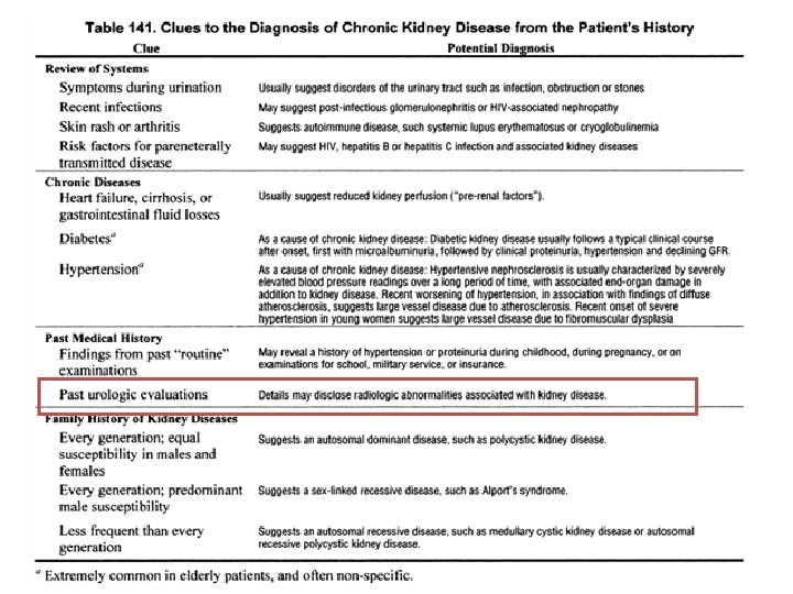
Screening for chronic kidney disease patients who are at risk for developing CKD should be screened with both • a urine test for proteinuria and • a blood test for creatinine to estimate glomerular filtration rate (GFR). Risk factors for CKD • History of diabetes, cardiovascular disease, hypertension, hyperlipidemia, obesity, metabolic syndrome, smoking, human immunodeficiency virus (HIV) or hepatitis C virus infection, and malignancy • Family history of kidney disease • Treatment with potentially nephrotoxic
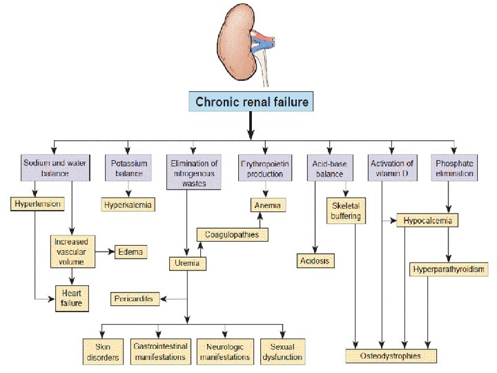
Clinical Abnormalities in Uremia • Fluid & electrolyte disturbances • Acid-Base disorders • Cardiovascular complications • Hematologic complications • Neurologic complications • Bone , phosphate & calcium abnormalities • Endocrine disorders
Fluid & Electrolyte Disturbances in CRF – Expansion of ECF – Hyponatremia • Dilutional • Impaired Na conservation – Hyperkalemia only when • GFR<10 ml/min • Oliguria /anuria develops • Potassium sparing diuretics used • ACEI and Beta blockers • Acidosis
METABOLIC ACIDOSIS IN CRF No change in arterial p. H/ plasma HCO 3 until GFR 30% of normal Plasma HCO 3 (24 m. Eq/L) Decreased (14 -18 m. Eq/L) Metabolic acidosis in CRF is due to: • Decreased nephron mass • Leading to limited NH 4 production and HCO 3 regeneration p. H and stable HCO 3 levels maintained at the expense of buffering by bone (Ca. PO 4 -Ca. HCO 3)
Cardiovascular complications in CRF • Hypertension – Salt and water retension – Hyperrenninemia • Pericarditis • Accelerated atherosclerosis – Coronary artery disease – Cerebrovascular disease – Peripheral vascular disease • Pulmonary edema
Hematologic complications in CRF Normochromic normocytic anemia › biosynthesis of erythropoetin › Bone-marrow depressive effect of uremic toxins › Hemolysis › GI loss of blood Abnormal hemostasis › bleeding time › Abnormal platelet aggregation &adhesiveness › activity of platelet factor 3 Enhanced susceptibility to infection
Neurologic complications Uremic encephalopathy Inability to concentrate, drowsiness Insomnia, behavioral changes Neuromuscular irritability › Hiccups, cramps, fasciculations › Asterixis, chorea, stupor, seizures Peripheral neuropathy Restless Legs
Bone phosphate & calcium abnormalities in CRF biosynthesis of 1, 25 -dihidroksikolekalsiferol Hypocalcemia Hyperphosphatemia Hyperparathyroidism Acidosis • Renal Osteodystrophy • Osteomalacia
Endocrine disorders in CRF Secondary hyperparathyroidism Glucose intolerance Disturbances of insulin metabolism › Hyperinsulinemia › Peripheral insulin resitance Pituitary, throid & adrenal are normal Libido and fertility
Essentials of Diagnosis • Decline in the GFR over months to years. • Persistent proteinuria or abnormal renal morphology may be present. • Hypertension in most cases. • Symptoms and signs of uremia when nearing end-stage disease. • Bilateral small or echogenic kidneys on ultrasound in advanced disease.
Estimation equations • Cockcroft-Gault • MDRD • CKD-EPI • Cockcroft-Gault equation (140 - age) x lean body weight [kg] • CCr (m. L/min) = ———————— Cr [mg/d. L] x 72
TREATMENT Slowing Progression • Treatment of the underlying cause of CKD is vital. Control of diabetes should be aggressive in early CKD; risk of hypoglycemia increases in advanced CKD, and glycemic targets may need to be relaxed to avoid this dangerous complication. • Blood pressure control is vital to slow progression of all forms of CKD; agents that block the renin-angiotensinaldosterone system are particularly important in proteinuric disease • Current guidelines suggest a blood pressure goal of 130/80 mm Hg for patients with CKD; a goal of 125/75 mm Hg is recommended for patients with proteinuria.
Dietary Management • Every patient with CKD should be evaluated by a renal nutritionist. Specific recommendations should be made concerning protein, salt, water, potassium, and phosphorus intake to help manage CKD progression and complications. • Protein restriction to 0. 6– 0. 8 g/kg/d may retard CKD progression • Salt and water restriction. A goal of 2 g/d of sodium is reasonable for most patients. A daily intake of 2 L of fluid maintains water balance. • Potassium restriction. Restriction is needed once the GFR has fallen below 10– 20 m. L/min/1. 73 m 2, or earlier if the patient is hyperkalemic. Patients should receive detailed lists describing potassium content of foods and should limit their intake to < 50– 60 m. Eq/d (2 g). • Phosphorus restriction. The phosphorus level should be kept in the ‘normal’ range (<4. 5 mg/d. L) predialysis,
Medication Management • Many drugs are excreted by the kidney; dosages should be adjusted for GFR. • Insulin doses may need to be adjusted. • Magnesium-containing medications, such as laxatives or antacids, should be avoided as should phosphorus-containing medicines, particularly cathartics. • Morphine metabolites are active and can accrue in advanced CKD; • Drugs with potential nephrotoxicity (NSAIDs, intravenous contrast) should be avoided • The anemia of CKD is primarily due to decreased erythropoietin production, which often becomes clinically significant during stage 3 CKD. Many patients are iron deficient as well due to impaired GI iron absorption.
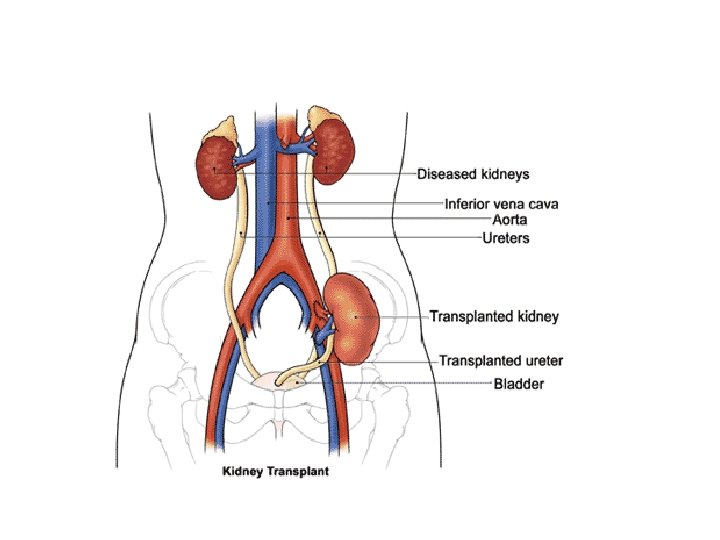
Treatment of End-Stage Renal Disease HEMODIALYSIS END STAGE RENAL FAILURE PERITONEAL DIALYSIS TRANSPLANTATION When GFR declines to 5– 10 m. L/min/1. 73 m 2 (with or without overt uremic symptoms), renal replacement therapy (hemodialysis, peritoneal dialysis, or kidney transplantation) is required to sustain life.
Kidney transplant sources • Living Related Unrelated q. Deceased ? n ? t? s a pl t o n Xe n ra
Vascular access for HD
Native AV Fistula
Vascular access for HD
Principle of hemodialysis cellophane sausage casings, a cooling system from an old Ford, parts from a crashed German fighter plane, and washing machine tubs.
Principle of PD
When to Refer • Patient education is important in understanding which mode of therapy is most suitable, as is timely preparation for treatment; therefore, referral to a nephrologist should take place in late stage 3 CKD, or when the GFR is declining rapidly. Such referral has been shown to improve mortality • A patient with other forms of CKD such as those with significant proteinuria (> 1 g/d) or polycystic kidney disease should be referred to a nephrologist at earlier stages.
Prognosis in ESRD • Compared with kidney transplant recipients and age-matched controls, mortality is higher for patients undergoing dialysis. There is likely little difference in survival for well-matched peritoneal versus hemodialysis patients. • Survival rates on dialysis depend on the underlying disease process. Five-year Kaplan. Meier survival rates vary from 36% for patients with diabetes to 53% for patients with glomerulonephritis.
Prognosis in ESRD • Overall 5 -year survival is currently estimated at 39%. Patients undergoing dialysis have an average life-expectancy of 3– 5 years, but survival for as long as 25 years may be achieved depending on comorbidities. • The most common cause of death is cardiac disease (50%). Other causes include infection, cerebrovascular disease, and malignancy.
Case 1 • 35 years old male • Nocturia since 2006 • He had a history of pyelonephritis with kidney stones • Since then he had no hospital admission
PHYSICAL EXAMINATION • BP: 186/100 mm. Hg P: 90/min/R • Weight 72 kg, Height 175 cm Temperature: 36. 4 0 C Raised JVP Dyspneic with crackles over the lung bases
Laboratory tests • • Hb: 10. 6 g/dl Htc: 31. 9% Na: 137 m. Eq/L Serum BUN: 78 mg/dl ; Kr: 3. 6 mg/d. L Urine specific gravity: 1012 Ca 8. 3 mg/d. L Pi 4. 6 mg/d. L Albumin 3. 6 g/d. L Fe 16 ug/d. L (59 - 158 ) TIBC 205 ug/d. L (228 - 428)
n. Estimated Cr. Cl (Cockcroft-Gault formula) 140 -age)x Wt 72 x p. Cr For Female= x 0. 85
http: //www. kidney. org/professionals/kdoqi/gfr_calculator. cfm
Stage 4 CKD
URINARY USG • RK 85 mm, 5 x 7 mm mid pole kidney stone • LK 88 mm, 7 x 6 mm apical pole kidney stone • GII Renal Parenchimal Dis. Proteinuria: 354 mg/day
• • Renal bx. ? Urinary CT? Urinary Ca? Stone analysis?
How would you manage this patient?
Treatment Strategies • • • Anemia EPO, Parenterally forms of iron Hyperphosphatemia Phosphate binders HT Do not use ACEIs+ARBs Do not use contrast agents Do not use NSAIDs
Case 2 • • 48 years old female Left flank pain, vomiting, fever History of PCKD ( diagnosed in 1999) Her mother died because of intracranial hemorage (AVM? and AVA? )
PHYSICAL EXAMINATION • • • BP: 100/60 mm. Hg P: 118/min/R Weight 59 kg, 38. 2 0 C Dyspne(+), tachipne (+) turgor , Pale face, Looks weak and unwell fullness of neck veins (+)
Laboratory tests • WBC: 12. 860/mm 3 • Hb: 12. 2 g/dl Htc: 37. 3% Na: 137 m. Eq/L • BUN: 156 mg/dl ; Kr: 10. 3 mg/d. L • ABG analysis PH 7. 26 HCO 3 12. 8 BE-12 • Urine specific gravity: 1010 • S. Na 133 m. Eq/l S. K 6. 7 m. Eq/l • Ca 8. 3 mg/d. L Pi 6. 8 mg/d. L • CRP 184
URINARY USG • RK 186 mm with multiple cystis and stones, • LK 167 mm with multiple cystis and stones • R ureter and pelvis dilatated
• • Culture of the urine ? Culture of blood? Urine analysis ? Urinary CT ? ( Contrast agent)
How would you manage this patient? ?
Case 3 • • 28 years old male dyspnea, vomiting, bad feutor History of urinary tract infections before age 12. His brother on dialysis because of VUR nephropathy
PHYSICAL EXAMINATION • • BP: 110/70 mm. Hg P: 88/min/R Weight 72 kg, 36. 5 0 C Dyspne(-), tachipne (-) turgor n, fale face,
Laboratory tests • Hb: 9. 6 g/dl Htc: 31% MCV 79 • Na: 135 m. Eq/L, K 3. 5 m. Eq/L Ca 7. 6 mg/d. L Pi 9. 6 mg/d. L Albumin 3. 6 g/d. L ; CRP 30 Fe 27 ug/d. L (59 - 158 ) TIBC 308 ug/d. L (228 - 428) • Serum BUN: 168 mg/dl ; Kr: 12. 3 mg/d. L • Urine specific gravity: 1010, • Urinary sediment: 8 -10 leucocytes, 2 -3 waxy casts in every field of microscopic areas
US • Solitery enlarged left kidney and proximal segments of ureter
Urinary bt( Without contrast)
• PTH 354 pg/ml • Uric acid 8. 9 mg/dl • Culture of urine : (-) What is your likely diagnosis ?
Nephropathy of VUR
Voiding cystouretrography
How would you manage this patient ?
HEMODIALYSIS END STAGE RENAL FAILURE PERITONEAL DIALYSIS TRANSPLANTATION
• • • Anemia EPO, Parenterally forms of iron Hyperphosphatemia Phosphate binders HT Do not use ACEIs+ARBs Do not use contrast agents Do not use NSAIDs Nephrectomy or Defflux inj. Befor renal transplantation
Case 4 • • • 57 years old female Dispnea and vomiting History of NIDDM ( diagnosed in 1985) Her father died because of CAD Her mother had the history of dialysis & died because of sepsis
PHYSICAL EXAMINATION • • • BP: 190/60 mm. Hg P: 92/min/R Weight 72 kg, BMI 30 edeama (+++/+++) turgor n, pale face, Dyspneic and orthopneic with crackles over the lung bases, tachipne (+)
LABORATORY FINDINGS • Hb: 8. 7 Htc: 25% WBC: 7200 • BUN: 58 mg/d. L Cr: 3. 2 mg/d. L K: 6. 7 m. Eq/L Na: 135 m. Eq/L • S. alb 3. 1 g/dl • Urinalysis = D 1010 35 -40 WBC
Chronic Kidney Failure (Due to diabetic nepropathy) Stage 5
HEMODIALYSIS END STAGE RENAL FAILURE PERITONEAL DIALYSIS TRANSPLANTATION