Approach to the patient with Back pain Presented

Approach to the patient with Back pain Presented By: Hesham Al. Ghofili & Gassan Almoqbel Supervised by: Dr. Amr Jamal

Objectives ▹ Diagnosis including history, Red Flags, and Examination ▹ Brief comment on Mechanical, Inflammatory, Root nerve compression, and Malignancy ▹ Common causes ▹ Role of primary health care in management ▹ When to refer to a specialist ▹ Prevention and Education

In USA it is the commonest cause of limitation of activity in those under the age of 45. (1) The lifetime prevalence of non-specific (common) low back pain is estimated at 60– 70% (1)

53. 2% 79. 2% In Saudi Arabia Seven studies were cross-sectional and found a prevalence and pattern ranging from 53. 2% to 79. 17%. (2)

Role play

MCQs!
")
1 - Which of the following is not indicative of inflammatory back pain? (3) a. Insidious onset b. Onset before 40 years of age c. Pain for more than 3 months d. Morning stiffness e. Aggravation of pain with activity https: //Poll. Ev. com/surveys/k. PSMW 0 SBL/web
 characteristic of a history of mechanical")
2 - Which of the following is (are) characteristic of a history of mechanical Lower Back Pain? (3) a. Relatively acute onset b. History of overuse or a precipitating injury c. Pain worse during the day d. All
 a \"red flag(s)\" or danger signal(s)")
3 - Which of the following is (are) a "red flag(s)" or danger signal(s) relative to the diagnosis of LBP? (3) a. Cough b. Impotence c. Chest pain d. Constipation

4 - What is the most cost-effective and crucial aspect of the treatment of chronic LBP? (3) a. Patient education b. Physiotherapy c. Bed rest d. Morning stiffness
")
5 - Which of the following is the most common cause of LBP? (3) a. Metastatic bone disease b. Inflammatory back pain c. Lumbosacral sprain or strain d. None of the above

What is the diagnostic approach to a patient with back pain? A thorough history and physical examination helps elucidate the diagnosis in most patients.
 Doctor I have PAIN!")
Hx taking (4) Doctor I have PAIN!

DDx Mechanical Systemic Referred Lumbar strain or sprain Malignancy Acute Aneurysm Multiple myeloma Pelvic disease Metastatic carcinoma Prostatitis/Endometriosis Infection Renal disease Compression fracture Osteomyelitis Stones / Pyelonephritis Spondylolysis TB GI disease Brucellosis Pancreatitis Inflammation Ankylosing spondylitis Cholecystitis Herniated disc and spinal stenosis Degenerative processes of disc and facet joint

Hx taking 1 Personal Data 2 SOCRATES 3 Neurological? 4 Red Flags! 5 PMH 6 Past Surgical Hx 7 Medications 8 Family, Social & Systemic Review

Hx taking 1 Personal Data Age? Residence? Occupation? 2 SOCRATES Site? Associated? Onset? Timing? Character? Exacerbating? Radiation? Severity?

W di ar ag ra no nt st ad ic di im tio ag na i n l g Hx taking 3 Neurological? 4 Red Flags!

Hx taking 5 PMH Trauma? Cancer? 6 Past Surgical Hx 7 Medications Psychatic? Steroid? 8 Family, Social & Systemic Review Inherited disease? Social? Smoking? Alcohol?

Hx taking One More thing Idea Concern Expectation How does it affect him/her Emotionally/Functionally

Physical Examination Will be explained by Gassan in latter slides Laboratory tests ▹ Not necessary in the evaluation of back pain unless the concerned about the possibility of malignancy or infection (5) ▹ FBC, ESR, C-reactive protein (CRP), and blood cultures ▹ Urinalysis and Culture. . Why?

Imaging ▹ High-risk patients ▹ Most patients with low back pain with or without sciatica do not routinely require imaging when presenting in a non-specialist setting. (5) ▹ Reassure. . that their symptoms will respond to conservative treatment. (5) ▹ If symptoms persist longer than 6 to 8 weeks, plain x-rays should be obtained at that time (5)

Imaging ▹ MRI or CT, only if: neurological compromise, infection, or tumors is considered (5) ▹ After discussion with a spinal surgeon. ▹ MRI is the preferred study. If contraindicated a CT myelogram is usually warranted. (5) ▹ In trauma situations, the standard AP pelvis and cervical spine radiographs should be obtained. (5)
 • Do not routinely offer imaging in a non-specialist setting. • Explain")
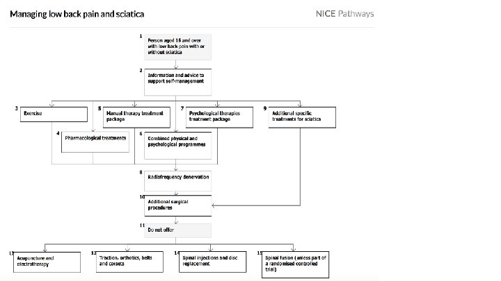
Imaging (6) • Do not routinely offer imaging in a non-specialist setting. • Explain to the patient that if she/he is being referred for specialist opinion, she/he may not need imaging. • Consider imaging for people with low back pain with or without sciatica only if the result is likely to change management.

Case A 38 -year-old man with no significant history of back pain developed acute LBP when lifting boxes 2 weeks ago. The pain is aching in nature, located in the left lumbar area, and associated with spasms. He describes previous similar episodes several years ago, which resolved without seeing a doctor. He denies any leg pain or weakness. He also denies fevers, chills, weight loss, and recent infections On examination, there is decreased lumbar flexion and extension secondary to pain, but a neurologic exam is unremarkable. Mechanical Inflammation Malignancy Root Nerve Compression

Case A 20 -year-old man presents to his primary care physician with low back pain and stiffness that has persisted for more than 3 months. There is no history of obvious injury but he is a very avid sportsman. His back symptoms are worse when he awakes in the morning, and the stiffness lasts more than 1 hour. His back symptoms improve with exercise. He has a desk job and finds that sitting for long periods of time exacerbates his symptoms. His back symptoms also wake him in the second half of the night. He normally takes an anti-inflammatory drug during the day, and finds his stiffness is worse when he misses a dose. He has had 2 bouts of iritis in the past. Mechanical Inflammation Malignancy Root Nerve Compression

Case 27 9 -year-old male patient who had a history of low back pain for about a year and was reporting severe, constant, and aching lower back pain of 8 -9 on a scale of 10. as well as radicular right leg pain, decreased sensation on the right, difficulty straightening his right leg making it difficult to walk. An MRI showed a rather large L 5 -S 1 herniated disc on the right side with severe degenerative disc disease. Mechanical Inflammation Malignancy Root Nerve Compression

Case 28 A man aged 78 years presented to his general practitioner with newonset low back pain. The patient had metastatic prostate cancer, which was diagnosed 2 years ago. He reported a 6 -week history of constant, burning pain in the lower lumbar region, which was worse at night and was 7/10 in severity on the Numeric Pain Rating Scale. He had no relief with regular paracetamol or ibuprofen. He had not had lower limb weakness, numbness, lower urinary tract symptoms or weight loss. Mechanical Inflammation Malignancy Root Nerve Compression

Mechanical 4 ▹ Tends to get better or worse depending on your position – for example, it may feel better when sitting or lying down. ▹ Typically feels worse when moving ▹ Can develop suddenly or gradually ▹ Might sometimes be the result of poor posture or lifting something awkwardly, but often occurs for no apparent reason ▹ May be due to a minor injury

Inflammation 4 ▹ ▹ ▹ ▹ Age at onset of back pain <45 years Back pain lasting > 3 months Night pain Early morning pain and stiffness lasting more than one hour Insidious onset Tenderness/inflammation over the joint Increased by Rest and Relived by activity

31

4 Root Nerve Compression ▹ Characterized by radicular pain arising from nerve root impingement due to herniated discs. ▹ Radicular pain: Pain that radiates into the lower extremity directly along the course of a spinal nerve root.

4 Malignancy ▹ Metastatic tumors are found mostly in patients older than 50 years. ▹ Metastatic disease is more common than primary tumors of the spine, and thoracic spine metastatic lesions are more common than lumbar ▹ Patient usually has constitutional symptoms such as fever , Wight loss, loss of appetite and NV

34 Others ▹ Pediatrics: ▹ In Primary care settings most of the cases are due to ▹ ▹ overloaded school backpacks. . (10) Also think about psychological causes to avoid going to school. Women: Ask about pregnancy. Psychiatric patients: 75% of Depressed patients present with pain. . One of the most common sites is Lower back pain (11)
 comes to")
1 Case A 28 -year-old man with chronic low back pain (LBP) comes to your office for renewal of his medication. He was injured at work 5 years ago while attempting to lift a box of heavy tools. Since that time, he has been off work, living on compensation insurance payments, and he has not been able to find a job that does not aggravate his back. On physical examination, the patient demonstrates some vague tenderness in the paravertebral area around L 3 to L 5. He has some limitations on both flexion and extension. What is the most likely diagnosis? What is the next step?

1 Lumbar muscular strain/sprain ▹ Stiffness, and/or soreness of the lumbosacral region (underneath the twelfth rib and above the gluteal folds) persisting for <12 weeks. (12) ▹ What is the difference between strain and sprain? Strains occur when a muscle is stretched too far and tears, damaging the muscle itself. Sprains happen when over-stretching and tearing affects ligaments, which connect the bones together.

1 Lumbar muscular strain/sprain ▹ The most common source of back pain but do not sound serious and do not typically cause long-lasting pain. ▹ Arises from any combination of pathology involving discs, vertebrae, facet joints, ligaments, and/or muscles.

1 Lumbar muscular strain/sprain ▹ Clinical presentation: Sharp intense pain for 1 to 2 days; muscle spasm; most patients recover within 3 months ▹ Risk factors? 1. Lifting a heavy object, or twisting the spine while lifting 2. Sudden movements that place too much stress on the low back, such as a fall 3. Poor posture over time 4. Sports injuries, especially in sports that involve twisting or large forces of impact
 <4 Weeks Clinical")
1 Lumbar muscular strain/sprain ▹ Benign physical examination. ▹ Investigations? (12) <4 Weeks Clinical diagnosis >6 Weeks Lumbar Spine X-Ray Lumbar Spine MRI Lumbar Spine CT Labs ▹ Management: ▹ What is the aim of the treatment? ▹ Reducing pain and restoring functional status.
 Acute (< 4 Weeks) 1 st Line:")
1 Lumbar muscular strain/sprain ▹ Management: (12) Acute (< 4 Weeks) 1 st Line: Patient Education + Normal Activity. Subacute (4 -12 Weeks) Chronic (> 12 Week) 1 st Line: Patient Education + 1 st Line: CBT Normal Activity. Evidence B +: Self care temp. treatment Heat Ice Evidence B Evidence C Adjunctive: Analgesics/Muscle relaxant. Adjunctive: Active physiotherapy and exercise therapy. Adjunctive: Rehabilitation/TCA/Opioi d/Surgery.

Lumbar muscular strain/sprain 1 ▹ ▹ • • Management: For the Muscle relaxant: NSAIDs if the first line. (Cochrane) Paracetamol (Evidence C)

2 Case 38 year old laborer who had the immediate onset of left leg burning pain and weakness after lifting a heavy load. He had onset of some bladder incontinence What is the most likely diagnosis? What is the next step?
 2 ▹ A complex, multi-factorial, clinical condition characterized by low")
Herniated nucleus pulposus (HNP) 2 ▹ A complex, multi-factorial, clinical condition characterized by low back pain with or without the concurrence of radicular lower limb symptoms in the presence of radiologically-confirmed degenerative disc disease. ▹ The pain is exacerbated by activity, but may be present in certain positions, such as sitting.
 ▹ Radiating lower extremity pain in a dermatomal distribution;")
2 Herniated nucleus pulposus (HNP) ▹ Radiating lower extremity pain in a dermatomal distribution; ▹ History of bowel or bladder dysfunction, bilateral sciatica, and saddle anesthesia may be symptoms of severe compression of the cauda equine ▹ Positive straight-leg raise or contralateral straight leg (reproduced below 60° of hip flexion); positive femoral stretch test may suggest upper lumbar disc herniation.
 ▹ Causes: ▹ Complex mechanical and inflammatory process. ▹")
2 Herniated nucleus pulposus (HNP) ▹ Causes: ▹ Complex mechanical and inflammatory process. ▹ Genetic influences have been found to be more important than the mechanical effects. ▹ Associated with increasing age, smoking, the presence of facet joint tropism and arthritis, abnormal pelvic morphology, and changes in sagittal alignment.
 ▹ Investigations: ▹ MRI: herniated disc ▹ Management: Saddle")
2 Herniated nucleus pulposus (HNP) ▹ Investigations: ▹ MRI: herniated disc ▹ Management: Saddle (perineal) anaesthesia, sphincter dysfunction, bladder retention, and leg weakness = Cauda Equina Syndrome (CES) Urgent referral to the hospital. Emergency decompression of the spinal canal within 48 hours after the onset of symptoms
 ▹ Management: < 3 Months > 3 Months 1")
2 Herniated nucleus pulposus (HNP) ▹ Management: < 3 Months > 3 Months 1 st Line: Paracetamol 1 st Line: Continue pain management Adjunctive: Topical analgesics/Opioid/Muscle Relaxant Refer to pain clinic. If with Axial back pain: +Physiotherapy.

3 Case A 63 -year-old woman presents with low back pain and cramping in both posterior thighs and numb- ness radiating into the feet with ambulation. It wors- ens with standing and walking and improves with sit- ting and bending forward. She has no bowel or bladder complaints. On examination, she has full strength, normal sensation, reflexes are symmetric, and she has 2+ peripheral pulses. Straight leg raise is negative. What is this patient’s most likely diagnosis? What is the most likely diagnosis? What is the next step?

3 Spinal stenosis ▹ Lumbar spondylosis refers to degenerative conditions of the lumbar spine that narrow the spinal canal, lateral recesses, and neural foramina. Facet joint and ligamentous hypertrophy, intervertebral disc protrusion, and spondylolisthesis may all contribute to the stenosis, and symptoms result from neural compression of the cauda equina, exiting nerve roots, or both.

3 Spinal stenosis ▹ Intermittent pain radiating to the thigh or legs. ▹ Worse with prolonged standing, activity, or lumbar extension. ▹ Pain is typically relieved by sitting, lying down, and/or lumbar flexion; patient may describe intermittent burning, numbness, heaviness, or weakness in their legs, unilateral or bilateral radicular pain, motor deficits, bowel and bladder dysfunction, and back and buttock pain with standing and ambulation

3 Spinal stenosis ▹ Patients walk with a forward flexed gait; patients with vascular claudication have diminished pulses and typical skin changes, such as mottled discoloration, thinning and shiny skin

3 Spinal stenosis ▹ Investigations: ▹ MRI:

3 Spinal stenosis ▹ Management: significant acute neurological deficit 1 st Line: surgical decompression No significant acute neurological deficit: pain affecting quality of life and/or functional activities 1 st Line: analgesics (NSAIDs) Adjunct: non-pharmaceutical measures/Oral corticosteroids 2 nd Line: epidural corticosteroid injection Chronic symptoms 1 st analgesics Adjunct: Non-pharmaceutical measures / Chronic pain agents / Surgery

3 Spinal stenosis ▹ Management: ▹ Non Pharmaceutical measures: ▹ Temporary reduction in physical activity is recommended; patients should be careful to avoid bending, lifting, or twisting movements until the pain subsides. Bed rest is not recommended. ▹ ▹ Prolonged bed rest (>4 days) is contra-indicated, especially in older patients as it may lead to rapid de-conditioning and increased risk of DVT.

55 Almost done

4 Case A 70 -year-old man, 6 months after renal transplantation and on corticosteroid treatment, presents with severe back pain. X-ray evaluation of the thoracic and lumbar spine discloses evidence of multiple vertebral compression fractures. What is the most likely diagnosis? What is the next step?

4 Compression fracture ▹ Most osteoporotic spinal compression fractures represent an isolated failure of the anterior spinal column due to a combination of flexion and axial compression loading. ▹ The stability of the spine is not compromised with this type of fracture. These fractures are traditionally considered benign injuries that heal without complications.

4 Compression fracture ▹ Typically history of trauma, although acute event not always recalled; pain at rest and at night, previous history of fractures (e. g. , distal radius, hip or other vertebral compression fractures) ▹ Tenderness to palpation over the midline; increased kyphosis, normal neurological examination unless there is retropulsion of bone into the neural elements, such as in burst fractures

4 Compression fracture ▹ plain x-rays: ▹ MRI? ▹ Useful in distinguishing between osteoporotic compression fractures and those caused by underlying tumour or infection.

4 Compression fracture ▹ Management: ▹ 1 st: Limited bed rest. ▹ Adjunctive: Analgesia (Paracetamol/NSAIDs)

Examination ▹ look, feel, move and test function. ▹ You should perform a physical examination to reproduce the patient’s symptoms and localized the level of lesion. (7)(14)(15)

Components of physical examination ▹ ü ü ü ü 1 - Inspection: Position Exposure Look for deformity, inspecting from both the back and the side. Note especially loss of the normal thoracic kyphosis and lumbar lordosis, which is typical of ankylosing spondylitis. Also note any evidence of scoliosis, a lateral curvature of the spine that may be simple (‘C’ shaped) or compound (‘S’ shaped) soft-tissue abnormalities like a hairy patch or lipoma that might overlie a congenital abnormality, e. g. spina bifida. Muscle wasting shoulders & pelvis level. (7)(14)(15)

Components of physical examination ▹ 2 - Palpation ü Patient should be in prone position. ü Palpation occurs: 1. centrally 2. unilateral 3. Soft tissues 4. After warning the patient, lightly percuss the spine with your closed fist and note any tenderness. (7)(14)(15)

Components of physical examination ▹ 3 - Active movements: ü There are three main movements of the lumbar spine: 1. Flexion 2. Extension 3. Lateral bending 4. Rotation (7)(14)(15)(16)

Components of physical examination ▹ 4 - provocative tests ü Femoral stretch test: 1. Knee flexion hip extension while the patient is lying in prone position. 2. Positive if pain felt in ipsilateral anterior thigh. 3. Positive test mean that the L 3 and L 4 nerve roots are involved. (7)(14)(15)
 test 1. Done while the")
Components of physical examination ü Straight leg raising (SLR) test 1. Done while the patient lying in supine position. 2. Tension increased by dorsiflexion of foot (Bragard’s test). Root tension relieved by flexion at the knee. 3. Pressure over centre of popliteal fossa causes pain locally and radiation into the back. 4. Positive test mean that the L 4, L 5 and S 1 nerve roots are involved. (7)(14)(15)

Components of physical examination ▹ 5 - Neurological testing of lower limbs ü Do Neurological examination if patient has any signs or symptoms of nerve root compression. ü If there is nerve roots compression patient will have pain, pareasthesia, anesthesia and weakness, extend into the leg. (7)(14)(15)
 ü Sensation")
Main nerve roots ▹ A- L 1: ü Motor Hip flexion(psoas major) ü Sensation around the groin and hip area ü Reflex cremasteric relfex (L 1, L 2) ▹ B- L 2: ü Motor Hip flexion ü Sensation anterior thigh ü Reflex knee jerk (L 2, L 3, L 4)(7)(14)(15)

Main nerve roots ▹ C- L 3 ü Motor ü Sensation ü Reflex extension of knee anterior thigh knee jerk (L 3, L 4) ▹ D- L 4 ü Motor ü Sensation ü Reflex ankle dorsiflexion and foot inversion. inner border of foot to great toe knee jerk(7)(14)(15)
,")
Main nerve roots ▹ E- L 5 ü Motor: walking on heels (ankle dorsiflexion), extension of great toe ü Sensation middle three toes (dorsum) ü Reflex nil ▹ F- S 1 ü Motor: walking on toes (ankle planter flexion), foot eversion ü Sensation little toe, most of sole ü Reflex ankle jerk (S 1, S 2)(7)(14)(15)

X-Ray ▹ Normal lumbar spine radiographs. A: AP projection. B: Lateral projection.

X-Ray ▹ Metastatic prostate cancer

MRI ▹ Normal MRI

MRI ▹ Degenerative disease

Role of PHC: v Educate patient about the natural history of back pain. v Ask about and address the patient’s concerns and goals. (patient centered care) v Maximize functional status. v Relief the pain. v Improve associated symptoms, such as sleep or mood disturbances or fatigue. v Referral of complicated cases. v Prevention heavy lifting, socio-demographic factors such as smoking and obesity v Primary care services have an approach to risk stratification for young people and adults presenting with a new episode of low back pain with or without sciatica. v Young people and adults with low back pain with or without sciatica are given advice and information to self-manage their condition. (6)

When to refer ? ▹ Urgent/Emergency referrals : 1. Cauda equina 2. Sever radiculopathy. 3. Fractures. ▹ 1. 2. 3. 4. 5. Other referrals Recalcitrant spinal canal stenosis Neoplasia or infection Undiagnosed back pain Paget disease Continuing pain of 3 months’ duration without a clearly definable cause(16)

Prevention and education ▹ ▹ Losing weight: too much upper body weight can strain the lower back . Posture: How you sit, stand lie down can have an important effect on your back. The following tips should help you maintain a good posture. Standing: Stand upright, with your head facing forward and your back straight. Balance your weight evenly on both feet and keep your legs straight

Prevention and education Sitting and driving: Sit up with your back straight and your shoulders back. Your knees and hips should be level and your feet should be flat on the floor.

Prevention and education ▹ Sleeping: Your mattress should be firm enough to support your body while supporting the weight of your shoulders and buttocks, keeping your spine straight.

Prevention and education ▹ Lifting and carrying: ü One of the biggest causes of back injury is lifting or handling objects incorrectly. ü Think before you lift: ü can you manage the lift? ü Push rather than pull – if you have to move a heavy object across the floor, it is better to push it rather than pull it.

Prevention and education ▹ Exercise: ü Exercise is both an excellent way of preventing back pain and of reducing it, but should seek medical advice before starting an exercise programs if you've had back pain for six weeks or more.

Role play

1 - Which of the following is not indicative of inflammatory back pain? a. Insidious onset b. Onset before 40 years of age c. Pain for more than 3 months d. Morning stiffness e. Aggravation of pain with activity https: //Poll. Ev. com/surveys/k. PSMW 0 SBL/web
 characteristic of a history of mechanical")
2 - Which of the following is (are) characteristic of a history of mechanical Lower Back Pain? a. Relatively acute onset b. History of overuse or a precipitating injury c. Pain worse during the day d. All
 a \"red flag(s)\" or danger signal(s)")
3 - Which of the following is (are) a "red flag(s)" or danger signal(s) relative to the diagnosis of LBP? a. Cough b. Impotence c. Chest pain d. Constipation

4 - What is the most cost-effective and crucial aspect of the treatment of chronic LBP? a. Patient education b. Physiotherapy c. Bed rest d. Morning stiffness

5 - Which of the following is the most common cause of LBP? a. Metastatic bone disease b. Inflammatory back pain c. Lumbosacral sprain or strain d. None of the above

References 1. Hengel KM, Visser B, Sluiter JK. The prevalence and incidence of musculoskeletal symptoms among hospital physicians: A systematic review. International Archives of Occupational and Environmental Health. 2011; 84(2): 115 -9. 2. Awaji M. Epidemiology of Low Back Pain in Saudi Arabia. J Adv Med Pharm Sci [Internet]. 2016; 6(4): 1– 9. Available from: http: //sciencedomain. org/abstract/13544 3. Swanson’s Family Medicine Review (7 th Ed. ) 4. Henderson M, Tierney L, Smetana G. The patient history. New York: Mc. Graw-Hill Medical; 2012. 5. Assessment of back pain [Internet]. BMJ Best Practice; 2018 [cited 4 September 2018]. Available from: https: //bestpractice. bmj. com/topics/en-gb/189/pdf/189. pdf

References 6. Low back pain and sciatica in over 16 s: assessment and management. NICE guideline; 2016. 7. Evaluation of low back pain in adults [Internet]. Uptodate. com. 2018 [cited 4 September 2018]. Available from: https: //www. uptodate. com/contents/evaluation-of-low-back-pain-inadults? search=evalu%20 ation-of-low-back-pain-in adults&source=search_result&selected. Title=1~150&usage_type=default&display_rank=1 8. Dog and Rooster I. Disc Herniation Case Studies| Spine Institute of San Diego : Center for Spinal [Internet]. Sdspineinstitute. com. 2018 [cited 6 September 2018]. Available from: http: //www. sdspineinstitute. com/case-studies/disc-herniations/lumbar. html 9. Practitioners T. RACGP - Back pain in a cancer patient: a case study [Internet]. Racgp. org. au. 2018 [cited 6 September 2018]. Available from: https: //www. racgp. org. au/afp/2014/august/back-pain-in-a-cancer-patient/
![References 10. Evaluation of the child with back pain [Internet]. Uptodate. com. 2018 [cited](http://slidetodoc.com/presentation_image_h/0b7c1f3c639936cf3bebf10cb4d6f061/image-90.jpg "References 10. Evaluation of the child with back pain [Internet]. Uptodate. com. 2018 [cited")
References 10. Evaluation of the child with back pain [Internet]. Uptodate. com. 2018 [cited 4 September 2018]. Available from: https: //www. uptodate. com/contents/evaluation-of-the-child-with-backpain 11. Kleiber B, Jain S, Trivedi MH. Depression and Pain: Implications for Symptomatic Presentation and Pharmacological Treatments. Psychiatry (Edgmont). 2005; 2(5): 12 -18. 12. Musculoskeletal lower back pain [Internet]. BMJ Best Practice. 2018 [cited 4 September 2018]. Available from: https: //bestpractice. bmj. com/topics/en-gb/778 13. Osteoporosis - Symptoms, diagnosis and treatment | BMJ Best Practice [Internet]. Bestpractice. bmj. com. 2018 [cited 6 September 2018]. Available from: https: //bestpractice. bmj. com/topics/en-us/85/case-history

References 14. Talley Clinical Examination - 7 th Ed 15. Macleod's Clinical Examination (13 th Ed. ) 16. Low back pain [Internet]. EBM Guidelines Available from: http: //www. ebmguidelines. com/ebmg/ltk. free? p_artikkeli=ebm 00435

Take Home Message ▹ ▹ ▹ ▹ Back pain mostly diagnosed clinically. Exclude the RED FLAGS Don’t forget ICE Imaging is not a must ﺍﺑﻨﻲ ﻳﺎ Medications is not everything even ﺍﺑﻨﻲ ﻳﺎ When you suspects Cauda Equine Refer!! Not every patient deserve a complete rest. . Some are contraindicated.

- Slides: 93