APPROACH TO PATIENT WITH THYROID NODULE By Reem

APPROACH TO PATIENT WITH THYROID NODULE By : Reem Theab , 6 th-year Medical Student

• Terms • A solitary or isolated thyroid nodule is one which clinically appears to be isolated without apparent pathology in the rest of the gland. • Dominant thyroid nodule for a similar swelling in a gland with clinical evidence of generalized abnormality in the form of a palpable contralateral lobe or generalized mild nodularity(e. g. multinodular goiter). • Discrete thyroid nodule refers to both solitary and dominant nodules. - 70% of discrete nodules are clinically isolated - 30% are dominant.
 • Cysts • Follicular Adenomas")
ETIOLOGY Causes of a solitary thyroid nodule Benign (85%) • Cysts • Follicular Adenomas Toxic Adenoma Non-toxic adenoma • thyroiditis Malignant (15 %) • Primary thyroid carcinomas • lymphoma or metastatic disease (Far less commonly) The importance of discrete swellings lies in the increased risk of neoplasia compared with other thyroid swellings. Some 15% of isolated swellings prove to be malignant, and an additional 30 -40% are follicular adenomas.
 shows that over 30% of clinically")
• Thyroid cysts Routine FNAC (or ultrasonography) shows that over 30% of clinically isolated swellings contain fluid and are cystic or partly cystic. Bleeding into a cyst : often presents with a history of sudden painful swelling, which resolves to a variable extent over a period of weeks if untreated. About 50% of cystic swellings are the result of colloid degeneration, or of uncertain aetiology because of absence of epithelial cells in the lining. • Although most of the remainder are the result of involution in follicular adenomas , some 10– 15% of cystic follicular swellings are histologically malignant (30% in men and 10% in women). • Papillary carcinoma is often associated with cyst formation.

• Ultrasound is the most useful tool for assessing cysts. If there is no discernable solid element then the cyst is almost certainly benign and does not need to be further investigated. • If there is an associated solid element then consideration should be given to targeting that area with an ultrasound guided FNAC An anechoic cyst in right lobe of thyroid.

• Thyroid adenomas -Follicular adenomas are the most common type of adenomas and arise from the follicular epithelium within the thyroid gland. -Follicular adenomas are further classified according to their cellular architecture and relative amounts of cellularity and colloid into fetal (microfollicular), colloid (macrofollicular), embryonal (atypical), and Hürthle (oxyphil) cell types. Colloid adenomas do not have any potential for microinvasion, while the fetal, embryonal, and Hürthle cell adenomas all have the potential for microinvasion.

Ultrasound features of follicular adenomas share many features with follicular carcinomas. absence of internal flow or predominantly Histopathology is required to peripheral flow indicates is associated with distinguish between a follicular reduced probability of thyroid follicular adenoma and follicular carcinoma( by malignancy looking for capsular or vascular invasion) Absence of these in adenoma

Papillary carcinoma is the most common malignancy of the thyroid gland with excellent prognosis. Presentation is usually with a solitary palpable thyroid mass. Papillary carcinoma has a tendency to metastasize early to local lymph nodes, with 50%of patients having nodal involvement at presentation. These metastases are usually to the ipsilateral jugular chain Increased risk with RET/PTC rearrengments and BRAF mutations , childhood radiation

• Ultrasound • usually appears as a solitary mass usually with an irregular outline, located in the subcapsular region and demonstrating vascularity. Small punctate regions of echogenicity representing microcalcifications (psammoma bodies) may present Empty appearing nuclie with central clearing “orphan Annie eyes “ , psammoma bodies , nuclear grooves

• Follicular carcinoma : Good prognosis , invades thyroid capsule and vasculature (unlike follicular adenoma), uniform follicles , hematogenous spread is common. Associated with RAS mutation and PAX 8 -PPAR- translocations. Medullary Carcinoma : from parafollicular “ C cells”, produces calcitonin , sheets of cells in an Amyloid stroma ( stains with congo red). Associated with MEN 2 A and 2 B (RET mutations). Undiffernetiated / Anaplastic carcinoma : older patients , presents with rapidly enlarging neck mass > compressive symptoms ( dyspnea , dysphagia ) , very poor prognosis. Associated with TP 53 mutation.

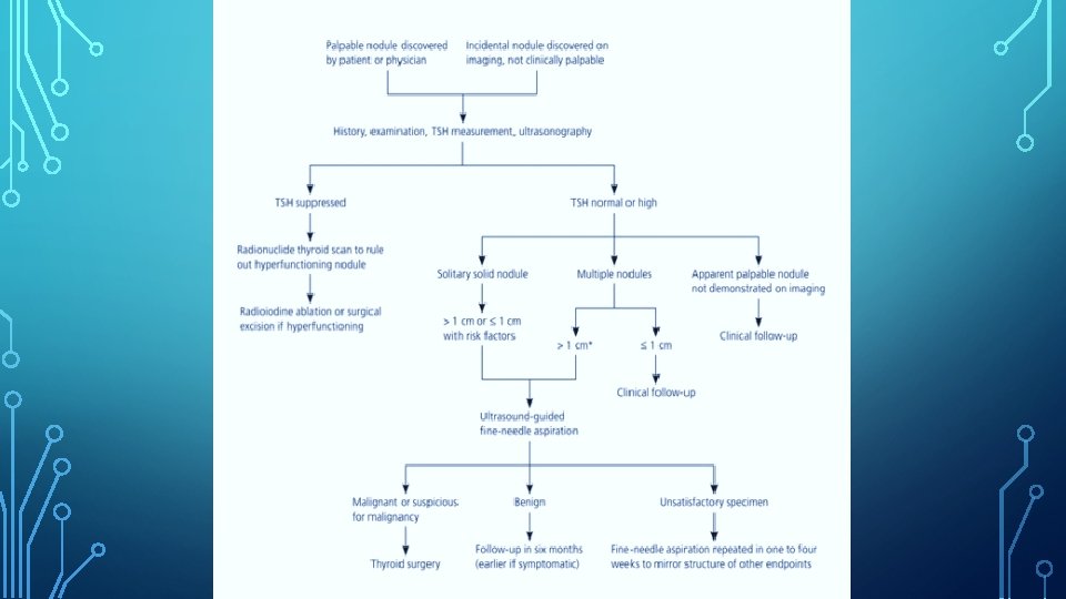
APPROACH TO THE PATIENT • The approach to the patient should focus on identifying which patients at risk of having malignancy and thus who might benefit from surgery. • HISTORY (Details regarding the nodule) - duration - change in size - associated symptoms, such as pain, dysphagia, dyspnea

• Features which might suggest malignancy include: -A nodule of recent origin that is Increasing in size rapidly. - A solitary thyroid nodule in a young patient less than 15 years or an old patient over 65 years -pain; Patients with medullary thyroid cancer may complain of a dull, aching sensation. -A history of hoarseness is worrisome because it may be secondary to malignant involvement of the recurrent laryngeal nerves. -History of exposure to ionizing radiation as it is associated with high risk of malignancy. - any member of the family has or had got thyroid cancer or any other endocrine cancer (MEN II in association with medullary thyroid cancer)
. The risk")
• The risk of malignancy in thyroid swellings (‘rule of twelve’). The risk is greater in isolated versus dominant swellings, solid versus cystic swellings and in men versus women.

PHYSICAL EXAMINATION • A thyroid nodule moves on swallowing. • Features of a thyroid nodule on examination which suggest malignancy include: -Firm, Fixed and irregular -Presence of cervical lymphadenopathy INVESTIGATIONS The objectives of investigation are as follows: • Determine if nodule is autonomously functioning and possibly causing hyperthyroidism. • Determine if nodule has high risk of malignancy.

DETERMINING NODULE FUNCTION DETERMINING THE RISK OF MALIGNANCY Thyroid function tests: -will direct further approach and should precede consideration of imaging studies and FNA biopsy. Patient with a low TSH level (toxic nodules) should be considered for radioisotope scans. Ultrasonography -Often reveals multinodular goiter rather than solitary thyroid nodule. -It can be used to guide needle placement during FNA biopsy as it Provides visualization of needle tip during procedure to ensure accurate sampling. -It may reveal sonographic features that may be suggestive of cancer: -Irregular margins -intranodular vascular spots -microcalcifications -hypoechogenicity within the nodule Radioisotope scans -in evaluation of solitary nodules should be limited to patients with a low TSH level to identify autonomously functioning nodules -Nodules with increased uptake (hot nodules) are toxic adenomas and almost never malignant. -Nodules that accumulate radioisotopes equal to surrounding tissue (warm nodules), or nodules with low uptake (cold nodule), are most often benign, But may be malignant and therefore require FNA biopsy. FNA CYTOLOGY is the most accurate test for the evaluation of thyroid nodules. Tissue samples are obtained for cytologic analysis using 22 - to 25 -gauge needles, with or without local anaesthesia. Specimen adequacy requires ≥ 2 slides **Radioisotope scans are unnecessary in setting showing ≥ 6 -8 cell clusters. False-positive and of a falsenegative results are uncommon when a high normal TSH level, and one may proceed directly

• MANAGEMENT • The management mainly depends on the result of the cytology sample from the FNA specimen. • Malignant lesions require surgical resection with the appropriate modality corresponding to the type of cancer • Benign lesions should be removed as indicated -usually if asymptomatic can be left alone and monitored periodically -Since the malignant nature of follicular neoplasms cannot be determined by cytology, surgical resection is indicated. • About 30% of FNA samples turns out to be thyroid cysts. Aspiration will resolve 25 -50% of cysts, but fluid re-accumulation is common. Surgical resection is indicated for growing or painful cysts. • Toxic adenoma can be treated with RAI ablation, surgery and antithyroid drugs.
- Slides: 17