APPROACH TO PATIENT WITH HEADACHE Dr Hana Albulaihe

APPROACH TO PATIENT WITH HEADACHE Dr. Hana Albulaihe Consultant Neurologist

■ Headache is pain in any part of the head, including the scalp, face, orbitotemporal area and interior of the head. ■ Headache is one of the most common reasons patients seek medical attention. ■ It is due to activation of pain-sensitive structures in or around the brain, skull, face, sinuses, or teeth.

■ Pain Sensitive structures in Brain : ■ Intracranial : 1. Cranial venous sinuses with afferent veins 2. Arteries at base of brain and arteries of dura including middle meningeal artery 3. Dura around venous sinuses and vessels 4. Falx cerebri

■ Extracranial : 1. Skin 2. Scalp appendages 3. Periosteum 4. Muscles 5. Arteries 6. Mucosa
 2. Facial (seventh cranial nerve) 3.")
■ Nerves : 1. Trigeminal (Fifth cranial nerve) 2. Facial (seventh cranial nerve) 3. Vagal (Tenth cranial nerve) 4. Glossopharyngeal (Ninth cranial nerve) 5. Second and Third cranial nerves

Classification ■ Primary headache. ■ Secondary headache.

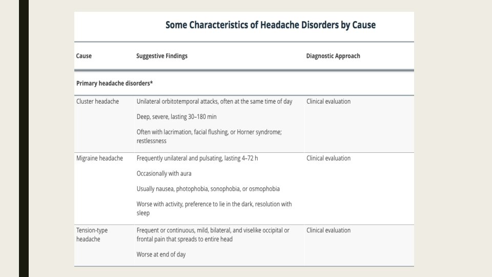
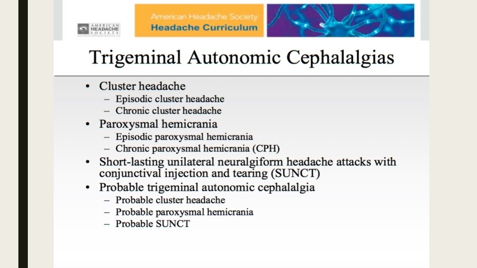
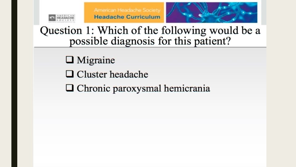
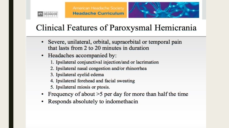
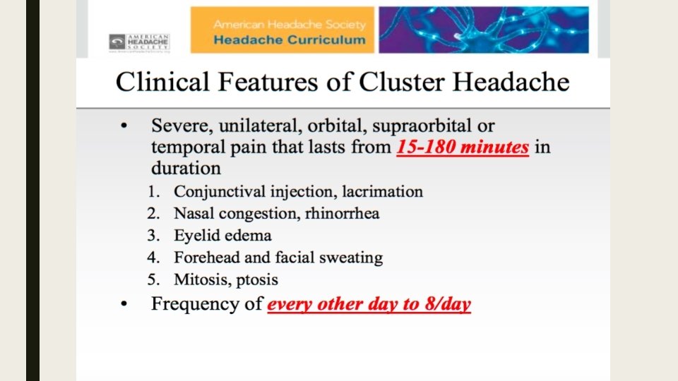
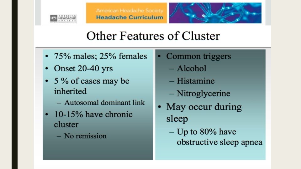
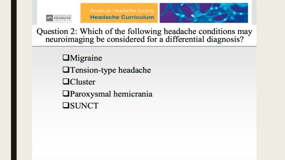
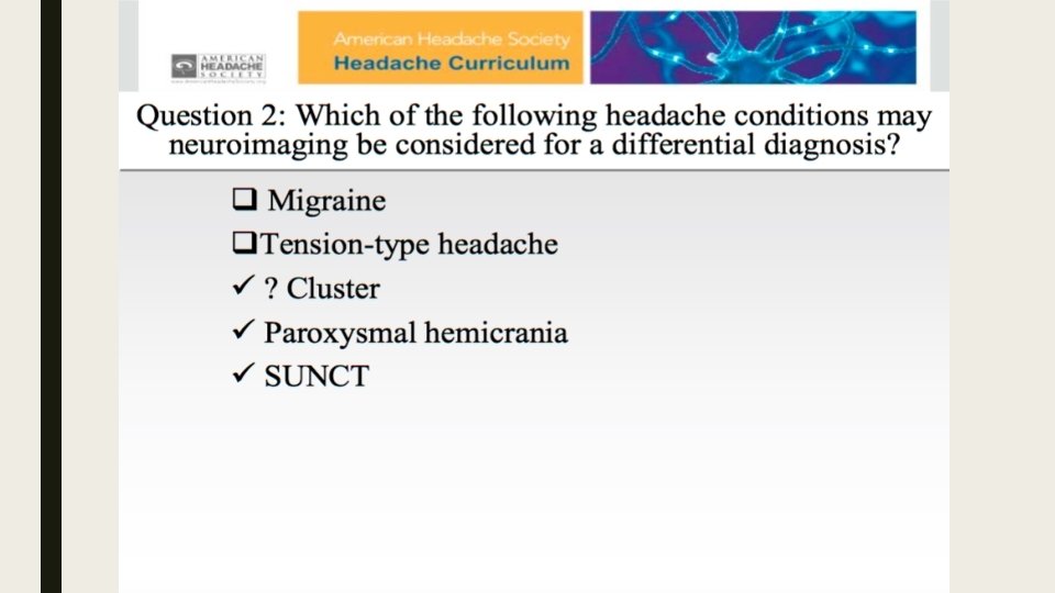
Classification Primary headache disorders: ■ Migraine. ■ Cluster headache. ■ Chronic paroxysmal hemicrania. ■ Hemicrania continua. ■ Tension-type headache.
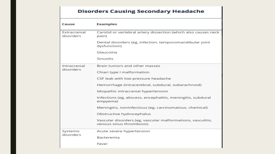

Evaluation ■ Determining whether a secondary headache is present. ■ Check for symptoms that suggest a serious cause. ■ If no cause or serious symptoms are identified, evaluation focuses on diagnosing primary headache disorders.

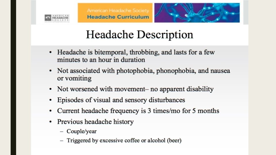
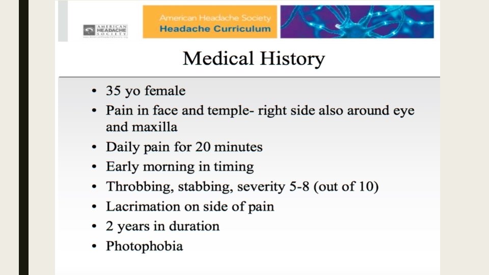
History of present illness: ■ Headache location. ■ Duration. ■ Severity. ■ Onset. ■ Quality ( throbbing, constant, intermittent, pressure-like). ■ Exacerbating and remitting factors ( head position, time of day, sleep, light, sounds, physical activity, odors, chewing).

History of present illness: ■ Previous or recurrent headaches. ■ The previous diagnosis (if any). ■ Whether the current headache is similar or different. ■ For recurrent headaches, age at onset, frequency of episodes, temporal pattern (including any relationship to phase of menstrual cycle), and response to treatments (including OTC treatments).

History ■ Headaches that have recurred since childhood or young adulthood suggest a primary headache disorder. ■ If headache type or pattern clearly changes in patients with a known primary headache disorder, secondary headache should be considered.

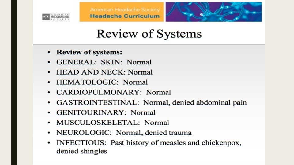
History Review of systems: ■ Vomiting: Migraine or increased intracranial pressure ■ Fever: Infection (eg, encephalitis, meningitis, sinusitis) ■ Red eye and/or visual symptoms (halos, blurring): Acute angle-closure glaucoma ■ Visual field deficits, diplopia, or blurring vision: Ocular migraine, brain mass lesion, or idiopathic intracranial hypertension ■ Lacrimation and facial flushing: Cluster headache ■ Rhinorrhea: Sinusitis ■ Pulsatile tinnitus: Idiopathic intracranial hypertension

History Review of systems: ■ Preceding aura: Migraine. ■ Focal neurologic deficit: Encephalitis, meningitis, intracerebral hemorrhage, subdural hematoma, tumor, or other mass lesion. ■ Seizures: Encephalitis, tumor, or other mass lesion. ■ Syncope at headache onset: Subarachnoid hemorrhage. ■ Myalgias and/or vision changes (in people > 55 yr): Giant cell arteritis.
, and toxins ■")
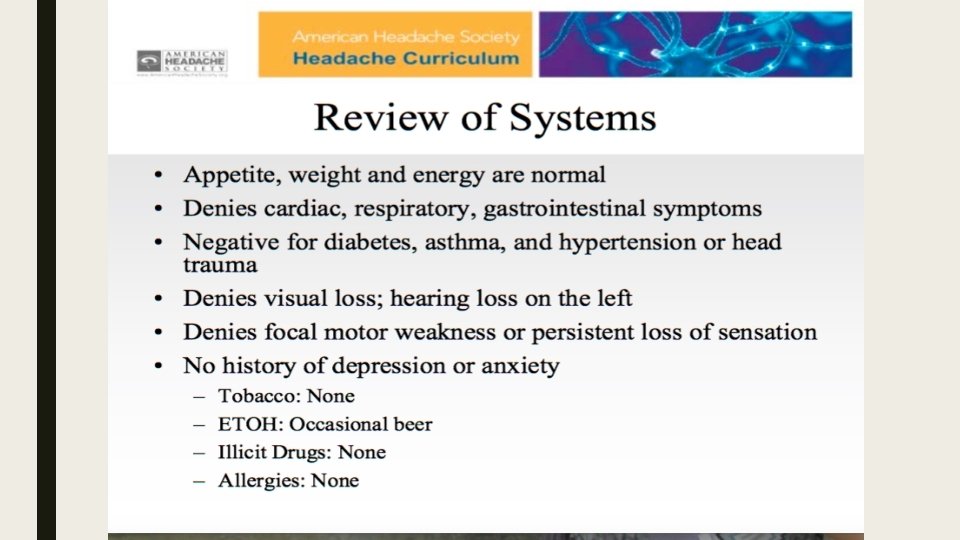
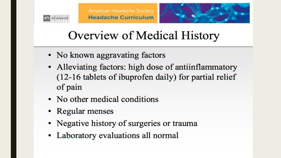
History Past medical history: ■ exposure to drugs, substances (particularly caffeine), and toxins ■ recent lumbar puncture; ■ immunosuppressive disorders or ■ IV drug use (risk of infection);
. ■ Cancer (risk of")
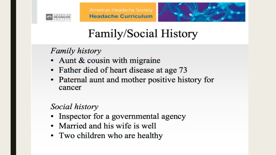
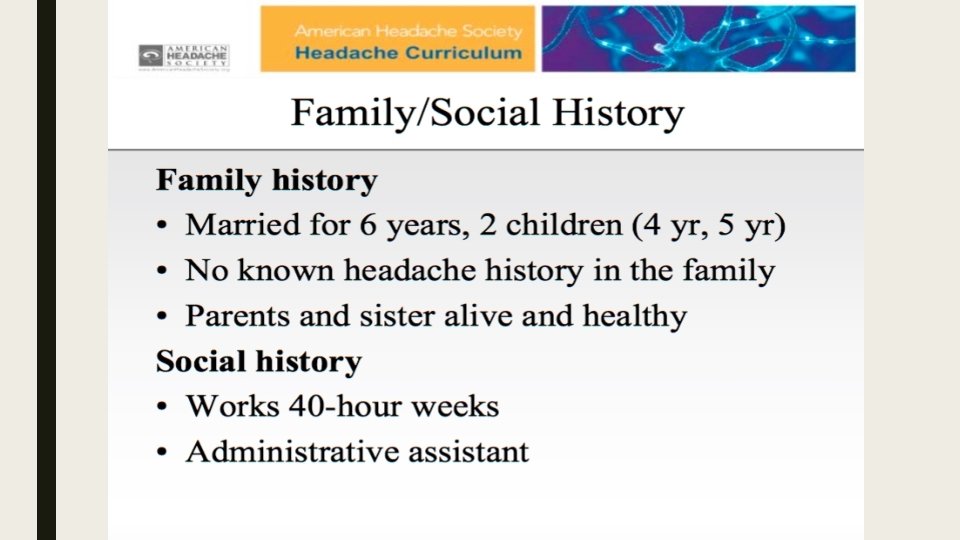
History Past medical history: ■ Hypertension (risk of brain hemorrhage). ■ Cancer (risk of brain metastases). ■ Trauma, coagulopathy, or use of anticoagulants or ethanol (risk of subdural hematoma). ■ Family and social history of headaches, particularly because migraine headache may be undiagnosed in family members.

Physical examination ■ Vital signs, including temperature. ■ General appearance (eg, whether restless or calm in a dark room) is noted. ■ Full neurologic examination are done. ■ The scalp is examined for areas of swelling and tenderness. ■ The ipsilateral temporal artery is palpated. ■ Temporomandibular joints are palpated for tenderness and crepitance while the patient opens and closes the jaw.

Physical examination ■ The eyes and periorbital area ( lacrimation, flushing, and conjunctival injection. ■ Pupillary size and light responses, extraocular movements, and visual fields. ■ The fundi for evidence of papilledema. If patients have visionrelated symptoms or eye abnormalities. ■ visual acuity. ■ The oropharynx for swellings, and the teeth for tenderness. ■ Neck flexion to detect discomfort or stiffness indicating meningismus. ■ The cervical spine for tenderness.

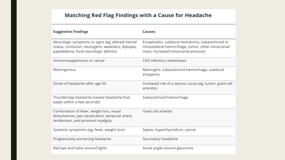
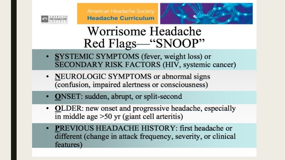
Red flags ■ Neurologic symptoms or signs ( altered mental status, weakness, diplopia, papilledema, focal neurologic deficits) ■ Immunosuppression or cancer. ■ Meningismus. ■ Onset of headache after age 50. ■ Thunderclap headache (severe headache that peaks within a few seconds)

Red flags ■ Symptoms of giant cell arteritis ( visual disturbances, jaw claudication, fever, weight loss, temporal artery tenderness, proximal myalgias). ■ Systemic symptoms ( fever, weight loss). ■ Progressively worsening headache. ■ Red eye and halos around lights.

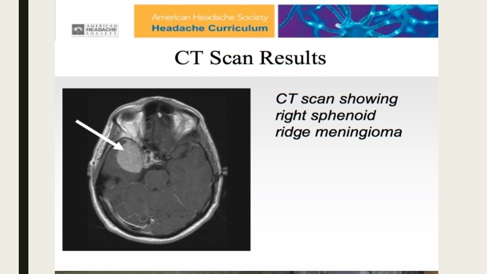
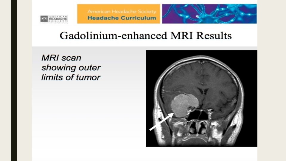
Diagnosis ■ Most patients can be diagnosed without testing. However, some serious disorders may require urgent or immediate testing. ■ ■ ■ ■ CT or MRI should be done in patients with any of the following findings: Age > 50 yr Weight loss Cancer Change in an established headache pattern Diplopia

Diagnosis ■ Thunderclap headache. ■ Altered mental status. ■ Meningismus. ■ Papilledema. ■ Signs of sepsis (eg, rash, shock). ■ Acute focal neurologic deficit. ■ Severe hypertension (eg, systolic > 220 mm Hg or diastolic > 120 mm Hg on consecutive readings).

Diagnosis ■ Lumbar puncture and CSF analysis should be done if * CNS infection ( meningitis, encephalitis). * Subarachnoid hemorrhage. * Headache is progressive and findings suggest idiopathic intracranial hypertension (transient obscuration of vision, diplopia, pulsatile tinnitus). ■ Tonometry should be done if findings suggest acute narrow-angle glaucoma (eg, visual halos, nausea, corneal edema).
. ■ CT of")
■ ESR (old age, visual symptoms, jaw claudication, temporal artery signs). ■ CT of the paranasal sinuses is done to rule out complicated sinusitis if patients have a moderately severe systemic illness (eg, high fever, dehydration, prostration, tachycardia) and findings suggesting sinusitis (eg, frontal, positional headache, epistaxis, purulent rhinorrhea).

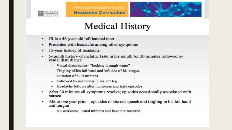
Case 1

Case 2


Thank You
- Slides: 60