APPROACH TO CORONARY BIFURCATION LESIONS Dr Santhosh Narayanan

APPROACH TO CORONARY BIFURCATION LESIONS Dr. Santhosh Narayanan SR Cardiology

Outline • Anatomy • Classification Systems – Medina System – MADS System • Important Stent Attributes for treating Bifurcation lesions • Bifurcation Treatment Techniques – Provisional Stenting – Two-stent • Clinical Evidence impacting current techniques • Summary
 to and/ or")
Any lesion with > 50% stenosis adjacent ( < 5 mm) to and/ or at the ostium of a side branch ( > 2 m m of diameter)

• Account for 16 -20 % of PCI's • Procedural complications – 9% • Restenosis as high as 36%

Bifurcation PCI Challenges Anatomical Variations • Vessel size variations • SB accessibility • SB takeoff angulations • Plaque distribution • Plaque volume • Plaque compliance • Peripheral Vascular Issues Procedural Options • Single vs double vs triple wire • Balloon predilation MB and SB • Plaque modification or debulking • Provisional vs multiple stents • DES vs BMS • Kiss? • Multiple Stent Strategies • Adjunctive Pharmacotherapy

SB angulation

The shear stress hypothesis • The outer walls of bifurcation points subjected to diastolic flow reversal, leads to oscillatory shear stress • Oscillatory (as versus laminar) shear stress is less efficient in stimulating e. NOS. • Monocytes bind more avidly to areas of oscillatory shear than to areas subjected to linear shear. • oscillatory shear stress is proatherogenic

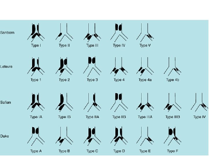
Classification • • Sanborn Lefevre Safian Duke • MEDINA • MOVAHED

MEDINA classification
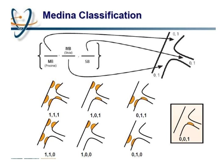

Limitations Does not take intoaccount 1. 2. 3. 4. 5. 6. Length of disease in the ostium of the SB Length of the LMCA before the bifurcation Trifurcation Vessel angulation No differentiation is madebetween a normal segment (lesion free segment) and a < 5 0 % lesion presenceof calcifications is not identified

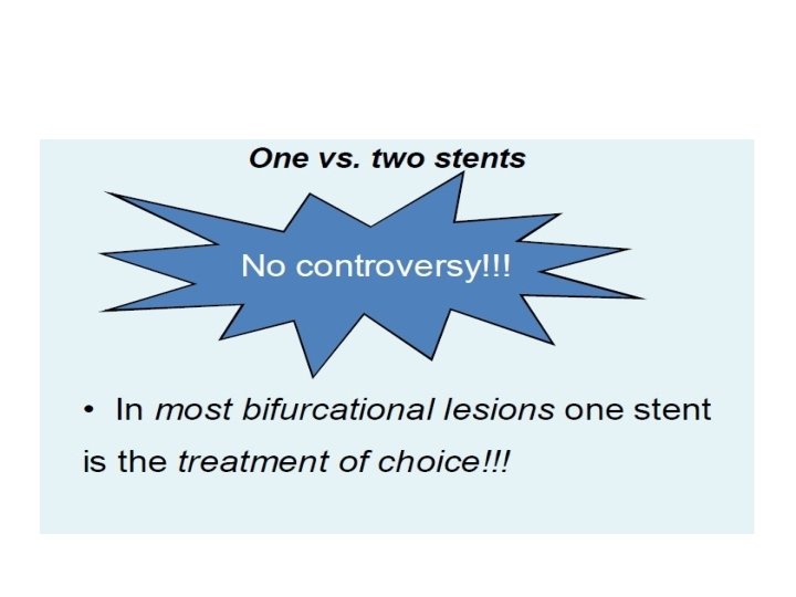
One vs two stents • • • Important trials NORDIC 2 BBC CACTUS

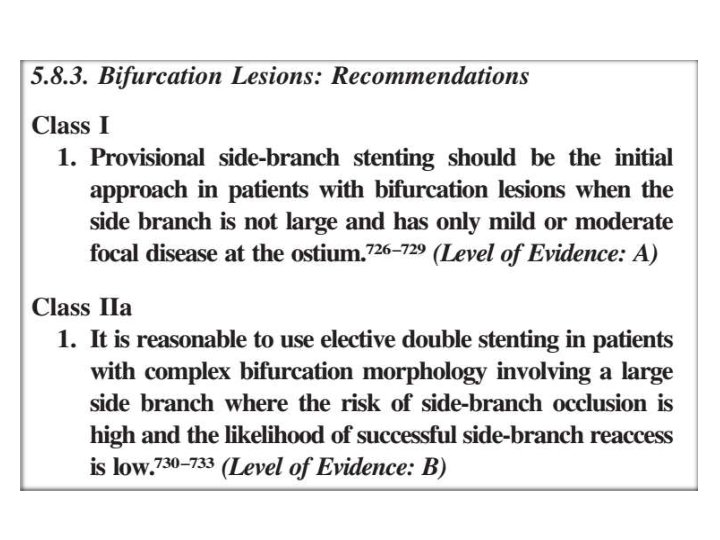
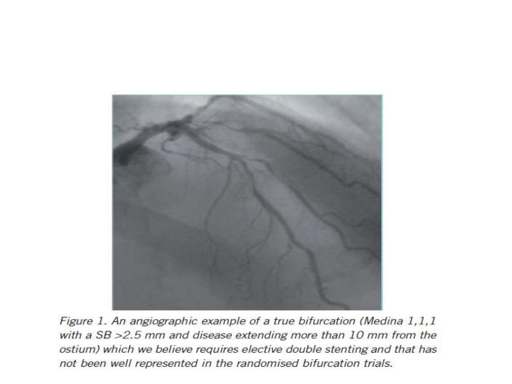
When to use elective two stents?

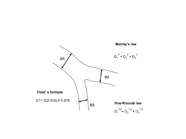
Ramifications of coronary tree follow minimal energy cost in providing myocardial blood flow Finet's law

OPTIMAL VIEW �For LMCA – RAO or LAO view with caudal inclination. �For LAD – D : AP with marked cranial angulations. �For LCx – OM : slight LAO or RAO with caudal angulations. �For distal RCA : AP with cranialangulations.
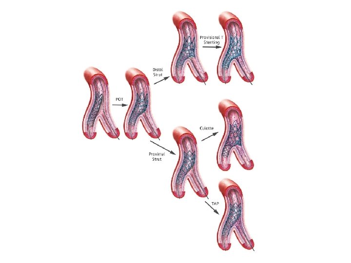
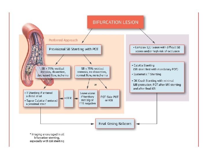
Provisional • Mainvessel stenting ± side branchangioplasty (Provisional) Tstenting, • TAP, •")
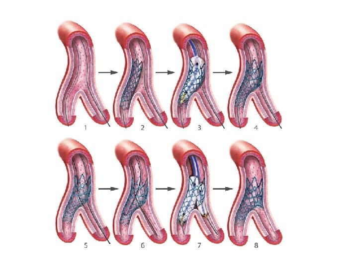
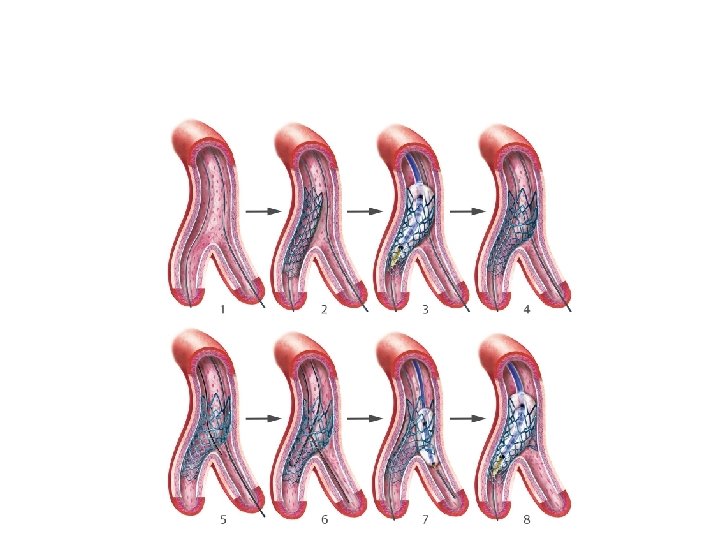
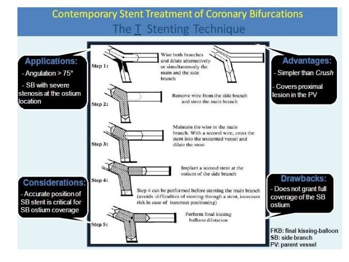
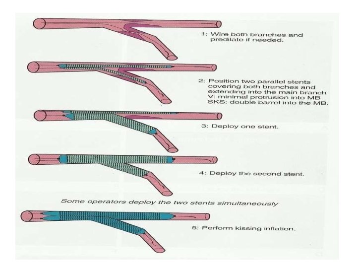
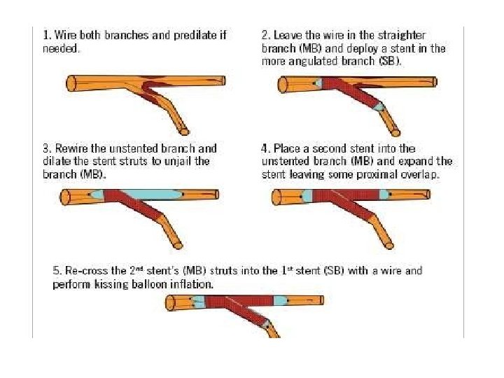
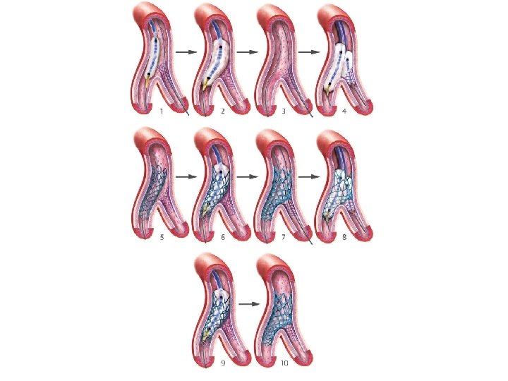
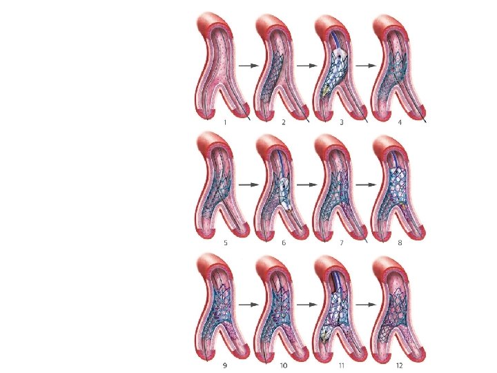
Stenting techniques 1)Provisional • Mainvessel stenting ± side branchangioplasty (Provisional) Tstenting, • TAP, • REVERSEINTERNAL CRUSH • REVERSECULOTTE. 2) elective • Culotte-stenting • Crush technique (reversecrush) T TECHNIQUE AND TAP • V STENTING • Y STENTING(SKS technique)

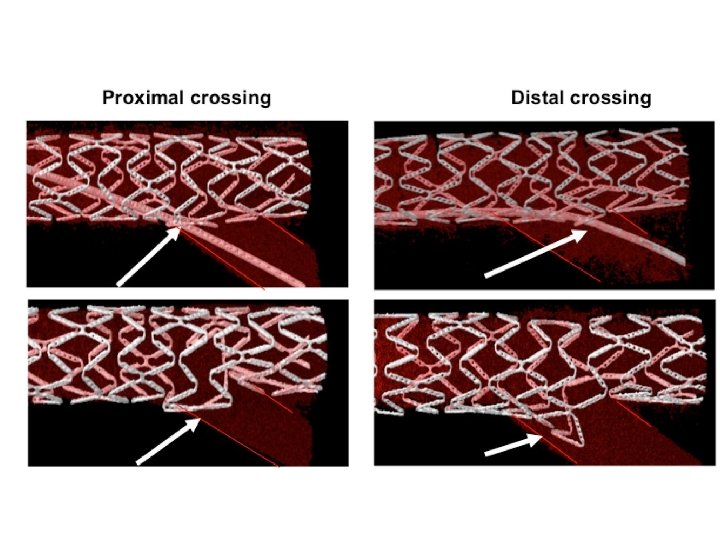
Provisional stenting strategy 1. Both branches are wired starting with the most difficult one 2. MB is stented (stent sized according to MB distal reference) and SB wire is jailed 3. The stent is post-dilated using the Proximal Optimization Technique (POT) to maximize stent apposition 4. Stent is now well apposed proximally, while the SB is partially covered by scaffolding 5. MB wire is pulled back and reinserted through the most distal strut of the SB opening scaffold 6. Jailed wire is removed and re-inserted in the distal MB (with a formed loop at the distal end) 7. The Kissing Balloon inflation is done to optimize side branch flow and access 8. Final result (if suboptimal, can then place additional stents)

Modified T technique

V technique

Culotte technique

� It provides near-perfect coverageof the carina &SBostium atthe expense of an excessof metal covering in proximal. MB. � Bestimmediate angiographic result &theoretically itmayguarantee a more homogeneous distribution of struts &drug. � Canbeused in all bifurcation lesions irrespective of bifurcationangle. � Open-cell stents arepreferred when the SBdiameter is >3 mm. � Disadvantages – � Complexity in the rewiring of both branches through the stentstruts, � Not advisable if both branches aredissected afterpredilatation.

DK crush

� immediate patency of both branches is assured &therefore it should be applied in conditions of instability or when the anatomy appears complex. � should be avoided in wide angle bifurcations. � Only SBhas to be re-wired ¬ both branches as in culotte technique. � The crush technique has evolved and is nowadays performed with less stent • protrusion into the MB(i. e. , mini-crush) &mandatory 2 -step FKI. � crush” technique can therefore be considered as a sort of simplified “culottes” technique � The mini-crush may be associated with more complete endothelialisation and • easier re-crossing of the crushed stent.

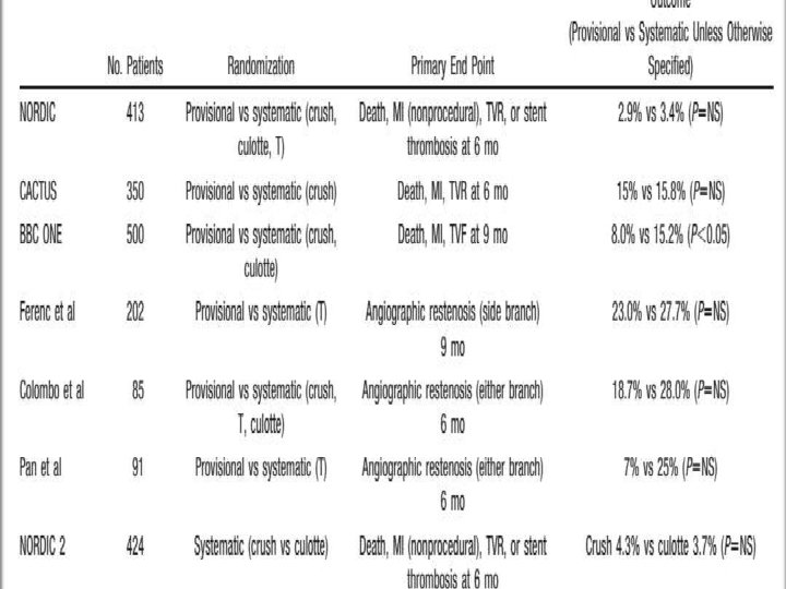
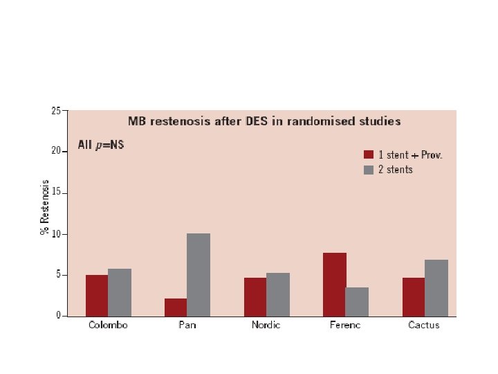
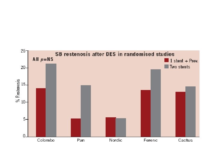
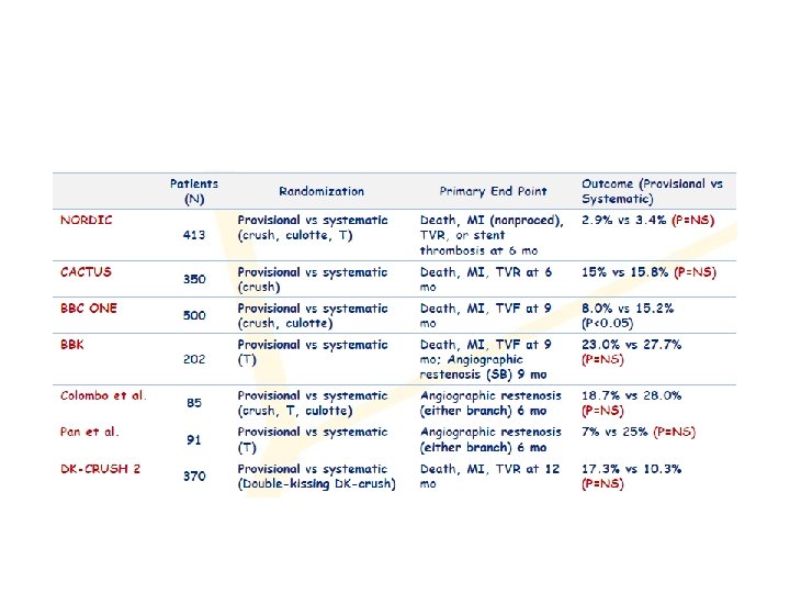
1. Nordic I: provisional T stenting as good as systematic side branch stenting 2. Nordic II: Culotte better than Crush 3. Cactus: provisional T stenting not worse than crush 4. BBC ONE: step wise approach with provisional T stenting better than initial complex procedures 5. Bad Krozingen: no difference provisional vs systematic T 6. Double Kiss Crush Study: DK Crush better than conv. crush Steigen Circulation 2006; 114: 1955; Erglis TCT 2008; Hildick-Smith TCT 200 Ferenc EHJ 2009; Chen JInterv Cardiol 2009; 22: 121 -27


Thank you
- Slides: 50