
Approach to Chronic Diarrhea Dr Nahla Ali Azzam
- Slides: 47
Approach to Chronic Diarrhea Dr Nahla Ali Azzam Assistant Professor And Consultant Gastroenterology and Hepatology King Saud University
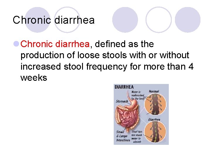
Chronic diarrhea l Chronic diarrhea, defined as the production of loose stools with or without increased stool frequency for more than 4 weeks
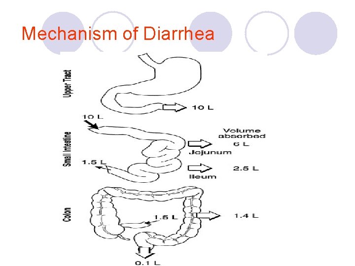
Mechanism of Diarrhea
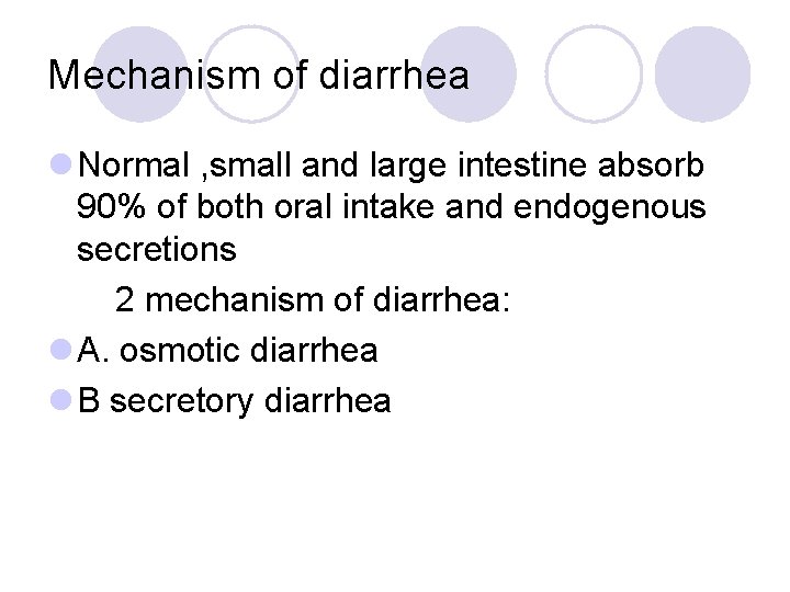
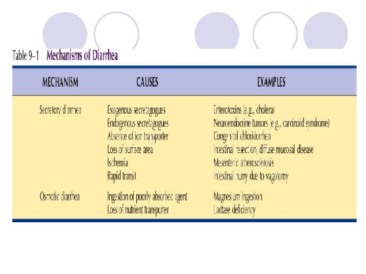
Mechanism of diarrhea l Normal , small and large intestine absorb 90% of both oral intake and endogenous secretions 2 mechanism of diarrhea: l A. osmotic diarrhea l B secretory diarrhea
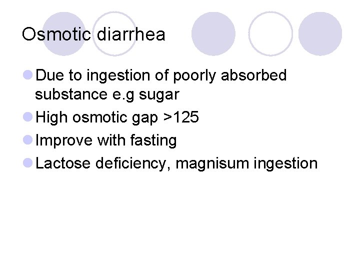
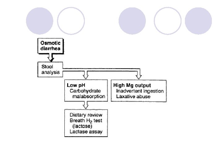
Osmotic diarrhea l Due to ingestion of poorly absorbed substance e. g sugar l High osmotic gap >125 l Improve with fasting l Lactose deficiency, magnisum ingestion
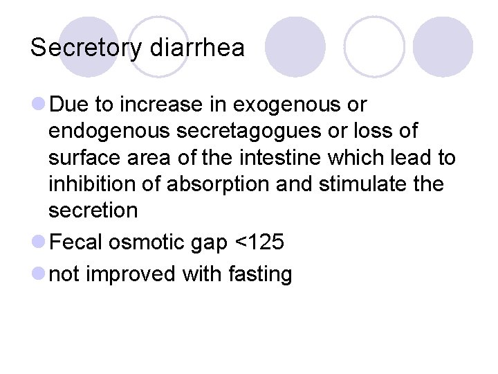
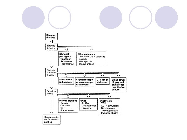
Secretory diarrhea l Due to increase in exogenous or endogenous secretagogues or loss of surface area of the intestine which lead to inhibition of absorption and stimulate the secretion l Fecal osmotic gap <125 l not improved with fasting
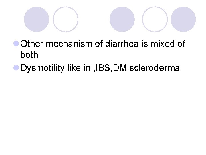
l Other mechanism of diarrhea is mixed of both l Dysmotility like in , IBS, DM scleroderma
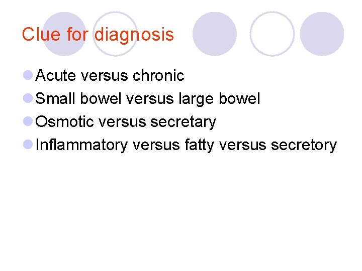
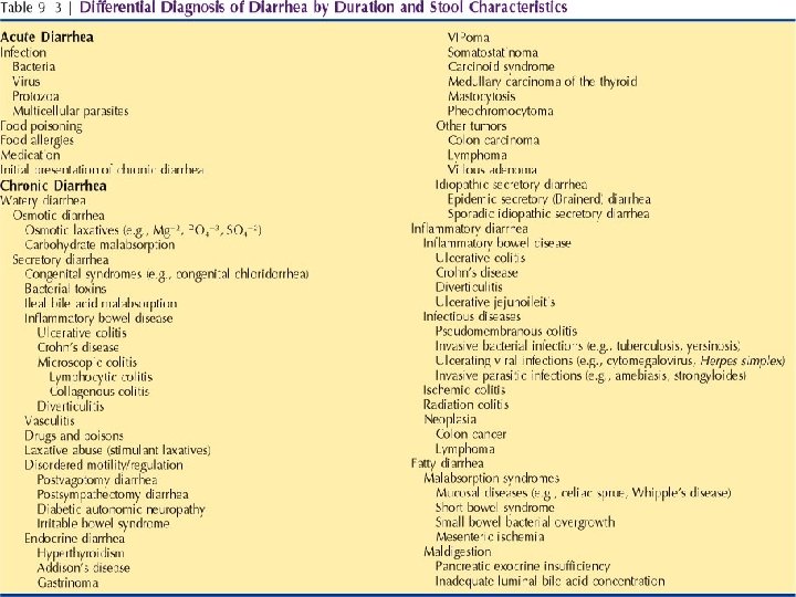
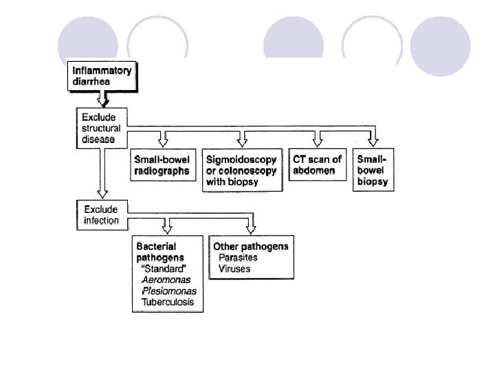
Clue for diagnosis l Acute versus chronic l Small bowel versus large bowel l Osmotic versus secretary l Inflammatory versus fatty versus secretory
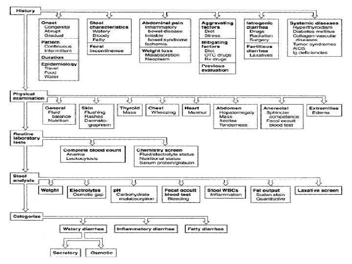
A careful history can provide clues to the cause of chronic diarrhea. The following 14 points should be assessed as part of a comprehensive history in a patient with chronic diarrhea : . 1 The characteristics of the onset of diarrhea congenital, abrupt, or gradual in onset. 2. The pattern of diarrhea should be recorded: Are loose stools continuous or intermittent. 3 The duration of symptoms should be identified clearly. 4 Epidemiological factors, such as travel before the onset of illness
Likely cause of diarrhea in certain epidemiologic classificatiuons l Travelers: bacterial, protozoal, tropical sprue l Diabetics patients l AIDS patients infections, drugs l Hospitalized patients : drugs, infections, ischemia , C. D toxin
l. 5. Stool characteristics stools are watery, bloody, or fatty. l 6. The presence or absence of fecal incontinence should be determined. l 7. The presence or absence of abdominal pain. Pain often is present in patients with inflammatory bowel disease, irritable bowel syndrome, and mesenteric ischemia. l 8. The presence of weight loss is more likely to be caused by nutrient malabsorption, neoplasm, or ischemia.
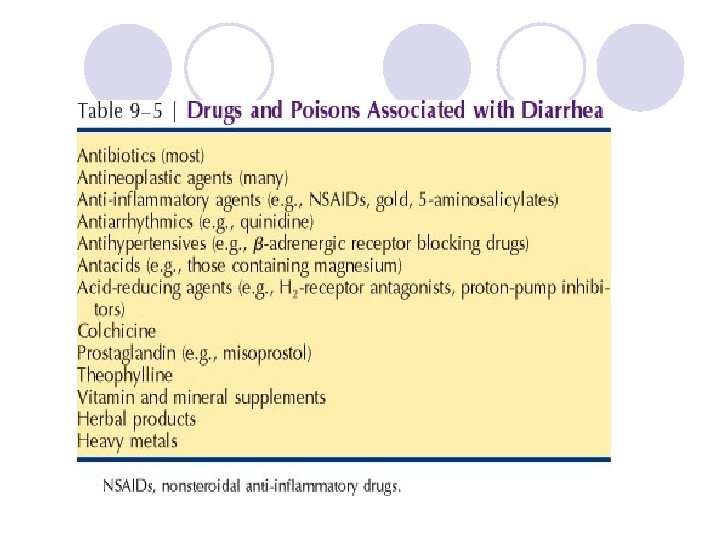
l 9. Aggravating factors, such as diet and stress, should be recorded. l 10. Mitigating factors, such as alteration of diet and use of both prescription and over-the-counter drugs. l 11. Previous evaluations should be reviewed whenever possible. . l 12. Iatrogenic causes of diarrhea , detailed medication history and a history of radiation therapy or surgery
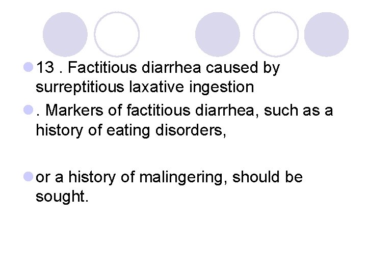
l 13. Factitious diarrhea caused by surreptitious laxative ingestion l. Markers of factitious diarrhea, such as a history of eating disorders, l or a history of malingering, should be sought.
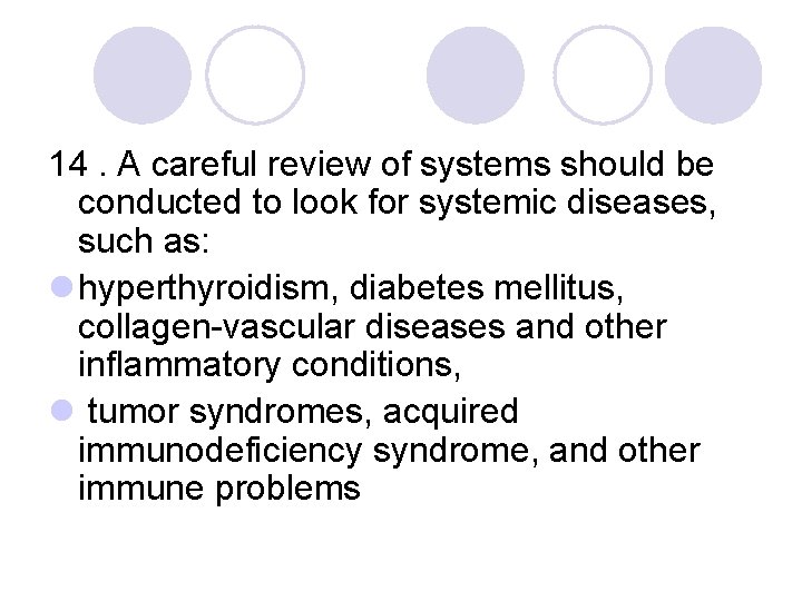
14. A careful review of systems should be conducted to look for systemic diseases, such as: l hyperthyroidism, diabetes mellitus, collagen-vascular diseases and other inflammatory conditions, l tumor syndromes, acquired immunodeficiency syndrome, and other immune problems
Evaluation of patient with dairrhea l History l Physical examination l Laboratory l Radiological l Endoscopy
Malabsorption
Pathophysiology . In general, the digestion and absorption of food. materials can be divided into 3 major phases -The luminal phase where dietary fats, proteins, and carbohydrates are hydrolyzed and solubilized by secreted digestive enzymes and bile. -The mucosal phase relies on the integrity of the brush-border membrane of intestinal epithelial cells to transport digested products from the lumen into the cells. -In the postabsorptive phase, nutrients are transported via lymphatics and portal circulation from. epithelial cells to other parts of the body
Causes l The best way to classify the numerous causes of malabsorption is to consider the 3 phases of digestion and absorption. l Luminal phase ¡ Impaired nutrient hydrolysis • The most common cause is pancreatic insufficiency due to chronic pancreatitis, pancreatic resection, pancreatic cancer, or cystic fibrosis. ¡ Inactivation of pancreatic enzymes by gastric hypersecretion, as seen in Zollinger-Ellison syndrome. .
¡ Impaired micelle formation • Impaired micelle formation causes lead to fat malabsorption. This impairment is due to different reasons, including • (1) decreased bile salt synthesis from severe parenchymal liver disease (eg, cirrhosis); • (2) impaired bile secretion from biliary obstruction or cholestatic jaundice (eg, primary biliary cirrhosis, primary sclerosing cholangitis); • (3) impaired enterohepatic bile circulation, as seen in small bowel resection or regional enteritis; • (4) bile salt deconjugation due to small bowel bacterial overgrowth. l. ¡ Luminal availability and processing • Luminal bacterial overgrowth can cause a decrease in the availability of substrates, including carbohydrates, proteins, and vitamins
l Mucosal phase l Impaired brush-border hydrolase activity ¡Disaccharidase deficiency. ¡Lactase deficiency ¡Immunoglobulin A (Ig. A) deficiency
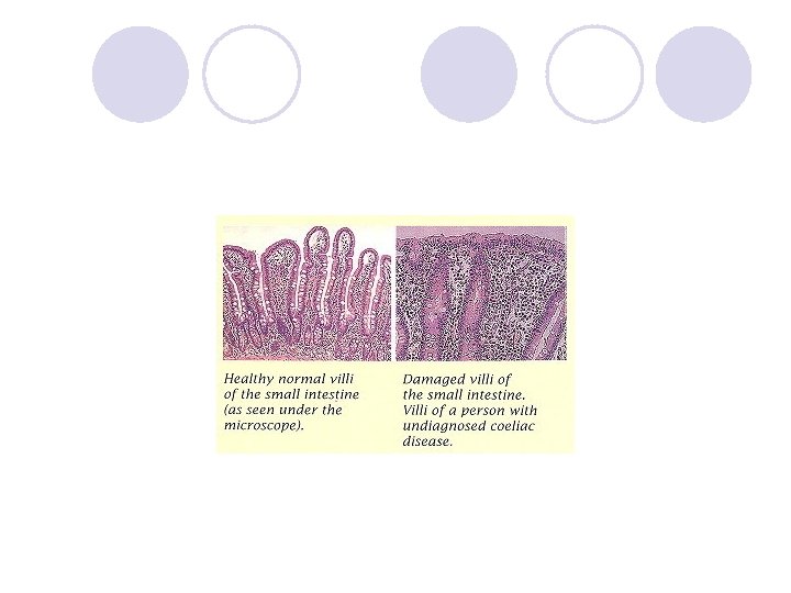
l Impaired nutrient absorption Acquired disorders are far more common and are caused by (1) decreased absorptive surface area, as seen in intestinal resection l (2) damaged absorbing surface, as seen in celiac sprue, tropical sprue, giardiasis, Crohn disease, AIDS enteropathy, chemotherapy, or radiation therapy; l (3) infiltrating disease of the intestinal wall, such as lymphoma and amyloidosis
l Postabsorptive phase: l Obstruction of the lymphatic system, both congenital (eg, intestinal lymphangiectasia) l acquired (eg, Whipple diseases , lymphoma, tuberculosis), l
Clinical presentation History : Diarrhea, Wt Loss, Steatorrhea , Flatulence and abdominal distention , Edema, anemia, Metabolic defects of bones
Physical l l l General Patients may have orthostatic hypotension. Signs of weight loss, muscle wasting, or both may be present. Patients may have signs of loss of subcutaneous fat Cheilosis, glossitis, or aphthous ulcers of the mouth. Abdominal examination The abdomen may be distended, and bowel sounds may be hyperactive. Ascites may be present in severe hypoproteinemia
Dermatological manifestations l Pale skin l Ecchymoses due to vitamin K deficiency l Dermatitis herpetiformis, erythema nodosum, and pyoderma gangrenosum may be present. l Pellagra, alopecia, or seborrheic dermatitis Neurological examination l Motor weakness, peripheral neuropathy, or ataxia may be present. l The Chvostek sign or the Trousseau sign may be evident due to hypocalcemia.
Lab Studies l Hematological tests ¡ A CBC ¡ Serum iron, vitamin B-12, and folate ¡ Prothrombin time. l Electrolytes and chemistries ¡ hypokalemia, hypocalcemia, hypomagnesemia, and metabolic acidosis. ¡ Protein malabsorption may cause hypoproteinemia and hypoalbuminemia. ¡ Fat malabsorption can lead to low serum levels of triglycerides, cholesterol ¡ ESR which is elevated in Crohn disease and Whipple disease
Stool analysis l Stool p. H may be assessed. Values of <5. 6 are consistent with carbohydrate malabsorption l Stool C/S l Pus cell in the stool e. g IBD
l Tests of fat malabsorption l For a quantitative measurement of fat absorption, a 72 -hour fecal fat collection l Qualitative test Sudan III stain of stool, less reliable.
l D-xylose test If the 72 -hour fecal fat collection results demonstrate fat malabsorption, then DO → the D-xylose test is used to document the integrity of the intestinal mucosa. proximal intestine mucosa primarily absorbs D-xylose. Approximately half of the absorbed D-xylose is excreted in urine, unmetabolized. If the absorption of D-xylose is impaired due to either a luminal factor (eg, bacterial overgrowth) or a reduced or damaged mucosal surface area
l Schilling test l Malabsorption of vitamin B-12 may occur as a consequence of deficiency of intrinsic factor (eg, pernicious anemia, gastric resection), pancreatic insufficiency, bacterial overgrowth, ileal resection, or disease.
3 STEPS SCHILLING TEST 1. oral VIT B 12 2. VIT B 12 orally +intrinsic factor 3. VIT B 12 orally +intrinsic factor+ oral antibiotics
Bacterial overgrowth l Bacterial overgrowth cause an early rise in breath hydrogen l JEUJENAL CULTURE l 14 c D–xylose breath test , high sensitivity and specificity
l Serology ¡No serologic tests are specific for malabsorption. ¡Serum antigliadin and antiendomysial antibodies can be used to help diagnose celiac sprue. ¡Serum Ig. A to rule out Ig. A deficiency ¡Determination of fecal elastase and chymotrypsin (2 proteases produced by the pancreas) can be used to try to distinguish between pancreatic causes and intestinal causes of malabsorption.
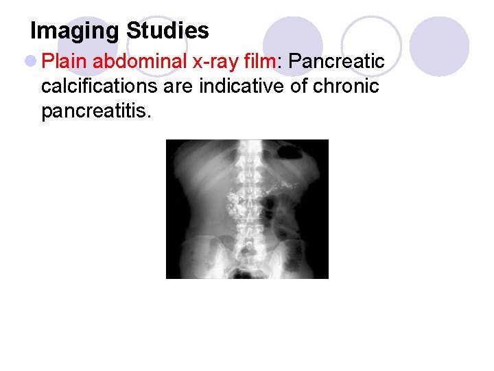
Imaging Studies l Plain abdominal x-ray film: Pancreatic calcifications are indicative of chronic pancreatitis.
l Small bowel barium studies . . ¡ Flocculation ¡ Small bowel dilation ¡ diverticulosis. ¡ stricture, ulceration, and fistula formation. l CT scan of the abdomen: l chronic pancreatitis l Enlarged lymph nodes are seen in Whipple disease and lymphoma l Luminal disease. l Endoscopic retrograde cholangiopancreatogram (ERCP): pancreatitis
Upper endoscopy with small bowel mucosal biopsy…. Examples l celiac sprue, l giardiasis, l Crohn disease, l Whipple disease, l amyloidosis, l abetalipoproteinemia, and lymphoma.
Treatment of causative diseases l A gluten-free diet helps treat celiac disease. l Similarly, a lactose-free diet l Protease and lipase supplements are therapy for pancreatic insufficiency. l Antibiotics are therapy for bacterial overgrowth. l Corticosteroids, anti-inflammatory agents, such as mesalamine, and otherapies are used to treat CD
l Nutritional support l Supplementing various minerals calcium, magnesium, iron, and vitamins l Caloric and protein replacement also is essential. l Medium-chain triglycerides can be used for lymphatic obstruction. l In severe intestinal disease, such as massive resection and extensive regional enteritis, parenteral nutrition may become necessary