APPROACH TO BRONCHIECTASIS LUNG ABSCESS DR PRAKHAR SHARMA

APPROACH TO BRONCHIECTASIS & LUNG ABSCESS DR. PRAKHAR SHARMA ASSISTANT PROFESSOR PULMONARY MEDICINE DEPARTMENT 03/09/2019

INTRODUCTION NOT COVERED QUICK RUN THROUGH THE LECTURE CYSTIC FIBROSIS CHILDREN ≤ 15 YEARS DIAGNOSIS ETIOLOGY INVESTIGATIONS AND MANAGEMENT

LEARNING OBJECTIVES – Definition – Etiology – Pathogenesis – Clinical manifestations – Diagnosis – Treatment

CASE 1 – A 42 -year-old man, gardener – Long history of respiratory problems starting in early childhood. – Previously diagnosed as asthma. – Frequent absence from work due to “recurrent chest infections”. – Unaware of any neonatal issues but believes that he was born at home without complications and is unsure of any previous tests he has had as he is now estranged from his parents. – Has a cousin with a “lung disease”. – Married but has “no kids”

INVESTIGATIONS – Sputum culture: P. aeruginosa – Sweat chloride = 73 meq/liter – Cystic fibrosis genetics: genotype was F 508 del/R 117 H – CYSTIC FIBROSIS: Multisystem disorder caused by mutations in the gene that encodes the CF transmembrane conductance regulator (CFTR) protein, a chloride channel expressed in epithelial cells. – More than 2000 CFTR mutations have been identified to date, but only the functional importance of a small number is known to cause the disease

HRCT THORAX – An upper lobe predominant distribution of cylindrical, cystic and varicose bronchiectasis associated with airway wall thickening, mucus plugging and parenchymal opacities on a HRCT scan should raise the suspicion of CF disease. – The presence of nasal polyposis and/or chronic rhinosinusitis, recurrent pancreatitis, malabsorption, diabetes, osteoporosis and male infertility are other typical features of CF

DIAGNOSIS Guidelines published by the Cystic Fibrosis Foundation in the USA allows diagnosis if: 1. Clinical presentation of the disease and evidence of biochemical and genetic markers of CFTR dysfunction. 2. Clinical features of the disease with concentration of chloride >60 mmol·L− 1 at the sweat test or a concentration in the intermediate range (30– 59 mmol·L− 1) but two disease-causing CFTR mutations. 3. CFTR genotype is undefined: CFTR physiologic tests, such as nasal potential difference and intestinal current measurement, should be performed.

MANAGEMENT 1. CFTR modulator therapies 2. Airway clearing techniques 3. Chest physical therapy 4. Humidification with sterile water or normal saline to facilitate airway clearance 5. Antibiotics 6. Mucus thinners 7. Lung transplantation

CASE 2 – 45 -year-old farmer with asthma since childhood. – Complaints: Decline in his exercise tolerance and an increase in cough which has become productive of purulent sputum with occasional thick/solid components. – Respiratory exacerbations not responding well to standard steroid antibiotic treatment. – He was noted to have variable pulmonary infiltrates on chest radiographs during these episodes

INVSETIGATIONS – Marked peripheral blood eosinophilia – Total Ig. E > 1000 IU/ ml – Aspergillus specific Ig. E > O. 35 ABPA: ABPA is an inflammatory disease caused by hypersensitivity to the ubiquitous fungus Aspergillus fumigatus – ABPA occurs most commonly in patients with asthma and CF – ABPA is the cause of 1– 10% of cases of bronchiectasis – Most ABPA cases occur in the third and fourth decade without a sex predilection.

DIAGNOSIS – Long standing uncontrolled asthma/ Cystic fibrosis – Brownish sputum – Peripheral eosinophilia > 500/ mm 3 – Total Ig. E > 1000 IU/ ml – Specific Ig. E for A. fumigatus > 0. 35 HRCT thorax: Ø Central bronchiectasis Ø High attenuation mucus Ø Finger in glove/ TIB Ø Tram track Ø Mosaic attenuation

MANAGEMENT 1. Corticosteroids 2. Antifungals 3. Airway clearing techniques 4. Chest physical therapy 5. Mucus thinners

CASE 3 – 77 -year-old retired librarian. – Cough for many years with new symptoms of fatigue, weight loss and fever. – A chest CT scan was performed looking for a possible occult malignancy and bronchiectasis was found.

DIAGNOSIS – HRCT thorax: cylindrical bronchiectasis and tree-in-bud pattern in middle and lower lobes – Sputum for M. Tuberculosis: negative – MGIT culture: MAC growth at 4 weeks – Repeat MGIT: Positive for MAC – Tests for immunodeficiency and ABPA: Negative

MANAGEMENT 1. Management of NTM as per the organism and clinical picture 2. Airway clearing techniques 3. Chest physical therapy 4. Mucus thinners

CASE 4 – A 66 -year-old woman with established idiopathic bronchiectasis had three to four exacerbations per year for the past 3 years despite performing daily chest physiotherapy. – Produces large volumes of sputum daily despite performing the active cycle of breathing technique. – Testing for NTM, ABPA and other complications were negative, but sputum shows persistent infection with P. aeruginosa.

– One of the most common presentations of bronchiectasis – Exacerbations are one of the most important manifestations of bronchiectasis and P. aeruginosa is the most frequent organism in severe bronchiectasis worldwide – Cylindrical bronchiectasis is the most common morphological pattern identified on CT scans

MANAGEMENT 1. Review current airway clearance regime. 2. Repeat sputum microbiology and repeat testing for NTM, ABPA and ensuring the all possible treatable causes and comorbidities have been identified. 3. First-line recommendation for P. aeruginosa with frequent exacerbations is an inhaled antibiotic.

ETIOLOGY

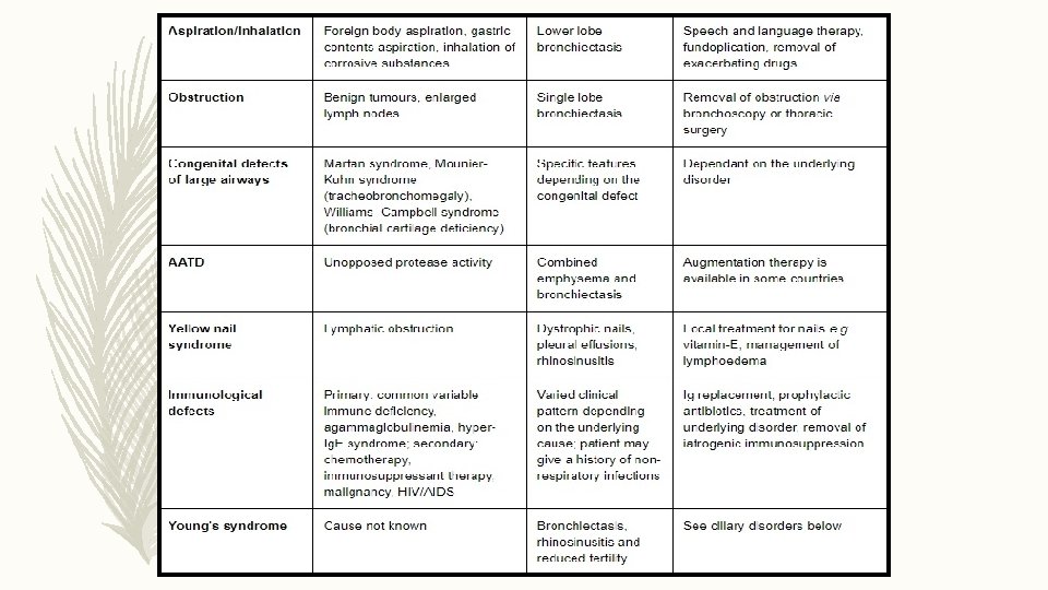
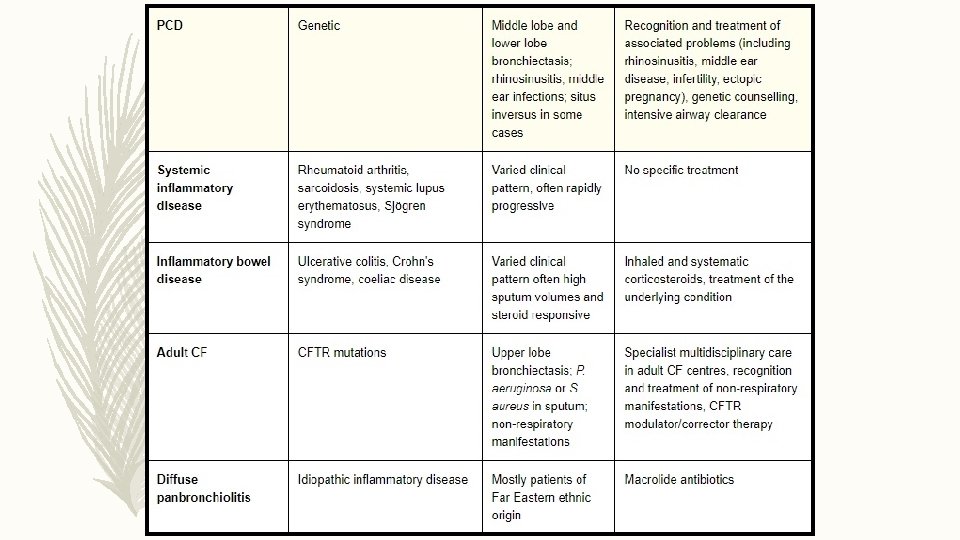

INVESTIGATIONS FOR CAUSE IN ALL CLINICALLY STABLE • COMORBIDITIES AND RELEVANT PAST HISTORY • FULL BLOOD COUNT/ SERUM TOTAL IGE/ SKIN PRICK TEST TO A. FUMIGATUS • SERUM Ig G/ Ig. A/ Ig. M • BASELINE SPECIFIC ANTIBODY LEVELS AGAINST CAPSULAR POLYSACCHRIDES OF STREPTOCOCCUS PNEUMONIAE • SPUTUM CULTURE : ROUTINE AND MYCOBACTERIAL • HIV • TEST FOR CYSTIC FIBROSIS/ PCD/ GERD • RA, ANTI CCP , ANCA, ANA CLINICALLY • ALPHA 1 AT SUSPECT • BRONCHIAL ASPIRATION OR WASH Hill A, Welham S, Sullivan A, Loebinger M. Updated BTS Adult Bronchiectasis Guideline 2018: a multidisciplinary approach to comprehensive care. Thorax. 2018; 74(1): 1 -3.

STEPWISE MANAGEMENT


AIRWAY CLEARANCE

Physiotherapy management-stepwise airway clearance.

ACBT

POSTURAL DRAINAGE

Airway clearance - exacerbations.

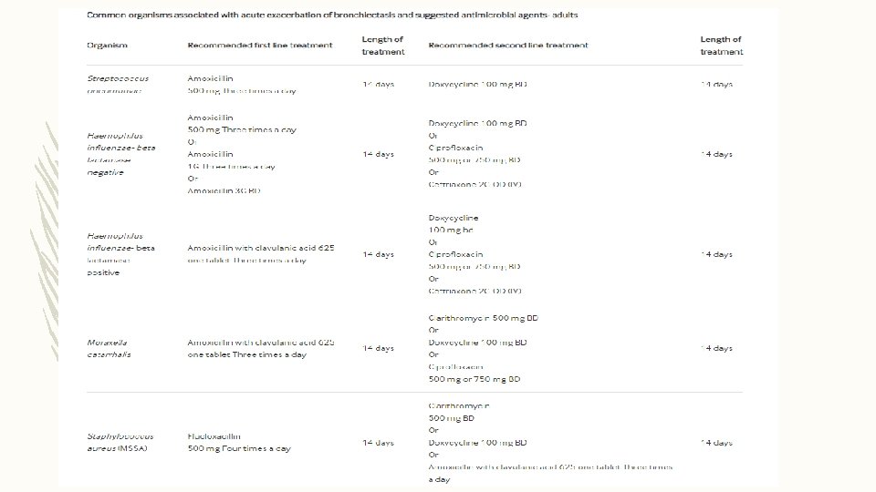
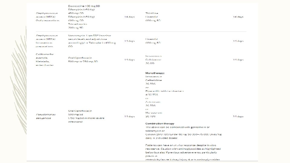
ANTIBIOTIC TREAMENT FOR EXACERBATION


WHAT IS THE ROLE OF SURGERY IN MANAGING BRONCHIECTASIS? RECOMMENDATIONS – Consider lung resection in patients with localized disease whose symptoms are not controlled by medical treatment optimized by a bronchiectasis specialist. (D) – Offer multidisciplinary assessment, including a bronchiectasis physician, a thoracic surgeon and an experienced anesthetist, of suitability for surgery and pre-operative assessment of cardiopulmonary reserve post resection. (D)

LUNG TRANSPLANTATION FOR BRONCHIECTASIS Recommendations – Consider transplant referral in bronchiectasis patients aged 65 years or less if the FEV 1 is <30% with significant clinical instability or if there is a rapid progressive respiratory deterioration despite optimal medical management. (D) – Consider earlier transplant referral in bronchiectasis patients with poor lung function and the following additional factors: massive haemoptysis, severe secondary pulmonary hypertension, ICU admissions or respiratory failure (particularly if requiring NIV). (D)

LUNG ABSCESS

DEFINITION Localized area of lung suppuration, leading to necrosis of the lung parenchyma with or without cavity formation. Type of liquefactive necrosis of the lung tissue and formation of cavities (more than 2 cm) containing necrotic debris or fluid caused by microbial infection.

ACUTE DURATION CHRONIC PRIMARY LUNG ABSCESS AETIOLOGY SECONDARY WAY OF SPREADING BRONCHOGENIC HEMATOGENIC
 • ❖ ACCORDING TO THE DURATION: • Acute (less than 6")
CLASSIFICATION (CONTD. ) • ❖ ACCORDING TO THE DURATION: • Acute (less than 6 weeks); • Chronic (more than 6 weeks) • ❖ BY ETIOLOGY: • Primary (aspiration of oropharyngeal secretions, necrotizing pneumonia, immunodeficiency); • Secondary (bronchial obstructions, haematogenic dissemination, direct spreading from mediastinal infection, from sub phrenic space, coexisting lung diseases) • ❖ WAY OF SPREADING: • Bronchogenic (aspiration of oropharyngeal secretions, bronchial obstruction by tumour, foreign body, enlarged lymph nodes, congenital malformation); • Haematogenic (abdominal sepsis, infective endocarditis, septic thromboembolisms)

DIFFERENTIATING ACUTE FROM CHRONIC ACUTE CIRCUMSCRIBED NOT WELL DEFINED FILLED WITH NECROTIC DEBRIS CHRONIC IRREGULAR WELL DEFINED FILLED WITH THICK DEBRIS
 § Excavating tuberculosis § Localized")
DIFFERENTIAL DIAGNOSIS § Excavating bronchial carcinoma (squamo-cellular or microcellular) § Excavating tuberculosis § Localized pleural empyema § Infected emphysematous bullae § Cavitary pneumoconiosis § Hiatus hernia § Pulmonary hematoma § Hydatid cyst of lung § Cavitary infarcts of lung § Wegener’s granulomatosis

DIAGNOSIS – Diagnostic bronchoscopy is a part of diagnostic protocol for taking the material for microbiological examination and to confirm intrabronchial cause of abscess-tumor or foreign body. – Sputum examination is useful for identification of microbiological agents or confirmation of bronchial carcinoma

MANAGEMENT STANDARD CONSERVATIVE THERAPY: MEDICAL MANAGEMENT § It is recommended to treat lung abscess with broad spectrum antibiotics, due to poly microbial flora, such as Clindamycin (600 mg IV on 8 h) and then 300 mg PO on 8 h or combination ampicilin/sulbactam (1. 5 -3 gr IV on 6 h). § Alternative therapy is piperacilin/tazobactam 3. 375 gr IV on 6 h or Meropenem 1 gr IV on 8 h. § For MRSA it is recommended to use linezolid 600 mg IV on 12 h or vancomycin 15 mg/kg BM on 12 h.

MANAGEMENT SURGICAL § Endoscopic drainage of lung abscesses is described as an alternative to chest tube drainage and is performed during the bronchoscopy with usage of laser. § Per cutaneous trans thoracic tube drainage § Surgical resection of lung abscess is therapy of choice for about 10% of patients. § Lobectomy is the resection of choice for large or central position of abscess. Atypical resection or segmentectomy are satisfactory procedures, if it is possible to remove complete abscess and if necessary surrounding lung tissue with necrotizing pneumonia

THANK YOU

BRONCHIECTASIS
 is a morphologic term used to describe")
DEFINITION – Bronchiectasis (broncos, airways; ectasia, dilatation) is a morphologic term used to describe abnormal, irreversibly dilated and thick walled bronchi. – This is an anatomic definition that evolved from Laennec’s original description in 1819 of ectatic bronchi in pathologic specimens. Laënnec RTH. On mediate auscultation, or a treatise on the diagnosis of diseases of the lungs and heart. Paris: J. -A. Brosson et J. -S. Chaudé; 1819.

PREVALENCE 1 US • Prevalence increased every year from 2000 to 2007 by an annual percentage change of 8. 74%. • Increased with age (peak= 80 -84 years) • Higher in women 2 UK • Prevalence in women 566/lakh ; men= 486/lakh • Women and age more than 60 years associated with higher rate of hospitalization INDIA • There is no good data on bronchiectasis in India • EMBARC INDIA REGISTRY (European Multi Centre Bronchiectasis Audit and Research Collaboration) 1. Mc. Shane P, Naureckas E, Tino G, Strek M. Non–Cystic Fibrosis Bronchiectasis. American Journal of Respiratory and Critical Care Medicine. 2013; 188(6): 647 -656. 2. Hill A, Welham S, Sullivan A, Loebinger M. Updated BTS Adult Bronchiectasis Guideline 2018: a multidisciplinary approach to comprehensive care. Thorax. 2018; 74(1): 1 -3.

BURDEN LONGER HOSPITAL STAY FREQUENT OPD VISITS MORTALITY RATE= 10 -16% INCREASED EXPENDITURE ON MEDICINES RISK FACTORS FOR MORTALITY LOW FEV 1 MALE INCREASED DYSPNOEA GRADE ADVANCED AGE COPD P. aeruginosa SPUTUM POSITIVITY • Goeminne PC, Scheers H, Decraene A, Seys S, Dupont LJ. Risk factors for morbidity and death in non–cystic fibrosis bronchiectasis: a retrospective cross-sectional analysis of CT diagnosed bronchiectatic patients. Respir Res 2012; 13: 21. • Weycker D, Edelsberg J, Oster G, Tino G. Prevalence and economic burden of bronchiectasis. Clin Pulm Med 2005; 12: 205– 209 .

PATHOGENESIS COLES’S VISCIOUS CYCLE MODEL

PULMONARY INFECTION/ TISSUE INJURY BACTERIAL COLONIZATION ABNORMAL MUCUS CLEARANCE ROBUST INFLAMMATORY RESPONSE AIRWAYS DESTRUCTION AND DISTORTION Cole PJ. Inflammation: a two-edged sword—the model of bronchiectasis. Eur J Respir Dis Suppl 1986; 147: 6– 15.

PATHOLOGY Hematoxylin and eosin stain of the bronchial wall in a patient with bronchiectasis (left) versus a normal subject (right). A: Pseudostratified columnar, ciliated epithelium B: thickened epithelium with intraepithelial lymphocytes C: submucosa with dense infiltrate of lymphocytes and plasma cells D: blood vessel with reactive endothelial cells. BRONCHIECTATIC NORMAL

TYPES REIDS CLASSIFICATION CYLINDRICAL VARICOSE SACULAR/CYSTIC Reid LM. Reduction in bronchial subdivision in bronchiectasis. Thorax 1950; 5: 233– 247.

TYPES OF BRONCHIECTASIS

ETIOLOGY


IN WHOM TO SUSPECT ?

PERSISTENT PURULENT OR MUCO PURULENT SPUTUM + COPD FREQUENT EXACERBATORS WITH SPUTUM CULTURE POSITIVE FOR P. AERUGINOSA WHEN STABLE HIV/ SOLID ORGAN OR BONE MARROW TRANSPLANT/ IMMUNOSUPRRESIVE THERAPY FOR LYMPHOMA OR VASCULITIS IN HEALTHY? ? 1 IBD/ GERD/ CTD/ RHINOSINUSITIS RA WITH RECURRENT CHEST INFECTIONS SEVERE/ POORLY CONTROLLED ASTHMA Hill A, Welham S, Sullivan A, Loebinger M. Updated BTS Adult Bronchiectasis Guideline 2018: a multidisciplinary approach to comprehensive care. Thorax. 2018; 74(1): 1 -3.

ALGORITHM FOR EVALUATION OF BRONCHIECTASIS
: W")
Cantin L, Bankier A, Eisenberg R. Bronchiectasis. American Journal of Roentgenology. 2009; 193(3): W 158 -W 171.
![INVESTIGATION: RADIOLOGY – BASELINE CHEST RADIOGRAPH – THIN SECTION CT [HRCT THORAX] CT FEATURES](http://slidetodoc.com/presentation_image_h/dd50923a631ced68233c2e92554ff69b/image-68.jpg "INVESTIGATION: RADIOLOGY – BASELINE CHEST RADIOGRAPH – THIN SECTION CT [HRCT THORAX] CT FEATURES")
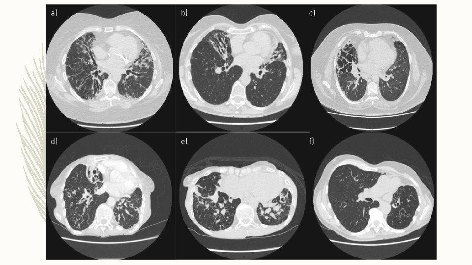
INVESTIGATION: RADIOLOGY – BASELINE CHEST RADIOGRAPH – THIN SECTION CT [HRCT THORAX] CT FEATURES OF BRONCHIECTASIS v. BRONCHIAL DILATATION SUGGESTED BY • BRONCHOARTERIAL RATIO >1 (INTERNAL AIRWAY LUMEN VS ADJACENT PULMONARY ARTERY) • LACK OF TAPERING • AIRWAY VISIBILITY WITHIN 1 CM OF COSTAL PLEURAL SURFACE OR TOUCHING MEDIASTINAL PLEURA. v. INDIRECT SIGNS • BRONCHIAL WALL THICKENING • MUCUS IMPACTION • MOSAIC PERFUSION / AIR TRAPPING ON EXPIRATORY CT

CHEST RADIOGRAPH

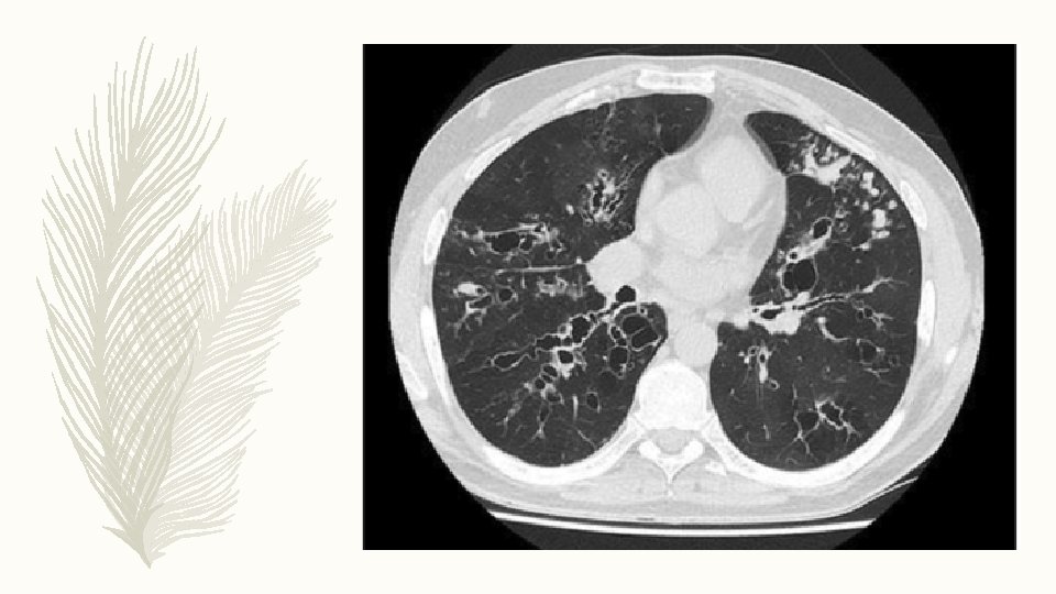
CYLINDRICAL BRONCHIECTASIS WITHIN I CM OF PLEURA TRAM TRACK SIGN

SIGNET RING SIGN

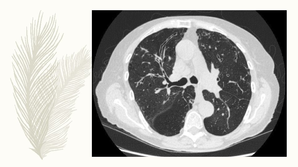
VARICOSE BRONCHIECTASIS VARICOSE

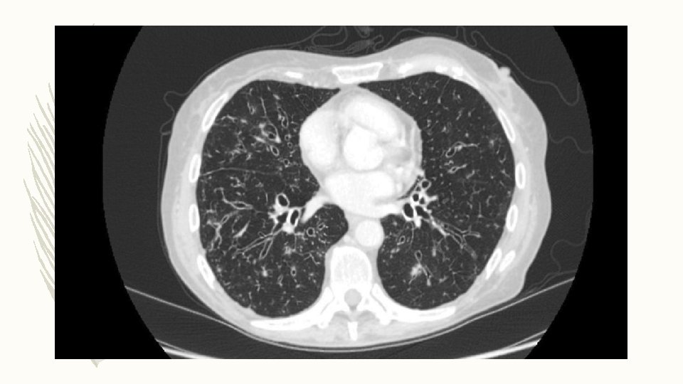
CYSTIC BRONCHIECTASIS CYSTIC CLUSTERS MUCUS IMPACTION

MOSAIC PERFUSION BLACK WHITE

INVESTIGATIONS FOR CAUSE IN ALL CLINICALLY STABLE • COMORBIDITIES AND RELEVANT PAST HISTORY • FULL BLOOD COUNT/ SERUM TOTAL IGE/ SKIN PRICK TEST TO A. FUMIGATUS • SERUM Ig G/ Ig. A/ Ig. M • BASELINE SPECIFIC ANTIBODY LEVELS AGAINST CAPSULAR POLYSACCHRIDES OF STREPTOCOCCUS PNEUMONIAE • SPUTUM CULTURE : ROUTINE AND MYCOBACTERIAL • HIV • TEST FOR CYSTIC FIBROSIS/ PCD/ GERD • RA, ANTI CCP , ANCA, ANA CLINICALLY • ALPHA 1 AT SUSPECT • BRONCHIAL ASPIRATION OR WASH Hill A, Welham S, Sullivan A, Loebinger M. Updated BTS Adult Bronchiectasis Guideline 2018: a multidisciplinary approach to comprehensive care. Thorax. 2018; 74(1): 1 -3.

STEPWISE MANAGEMENT
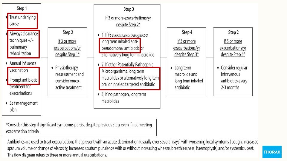

AIRWAY CLEARANCE

Physiotherapy management-stepwise airway clearance.

ACBT

POSTURAL DRAINAGE

Airway clearance - exacerbations.

ANTIBIOTIC TREAMENT FOR EXACERBATION

WHAT IS THE ROLE OF SURGERY IN MANAGING BRONCHIECTASIS? RECOMMENDATIONS – Consider lung resection in patients with localized disease whose symptoms are not controlled by medical treatment optimized by a bronchiectasis specialist. (D) – Offer multidisciplinary assessment, including a bronchiectasis physician, a thoracic surgeon and an experienced anesthetist, of suitability for surgery and pre-operative assessment of cardiopulmonary reserve post resection. (D)

LUNG TRANSPLANTATION FOR BRONCHIECTASIS Recommendations – Consider transplant referral in bronchiectasis patients aged 65 years or less if the FEV 1 is <30% with significant clinical instability or if there is a rapid progressive respiratory deterioration despite optimal medical management. (D) – Consider earlier transplant referral in bronchiectasis patients with poor lung function and the following additional factors: massive haemoptysis, severe secondary pulmonary hypertension, ICU admissions or respiratory failure (particularly if requiring NIV). (D)

LUNG ABSCESS

DEFINITION Localized area of lung suppuration, leading to necrosis of the lung parenchyma with or without cavity formation. Type of liquefactive necrosis of the lung tissue and formation of cavities (more than 2 cm) containing necrotic debris or fluid caused by microbial infection.

ACUTE DURATION CHRONIC PRIMARY LUNG ABSCESS AETIOLOGY SECONDARY WAY OF SPREADING BRONCHOGENIC HEMATOGENIC
 • ❖ ACCORDING TO THE DURATION: • Acute (less than 6")
CLASSIFICATION (CONTD. ) • ❖ ACCORDING TO THE DURATION: • Acute (less than 6 weeks); • Chronic (more than 6 weeks) • ❖ BY ETIOLOGY: • Primary (aspiration of oropharyngeal secretions, necrotizing pneumonia, immunodeficiency); • Secondary (bronchial obstructions, haematogenic dissemination, direct spreading from mediastinal infection, from sub phrenic space, coexisting lung diseases) • ❖ WAY OF SPREADING: • Bronchogenic (aspiration of oropharyngeal secretions, bronchial obstruction by tumour, foreign body, enlarged lymph nodes, congenital malformation); • Haematogenic (abdominal sepsis, infective endocarditis, septic thromboembolisms)

DIFFERENTIATING ACUTE FROM CHRONIC ACUTE CIRCUMSCRIBED NOT WELL DEFINED FILLED WITH NECROTIC DEBRIS CHRONIC IRREGULAR WELL DEFINED FILLED WITH THICK DEBRIS

 § Excavating tuberculosis § Localized")
DIFFERENTIAL DIAGNOSIS § Excavating bronchial carcinoma (squamo-cellular or microcellular) § Excavating tuberculosis § Localized pleural empyema § Infected emphysematous bullae § Cavitary pneumoconiosis § Hiatus hernia § Pulmonary hematoma § Hydatid cyst of lung § Cavitary infarcts of lung § Wegener’s granulomatosis

DIAGNOSIS – Diagnostic bronchoscopy is a part of diagnostic protocol for taking the material for microbiological examination and to confirm intrabronchial cause of abscess-tumor or foreign body. – Sputum examination is useful for identification of microbiological agents or confirmation of bronchial carcinoma

MANAGEMENT STANDARD CONSERVATIVE THERAPY: MEDICAL MANAGEMENT § It is recommended to treat lung abscess with broad spectrum antibiotics, due to poly microbial flora, such as Clindamycin (600 mg IV on 8 h) and then 300 mg PO on 8 h or combination ampicilin/sulbactam (1. 5 -3 gr IV on 6 h). § Alternative therapy is piperacilin/tazobactam 3. 375 gr IV on 6 h or Meropenem 1 gr IV on 8 h. § For MRSA it is recommended to use linezolid 600 mg IV on 12 h or vancomycin 15 mg/kg BM on 12 h.

MANAGEMENT SURGICAL § Endoscopic drainage of lung abscesses is described as an alternative to chest tube drainage and is performed during the bronchoscopy with usage of laser. § Per cutaneous trans thoracic tube drainage § Surgical resection of lung abscess is therapy of choice for about 10% of patients. § Lobectomy is the resection of choice for large or central position of abscess. Atypical resection or segmentectomy are satisfactory procedures, if it is possible to remove complete abscess and if necessary surrounding lung tissue with necrotizing pneumonia

THANK YOU

CASE 1 – A 42 -year-old man, gardener – Long history of respiratory problems starting in early childhood. – Previously diagnosed as asthma. – Frequent absence from work due to “recurrent chest infections”. – Unaware of any neonatal issues but believes that he was born at home without complications and is unsure of any previous tests he has had as he is now estranged from his parents. – Has a cousin with a “lung disease”. – Married but has “no kids”


INVESTIGATIONS – Sputum culture: P. aeruginosa – Sweat chloride = 73 meq/liter – Cystic fibrosis genetics: genotype was F 508 del/R 117 H – CYSTIC FIBROSIS: Multisystem disorder caused by mutations in the gene that encodes the CF transmembrane conductance regulator (CFTR) protein, a chloride channel expressed in epithelial cells. – More than 2000 CFTR mutations have been identified to date, but only the functional importance of a small number is known to cause the disease

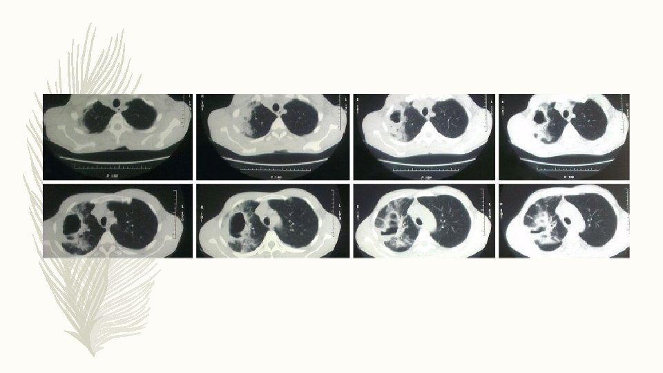
HRCT THORAX – An upper lobe predominant distribution of cylindrical, cystic and varicose bronchiectasis associated with airway wall thickening, mucus plugging and parenchymal opacities on a HRCT scan should raise the suspicion of CF disease. – The presence of nasal polyposis and/or chronic rhinosinusitis, recurrent pancreatitis, malabsorption, diabetes, osteoporosis and male infertility are other typical features of CF

DIAGNOSIS Guidelines published by the Cystic Fibrosis Foundation in the USA allows diagnosis if: 1. Clinical presentation of the disease and evidence of biochemical and genetic markers of CFTR dysfunction. 2. Clinical features of the disease with concentration of chloride >60 mmol·L− 1 at the sweat test or a concentration in the intermediate range (30– 59 mmol·L− 1) but two disease-causing CFTR mutations. 3. CFTR genotype is undefined: CFTR physiologic tests, such as nasal potential difference and intestinal current measurement, should be performed.

MANAGEMENT 1. CFTR modulator therapies 2. Airway clearing techniques 3. Chest physical therapy 4. Humidification with sterile water or normal saline to facilitate airway clearance 5. Antibiotics 6. Mucus thinners 7. Lung transplantation

CASE 2 – 45 -year-old farmer with asthma since childhood. – Complaints: Decline in his exercise tolerance and an increase in cough which has become productive of purulent sputum with occasional thick/solid components. – Respiratory exacerbations not responding well to standard steroid antibiotic treatment. – He was noted to have variable pulmonary infiltrates on chest radiographs during these episodes


INVSETIGATIONS – Marked peripheral blood eosinophilia – Total Ig. E > 1000 IU/ ml – Aspergillus specific Ig. E > O. 35 ABPA: ABPA is an inflammatory disease caused by hypersensitivity to the ubiquitous fungus Aspergillus fumigatus – ABPA occurs most commonly in patients with asthma and CF – ABPA is the cause of 1– 10% of cases of bronchiectasis – Most ABPA cases occur in the third and fourth decade without a sex predilection.

DIAGNOSIS – Long standing uncontrolled asthma/ Cystic fibrosis – Brownish sputum – Peripheral eosinophilia > 500/ mm 3 – Total Ig. E > 1000 IU/ ml – Specific Ig. E for A. fumigatus > 0. 35 HRCT thorax: Ø Central bronchiectasis Ø High attenuation mucus Ø Finger in glove/ TIB Ø Tram track Ø Mosaic attenuation

MANAGEMENT 1. Corticosteroids 2. Antifungals 3. Airway clearing techniques 4. Chest physical therapy 5. Mucus thinners

CASE 3 – 77 -year-old retired librarian. – Cough for many years with new symptoms of fatigue, weight loss and fever. – A chest CT scan was performed looking for a possible occult malignancy and bronchiectasis was found.


DIAGNOSIS – HRCT thorax: cylindrical bronchiectasis and tree-in-bud pattern in middle and lower lobes – Sputum for M. Tuberculosis: negative – MGIT culture: MAC growth at 4 weeks – Repeat MGIT: Positive for MAC – Tests for immunodeficiency and ABPA: Negative

MANAGEMENT 1. Management of NTM as per the organism and clinical picture 2. Airway clearing techniques 3. Chest physical therapy 4. Mucus thinners

CASE 4 – A 66 -year-old woman with established idiopathic bronchiectasis had three to four exacerbations per year for the past 3 years despite performing daily chest physiotherapy. – Produces large volumes of sputum daily despite performing the active cycle of breathing technique. – Testing for NTM, ABPA and other complications were negative, but sputum shows persistent infection with P. aeruginosa.


– One of the most common presentations of bronchiectasis – Exacerbations are one of the most important manifestations of bronchiectasis and P. aeruginosa is the most frequent organism in severe bronchiectasis worldwide – Cylindrical bronchiectasis is the most common morphological pattern identified on CT scans

MANAGEMENT 1. Review current airway clearance regime. 2. Repeat sputum microbiology and repeat testing for NTM, ABPA and ensuring the all possible treatable causes and comorbidities have been identified. 3. First-line recommendation for P. aeruginosa with frequent exacerbations is an inhaled antibiotic.

THANK YOU
- Slides: 118