
Applied physics in the clinic monitoring radiation doses



- Slides: 54
Applied physics in the clinic: monitoring radiation doses delivered to cancer patients Louis Archambault, Ph. D Assistant professor, Department of Physics, Laval University, Quebec city, QC, Canada Medical Physicist, Centre Hospitalier Universitaire de Québec
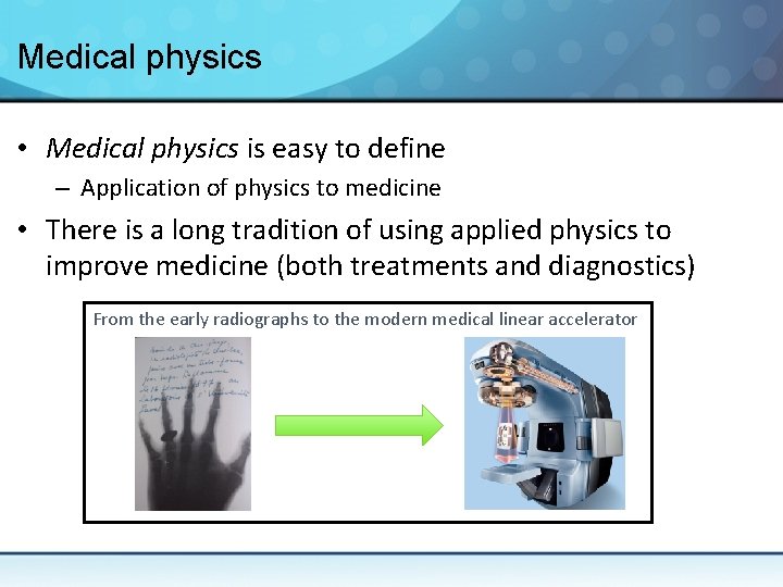
Medical physics • Medical physics is easy to define – Application of physics to medicine • There is a long tradition of using applied physics to improve medicine (both treatments and diagnostics) From the early radiographs to the modern medical linear accelerator
Medical physics • However, the clinic is much different from the lab • Despite numerous opportunities, crossing the clinical barrier can be challenging – Even for those with a biophysics/biomedical backgrounds • Nevertheless, several hospitals hire physicists – Clinical medical physics is a profession “The Practice of Medical Physics means the use of principles and accepted protocols of physics to assure the correct quality, quantity, and placement of radiation during the performance of a radiological procedure. ” - American Association of Physicists in Medicine
Medical physics • Medical physicists background: – – BSc in physics or engineering physics MS and/or Ph. D in medical physics Residency Certification (Canadian College of Medical Physicists) • Most medical physicists have some time for research – They can act as: • • Principal investigators Collaborators Evaluators of new technologies Facilitators for clinical trials
Medical physics • Where to find clinical medical physicists? – – Radiation oncology Diagnostic imaging Nuclear medicine Health/radiation protection
Medical physics • Where to find clinical medical physicists? – – Radiation oncology (~75%) Diagnostic imaging Nuclear medicine Health/radiation protection
Medical physics • What do a medical physicist do? – Radiation beam calibration and characterization – Image quality assessment – Consultation and treatment planning with practitioners to determine dose to be delivered – Validate the radiation delivery plans of (nearly) every patient Acceptance testing and commissioning – Radiation shielding design –. . . and much more In radiation oncology, the medical physicist’s job is to make sure that the patient receive precisely the intended dose of radiation
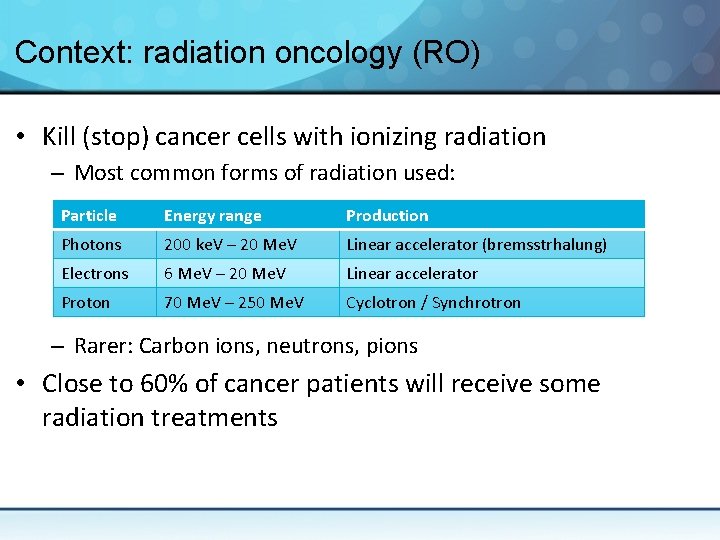
Context: radiation oncology (RO) • Kill (stop) cancer cells with ionizing radiation – Most common forms of radiation used: Particle Energy range Production Photons 200 ke. V – 20 Me. V Linear accelerator (bremsstrhalung) Electrons 6 Me. V – 20 Me. V Linear accelerator Proton 70 Me. V – 250 Me. V Cyclotron / Synchrotron – Rarer: Carbon ions, neutrons, pions • Close to 60% of cancer patients will receive some radiation treatments
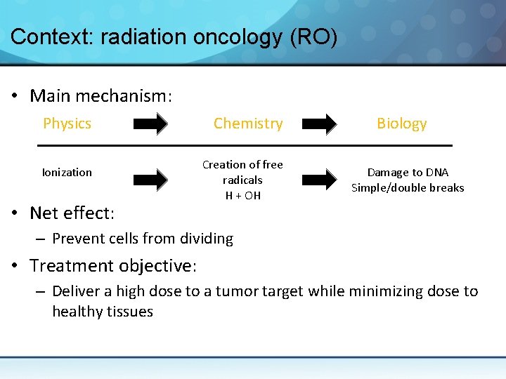
Context: radiation oncology (RO) • Main mechanism: Physics Ionization • Net effect: Chemistry Creation of free radicals H + OH Biology Damage to DNA Simple/double breaks – Prevent cells from dividing • Treatment objective: – Deliver a high dose to a tumor target while minimizing dose to healthy tissues
Advances in radiation oncology • The basic principles of RO are the same since the 1900 s – Point a tumor target and ‘shoot’
Advances in radiation oncology • Most recent advances aim to: – Better see the tumor target – Better focus radiation doses on small targets – Improve sparing of surrounding tissues to reduce side effects See the presentation by John Schreiner at 13 h 30 (Botterel B 143) for an excellent overview of modern radiation oncology
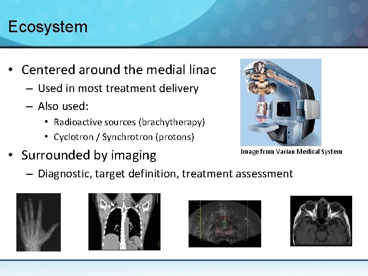
Ecosystem • Centered around the medial linac – Used in most treatment delivery – Also used: • Radioactive sources (brachytherapy) • Cyclotron / Synchrotron (protons) • Surrounded by imaging Image from Varian Medical System – Diagnostic, target definition, treatment assessment
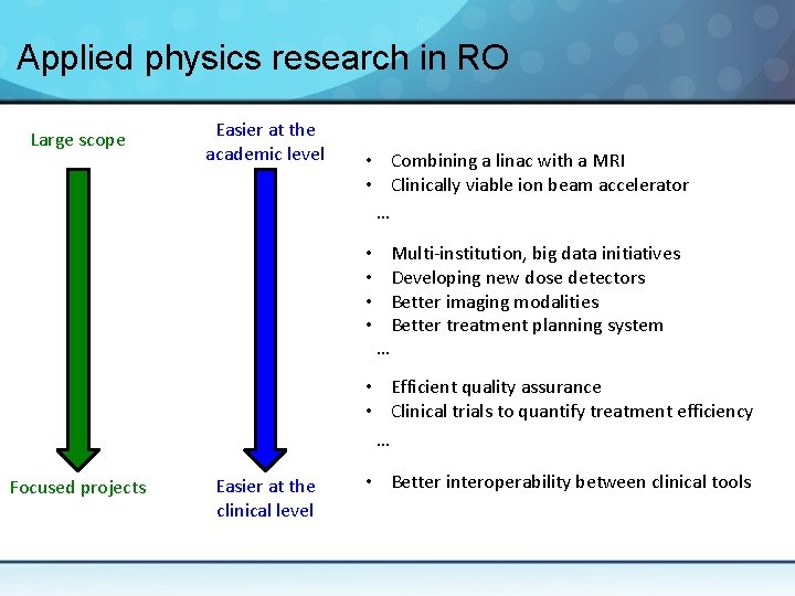
Applied physics research in RO • Different spectra of research – From colossal endeavor to tiny tweak to the clinical workflow – From purely academic research to assessment of new products • Often a collaboration between all the actors: – – – Academia Industry Clinical medical physicists Physicians Clinical professionals (e. g. therapists, dosimetrists, nurses)
Applied physics research in RO Large scope Easier at the academic level • Combining a linac with a MRI • Clinically viable ion beam accelerator … • • … Multi-institution, big data initiatives Developing new dose detectors Better imaging modalities Better treatment planning system • Efficient quality assurance • Clinical trials to quantify treatment efficiency … Focused projects Easier at the clinical level • Better interoperability between clinical tools
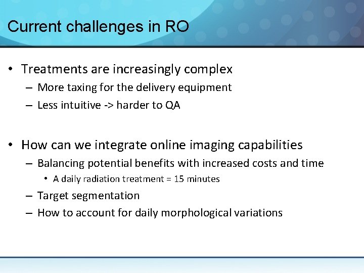
Current challenges in RO • Treatments are increasingly complex – More taxing for the delivery equipment – Less intuitive -> harder to QA • How can we integrate online imaging capabilities – Balancing potential benefits with increased costs and time • A daily radiation treatment = 15 minutes – Target segmentation – How to account for daily morphological variations
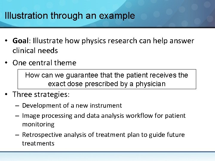
Illustration through an example • Goal: Illustrate how physics research can help answer clinical needs • One central theme How can we guarantee that the patient receives the exact dose prescribed by a physician • Three strategies: – Development of a new instrument – Image processing and data analysis workflow for patient monitoring – Retrospective analysis of treatment plan to guide future treatments
Development of a new instrument A multipoint scintillation detector
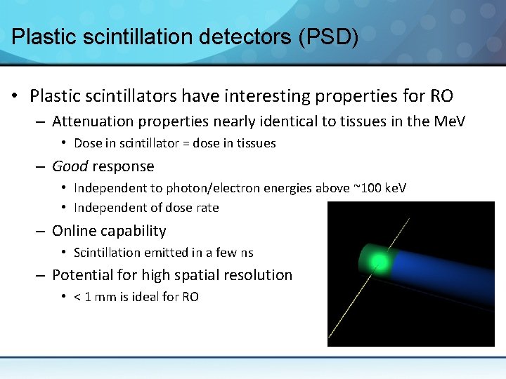
Plastic scintillation detectors (PSD) • Plastic scintillators have interesting properties for RO – Attenuation properties nearly identical to tissues in the Me. V • Dose in scintillator = dose in tissues – Good response • Independent to photon/electron energies above ~100 ke. V • Independent of dose rate – Online capability • Scintillation emitted in a few ns – Potential for high spatial resolution • < 1 mm is ideal for RO
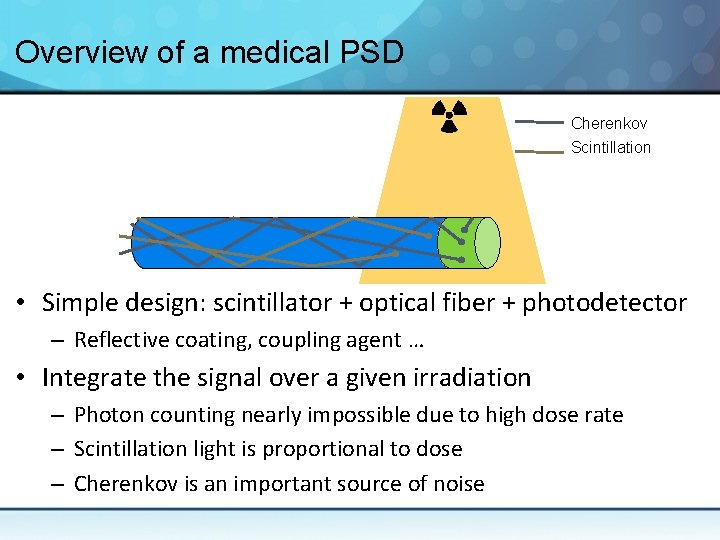
Overview of a medical PSD Cherenkov Scintillation • Simple design: scintillator + optical fiber + photodetector – Reflective coating, coupling agent … • Integrate the signal over a given irradiation – Photon counting nearly impossible due to high dose rate – Scintillation light is proportional to dose – Cherenkov is an important source of noise
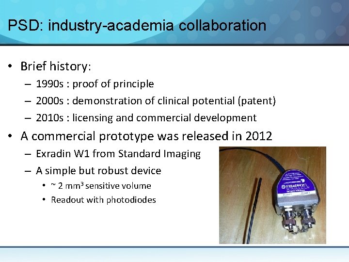
PSD: industry-academia collaboration • Brief history: – 1990 s : proof of principle – 2000 s : demonstration of clinical potential (patent) – 2010 s : licensing and commercial development • A commercial prototype was released in 2012 – Exradin W 1 from Standard Imaging – A simple but robust device • ~ 2 mm 3 sensitive volume • Readout with photodiodes
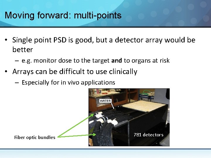
Moving forward: multi-points • Single point PSD is good, but a detector array would be better – e. g. monitor dose to the target and to organs at risk • Arrays can be difficult to use clinically – Especially for in vivo applications Fiber optic bundles 781 detectors
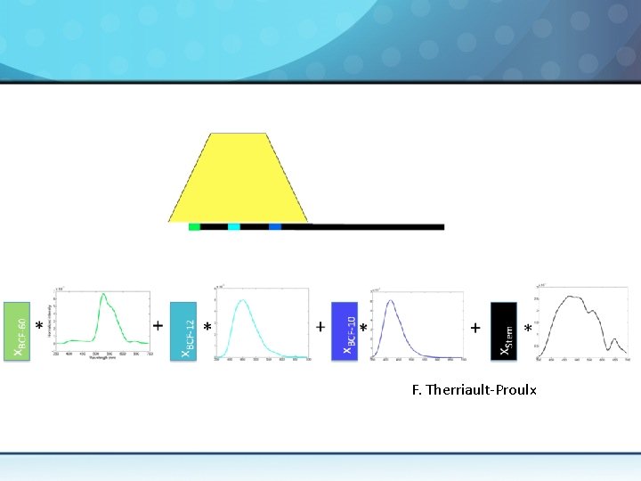
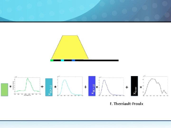
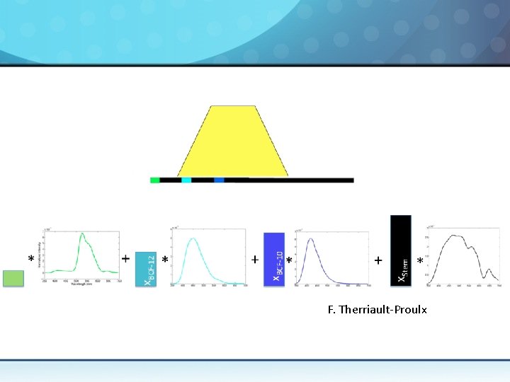
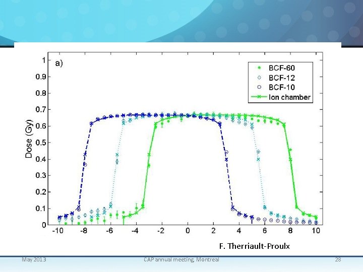
Multi-points PSD • To build efficient arrays, we can’t simply stack more detectors together • Alternative: – Multiple scintillating element along a single optical fiber – If we can decouple scintillation and Cherenkov, we can decouple multiple scintillation signal • Thus, spatial information can be encoded in the emission spectrum of the scintillating elements
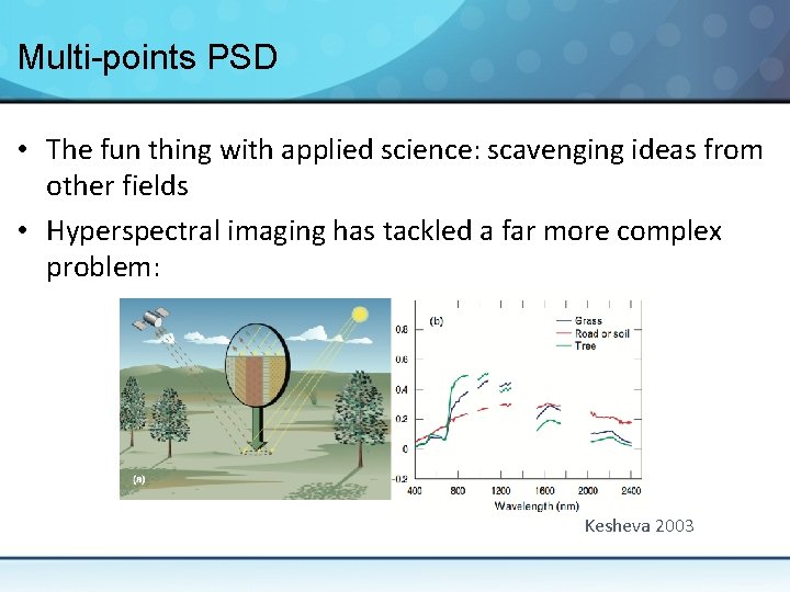
Multi-points PSD • The fun thing with applied science: scavenging ideas from other fields • Hyperspectral imaging has tackled a far more complex problem: Kesheva 2003
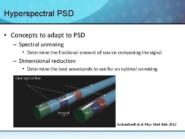
Hyperspectral PSD • Concepts to adapt to PSD – Spectral unmixing • Determine the fractional amount of source composing the signal – Dimensional reduction • Determine the best wavebands to use for an optimal unmixing Archambault et al. Phys. Med. Biol. 2012
F. Therriault-Proulx
F. Therriault-Proulx
F. Therriault-Proulx
F. Therriault-Proulx May 2013 CAP annual meeting, Montreal 28
Bringing hyperspectral PSD to the clinic • What remain to be done to bring this new instrument in the clinic? – Optimize the design to improve precision • Uncertainty < 2% ideal for a clinical dosimeter – Demonstrate clinical benefits – Commercialize • Partnership with the industry or with a new ‘startup’
Image processing and data analysis workflow for patient monitoring Patient classification during radiotherapy
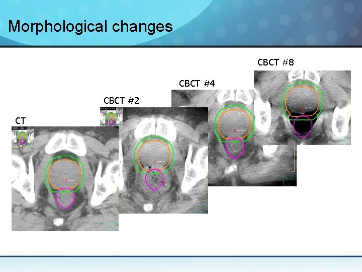
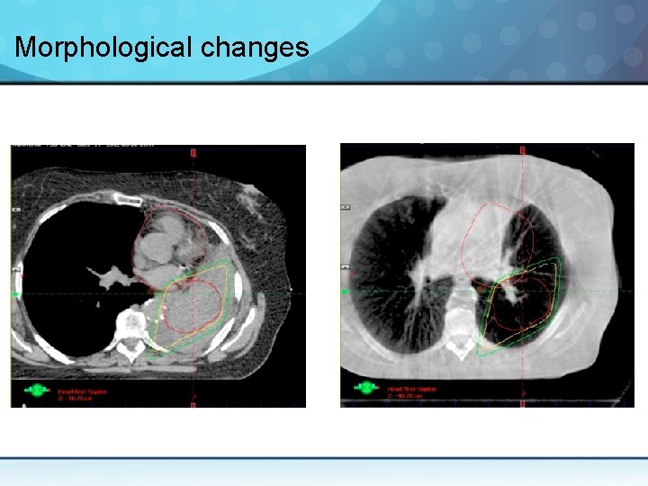
During radiation therapy (RT) • RT is a long process (up to 6 -8 weeks of treatments) – Treatment fractionation let us exploit biological differences between cancer and healthy cells • Morphological changes are frequent during RT – However, patients often have a single treatment plan • Based on the anatomy at the beginning of RT • For the rest of treatment:
Morphological changes CBCT #8 CBCT #4 CBCT #2 CT
Morphological changes
Adaptive RT • Adaptive RT: treating the anatomy of the day – Desirable, but resource intensive – Clinically impossible to implement in most cases • Shortcuts must be developed – Algorithms must do some of the work • Add an additional layer of complexity to RT • However, not all patient changes – Can we spot those most at risk?
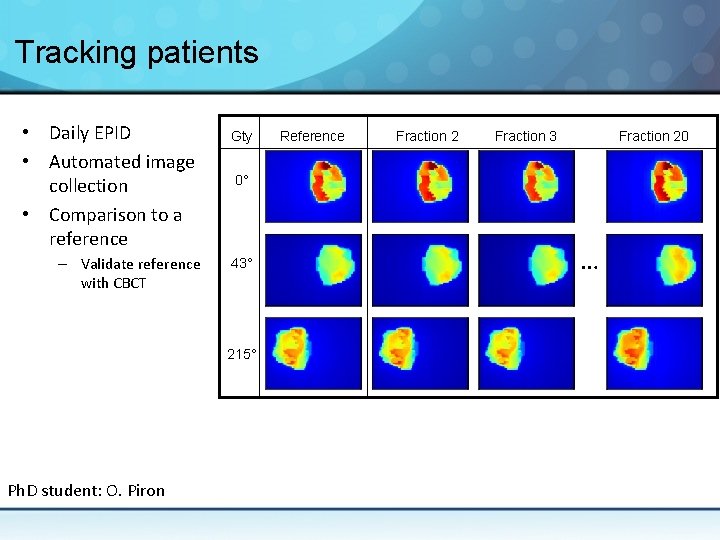
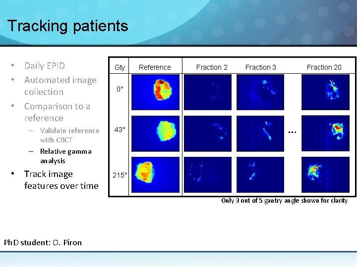
Automated patient monitoring • Our goal: deploy an automated patient safety net – Using exit (i. e. portal) dose (EPID) – EPID contains information on the dose delivered and the anatomy • Our strategy – Track relative changes – Classify treatment fractions through machine learning – Flag patients for more careful evaluation by a clinical physicist
Tracking patients • Daily EPID • Automated image collection • Comparison to a reference – Validate reference with CBCT Gty Fraction 2 Fraction 3 Fraction 20 0° 43° 215° Ph. D student: O. Piron Reference …
Tracking patients • Daily EPID • Automated image collection • Comparison to a reference – Validate reference with CBCT – Relative gamma analysis • Track image features over time Gty Reference Fraction 2 Fraction 3 Fraction 20 0° 43° … 215° Only 3 out of 5 gantry angle shown for clarity Ph. D student: O. Piron
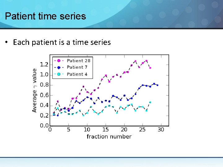
Patient time series • Each patient is a time series
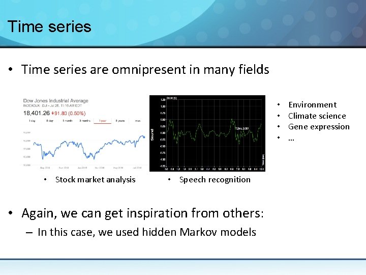
Time series • Time series are omnipresent in many fields • • • Stock market analysis • Speech recognition • Again, we can get inspiration from others: – In this case, we used hidden Markov models Environment Climate science Gene expression …
Hidden Markov models (HMM) • Unsupervised machine learning approach • Assume a system is composed of N hidden states – Each state is a Markov process – N given as an input • Useful for time series – Markov process: step k+1 transition determined by step k – Used to classify each fraction
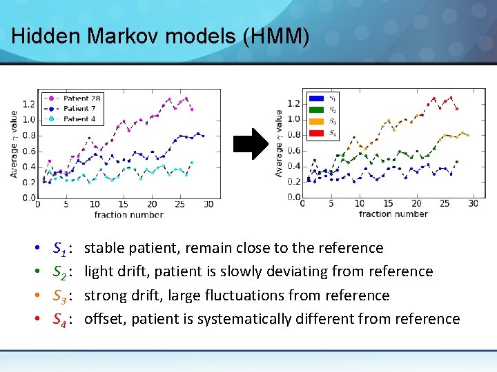
Hidden Markov models (HMM) • • S 1 : S 2 : S 3 : S 4 : stable patient, remain close to the reference light drift, patient is slowly deviating from reference strong drift, large fluctuations from reference offset, patient is systematically different from reference
Tracking patients • EPID is a rich (and mostly free) source of information • Machine learning models can be trained to: – Classify patient states – Flag patients likely to deviate from their plan • Thus automated and unbiased tracking of patient appears feasible • A prospective trial is underway to validate this workflow
Retrospective analysis of treatment plans to guide future treatments A stochastic frontier analysis to improve treatment planning
Treatment planning • Once a physician decide to treat a patient: – CT imaging for treatment planning – Tumor and organs at risk segmentation (contours) • Manually by the physician and dosimetrists – Selection of a treatment modality • Photon or electron, static or dynamic … – Dose optimization • Try to find the best possible dose distribution – Final dose calculation This whole process typically takes 1 to 2 weeks
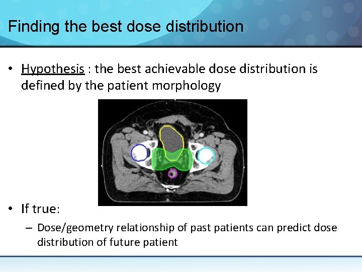
Finding the best dose distribution • Hypothesis : the best achievable dose distribution is defined by the patient morphology • If true: – Dose/geometry relationship of past patients can predict dose distribution of future patient
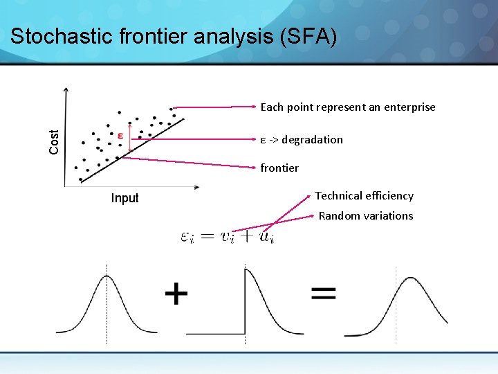
Stochastic frontier analysis (SFA) • This time, we get our inspiration from economy: – The stochastic frontier analysis is used to model the productivity of enterprises • In SFA, the output of an enterprise is a combination of – Technical efficiency – Random ‘shocks’ • Can be used to model outputs or costs
Stochastic frontier analysis (SFA) Cost Each point represent an enterprise ε -> degradation frontier Input Technical efficiency Random variations
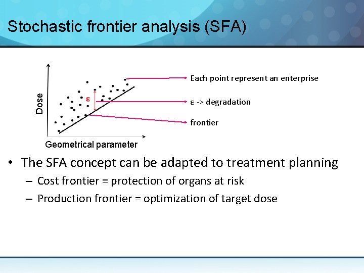
Stochastic frontier analysis (SFA) Dose Cost Each point represent an enterprise ε -> degradation frontier Geometrical Input parameter • The SFA concept can be adapted to treatment planning – Cost frontier = protection of organs at risk – Production frontier = optimization of target dose
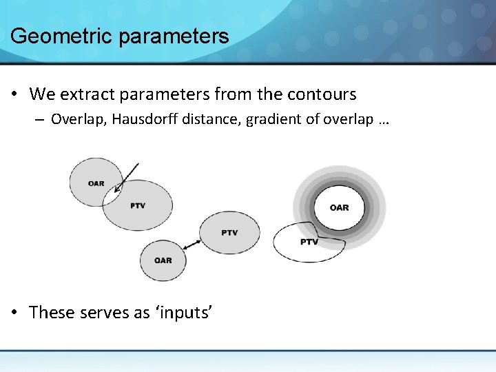
Geometric parameters • We extract parameters from the contours – Overlap, Hausdorff distance, gradient of overlap … • These serves as ‘inputs’
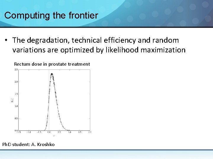
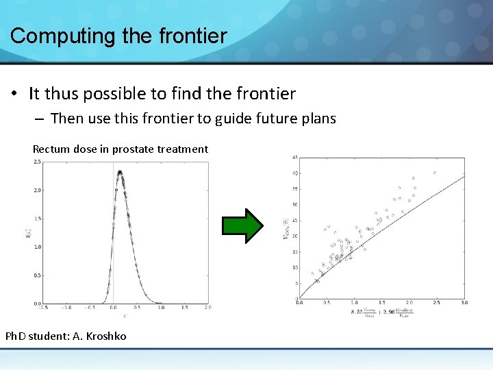
Computing the frontier • The degradation, technical efficiency and random variations are optimized by likelihood maximization Rectum dose in prostate treatment Ph. D student: A. Kroshko
Computing the frontier • It thus possible to find the frontier – Then use this frontier to guide future plans Rectum dose in prostate treatment Ph. D student: A. Kroshko
How to use the frontier • Using SFA, it is possible to harness information from past plans to improve current treatment quality – Help make sure patients receive the best possible dose distribution – Planning guidance saves valuable time in the clinic • Falls in the family of ‘knowledge-based planning’ – However our approach is not dependent on initial plan selection
Conclusion
Conclusion • Technology in RO is rapidly evolving (and complexifying) – Several opportunity for research • I hope these 3 brief examples showed the diversity of applied physics in the clinic – Hardware, imaging, software – Feel free to adapt great ideas from other fields • Clinical medical physicists can help you – Explain the needs for new tools and methods – They are often eager to contribute to research