Applied Nerve Muscle Physiology Nerve Conduction Study NCS
 )and Electromyography (")
 and electromyography ( emg). •")
 • A nerve conduction study (NCS) is an electrophysiology")


 L 2 L 1 NCV")


")
 is a technique for evaluating and recording physiologic properties of")

 – 5 m. V (")
 During Mild Effort During Moderate Effort note recruitment of additional motoneurons During")


- Slides: 19
Applied Nerve & Muscle Physiology : Nerve Conduction Study ( NCS) )and Electromyography ( EMG)
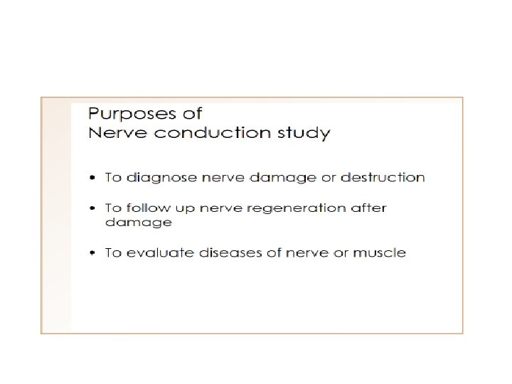
Objectives • Define what is nerve conduction study (NCS) and electromyography ( emg). • Explain the procedure of NCS using Abductor Pollicicis Brevis muscle. • Define the normal conduction velocity in upper limb and lower limb nerves. • Define the motor unit potentials ( MUPs) and how they are changed in muscle and nerve diseases.
Nerve Conduction Study ( NCS) • A nerve conduction study (NCS) is an electrophysiology test commonly used to evaluate the function of peripheral nerves of the human body. • It could be motor nerve conduction study ( motor NCS) , sensory nerve conduction study or mixed nerve conduction study. • In this lecture, because of time constraint, only motor nerve conduction study will be discussed • In the motor test the recorded response is the muscle CMAP ( compound muscle action potential ) 3
Procedure • An electrical stimulus is applied over a nerve ( e. g. , median nerve ) and a recording electrode is place over the muscle suppllied by that motor nerve. • The stimulus is applied at two sites : a distal site ( wrist ) and a proximal one ( antecubital fossa , elbow). • The muscle usually chosen in this routine test is the Abductor Pollicis Brevis • The active recording electrode (G 1) is place over thenar eminence which overlies the muscle. • And the reference recording electrode (G 2) about 3 cm away. • The oscilloscope ( CRO) sweep speed is adjusted to 2 ms / cm. 5
• The stimulus duration used is 0. 2 ms and stimulus frequency to 1 / sec. • Apply the stimulus and record the response from stimulation at the wrist. • Store the CMAP ( compound muscle action potential ) in the first channel of the oscilloscope. • Change the stimulating site from wrist to antecubital fossa ( elbow ). • Stimulate the nerve & record the CMAP for median nerve stimulation at the elbow.
Distance d = 284 mm L 1 Latency At wrist = 3. 5 ms L 2 Latency At elbow = 8. 5 ms 7
Nerve conduction velocity D 2 D 1 Latency (s) L 2 L 1 NCV = D 1 -D 2 L 1 -L 2
L 1 L 2
• Measure the distance from elbow to wrist with a measuring tape. • Measure the latency in first CMAP & in the next CAMP. • Enter the distance between the elbow and wrist. 10
MNCV • MNCV will appear. • It can also be calculated by formula • MNCV (m/sec)= Distance (mm) -------------L 2 -L 1 (ms) • L 1 = latency at wrist • L 2 = latency at elbow 11
Normal values for conduction velocity ü In arm – 50 – 70 m / sec. ü In leg – 40 – 60 m / sec. 12
Electromyography ( EMG)
• Electromyography (EMG) is a technique for evaluating and recording physiologic properties of muscles at rest and while contracting. • It’s a recording of electrical activity of the muscle by inserting needle electrode in the belly of the muscles ( needle emg ) or by applying the surface electrodes ( surface emg ) • The potentials recorded in needle emg are derived from motor units of the muscle, hence known as motor unit potentials (MUPs). • Q: Define what is a “ motor unit ”? 14
• A motor unit is defined as one motor neuron and all of the muscle fibers it innervates. 15
Normal MUPs • Amplitude : 300 μV ( microvolt) – 5 m. V ( millivolts) • Duration : 3 – 15 ms(milliseconds ) 16
MUPs (2) During Mild Effort During Moderate Effort note recruitment of additional motoneurons During Full Voluntary Effort. There is full recruitment ( you can not see the baseline )
Examples of Abnormalities of MUPs • In nerve diseases : Giant MUPs due to reinnervation > 5 m. V • In muscle disease : Small MUPs < 300μV
Thanks