Applications of Nanobiotechnology nanomedicine NANOBIOSENSORS What are biosensors

Applications of Nanobiotechnology • & nanomedicine

NANOBIOSENSORS •

What are biosensors? • A biosensor is an analytical device which converts a biological response into an electrical signal (Figure 6. 1).

• biological systems may be utilised by biosensors, for example, • whole cell metabolism, ligand binding and the antibody-antigen reaction.

• Biosensors represent a rapidly expanding field, at the present time, with an estimated 60% annual growth rate; • the major impetus coming from the health-care industry • (e. g. 6% of the western world are diabetic and would benefit from the availability of a rapid, accurate and simple biosensor for glucose)

• In recent years, a wide varietyof nanoparticles with different properties, such as • small size, • high speeds, • smaller distances for electrons to travel, lower power, and lower voltages Important advances in the field of nanotechnology have led to the utilization of nanomaterials such as metal nanoparticles, oxide nanoparticles, magnetic nanomaterials, carbon materials

• Functional nanoparticles that bound to biological molecules (e. g. peptides, proteins, nucleic acids) • • have been developed for use in biosensors to detect and amplyfi various signals.

NANOSTRUCTURED MATERIALS FOR BIOSENSING DEVICES • Nano structuredmaterials are interesting tools with specific physical and chemical properties because of their • quantum-size effects when compared to bulk materials • The exploration of these different characteristics provides the possibility to improve the sensitivity of biosensors

• These include the utilization of nanostructured materials with specific forms like • • • quantum dots nanoparticles (0 D), Nanowires and carbon nanotubes (1 D), and metallic platelets These devices offer improved sensitivities, due to their large surface-to-volume ratios, • .

NANOPARTICLES-BASED BIOSENSORS • Metallic nanoparticles are very interesting materials • with unique • electronic and electrocatalytic properties depending on their size and morphology • Nanoparticle-based biosensors are particularly

Nanoparticle-based biosensors are • - Particularly attractive because they can be easily • synthesized in bulk using standard chemical techniques, -do not require advanced fabrication approaches. • - They also offer particularly high surface areas due • to their extremely small size and are typically used • as suspensions in solutions (during the time when • they interact with the analyse). •

Most biological • molecules can be labelled withmetal • nanoparticleswithout compromising their biological activities [22]. In particular, gold nanoparticles are much explored • materials as components for biosensors, • due to their • capability to increase an electronic signal when a • biological component is maintained in contact with • its nanostructured surface [23]. •

• The exploration of gold nanostructured materials has provided new paths • for enzymatic biosensor development.

• This observationis especially useful in the development of electroluminescence • based biosensors. • Apart from gold, silver, platinum, palladium, copper, cobalt and other nanoparticles are also extensively explored in the development of biosensors

The research on • nanobioelectronics & biosensors aims at the • integration of nanoelectronics, tools and materials • into • lowcost, user • friendly • -interest in several fields such as • diagnostics, food analysis, environmentmonitoring • and other industries. •

Functionalization of nanoparticles using small molecule ligands • A wide variety of ligands have been incorporated onto nanoparticle to be used in intracellular delivery. For example, nanoparticles functionalized diagnosis of diseases, and surfaces.

• Nanotechnology plays an important role in advanced biology and medical research particularly in the development of potential site specific delivery systems with • lower drug toxicities • greater efficiencies. [5]

• The era of nanotechnology has allowed novel research strategies to flourish in the field of drug delivery. Nanotechnology designed drug delivery systems have been seen to be suitable for treating chronic intracellular infections.

• Recent progress in cancer nanotechnology gives rise to exciting opportunities in which diagnosis and treatment are based on the molecular profiles of individual patients.

• Nanotechnology" was first defined by Tokyo Science University, Norio Taniguchi in 1974. • Although the application of nanotechnology to medicine appears to be a relatively recent trend, the basic nanotechnology approaches for medical application dates back to several decades.

• Lipid vesicles which were named as liposomes, were described in 1965, • in 1976 the description about the first controlled release polymer system of macromolecules was given, the first quantum dot bioconjugate was described in 1998, and the first nanowire nanosenser was described in 2001. liposomes polymer system of macromolecules • quantum dot bioconjugate nanowire nanosenser

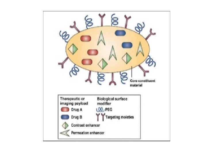
One of the major applications of nanotechnology in relation to medicine is drug delivery. • The problems with the new chemical entities such as • • • insolubility, degradation, bioavailability, toxicologic effects, targeted drug delivery, and controlled drug release • are solved by nanotechnology.

• For example, encapsulated drugs can be protected from degradation. • Specific nanosized receptors present on the surface of the cell can recognize the drug and elicit appropriate response by delivering and releasing therapy exactly wherever needed

• . Because of their small size and large surface area relative to their volume,

• nanoscale devices can readily interact with biomolecules. • Nanoscale devices include: nanoparticles [Figure 1], nanotubes, cantilevers, semiconductor nanocrystals, and liposomes.
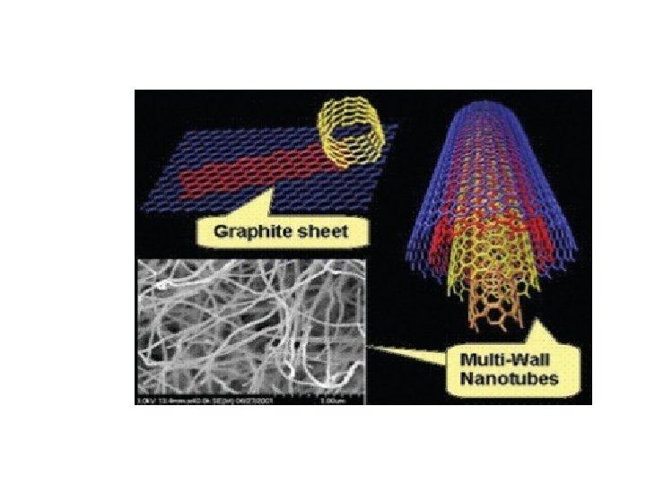
![Nanotubes • Nanotubes are smaller than nanopores [Figure 2]. Nanotubes help to identify Dioxyribonucleic](http://slidetodoc.com/presentation_image_h/06d3febde0afe295d230bf0423ee93e8/image-27.jpg "Nanotubes • Nanotubes are smaller than nanopores [Figure 2]. Nanotubes help to identify Dioxyribonucleic")
Nanotubes • Nanotubes are smaller than nanopores [Figure 2]. Nanotubes help to identify Dioxyribonucleic acid (DNA) changes associated with cancer cells. They are about half the diameter of a molecule of DNA. It helps to exactly pin point location of the changes.

• physical shape of the DNA can be traced with the help of the nano tube tip. A computer translates the information into topographical map.

• The bulky molecules identify the regions on the map where mutations are present. Since the location of mutations can influence the effects they have on a cell, these techniques are important in predicting disease

Quantum dotes • These are tiny crystals that glow when these are stimulated by ultraviolet light [Figure 3]. The latex beads filled with these crystals when stimulated by light, emit the color that lights up the sequence of interest.

• When the crystals are stimulated by light, the colors they emit serve as dyes and light up the sequences of interest
![Antibody conjugated quantum dotes[39]](http://slidetodoc.com/presentation_image_h/06d3febde0afe295d230bf0423ee93e8/image-33.jpg "Antibody conjugated quantum dotes[39]")
Antibody conjugated quantum dotes[39]
![Nanoshells (NS) are gold coated miniscule beads [Figure 4]. The wavelength of light related](http://slidetodoc.com/presentation_image_h/06d3febde0afe295d230bf0423ee93e8/image-34.jpg "Nanoshells (NS) are gold coated miniscule beads [Figure 4]. The wavelength of light related")
Nanoshells (NS) are gold coated miniscule beads [Figure 4]. The wavelength of light related to the thickness of the coatings. Thus, by manipulating the thickness of the layers making up the NS, the beads can be designed that absorb specific wavelength of light. which the beads absorb is

• The most useful NS are those that absorb near infrared light that can easily penetrate several centimeters in human tissues. Absorption of light by NS creates an intense heat that is lethal to cells. Metal NS which are intense near-infrared absorbers are effective both in-vivo and invitro on human breast carcinoma cells.

Nanoshells

Liposomes • Liposomes spherical, closed colloidal structures composed of lipid bilayers that surround a central aqueous space. Liposomal formulations have shown an ability to improve the pharmacokinetics and pharmacodynamics of associated drugs. Liposome based formulations of several anticancer agents have been approved for the treatment of metastatic breast cancer and Kaposi's sarcoma.

Cantilevers • Tiny bars anchored at one end can be engineered to bind to molecules associated with cancer [Figure 5]. These molecules may bind to altered DNA proteins that are present in certain types of cancer monitoring the bending of cantilevers; it would be possible to tell whether the cancer molecules are present and hence detect early molecular events in the development of cancer cells
![Nanocantilever array[26]](http://slidetodoc.com/presentation_image_h/06d3febde0afe295d230bf0423ee93e8/image-39.jpg "Nanocantilever array[26]")
Nanocantilever array[26]

Dendrimers • Dendrimers are new class of macromolecules which have a symmetric core and form the 3 -D spherical structure [Figure 6]. These have branching shape which gives them vast amounts of surface area to which therapeutic agents or other biologically active molecules can be attached.

• A single dendrimer can carry a molecule that recognizes cancer cells, a therapeutic agent to kill those cells, and a molecule that recognizes the signals of cell death. It is said that dendrimers can be manipulated to release their contents only in the presence of certain trigger molecules associated with cancer
![Dendrimer[41]](http://slidetodoc.com/presentation_image_h/06d3febde0afe295d230bf0423ee93e8/image-42.jpg "Dendrimer[41]")
Dendrimer[41]

Thanks for your • attention
- Slides: 43