APPENDICULAR SKELETON HONORS ANATOMY CHAPTER 7 PART II


 � 2 pectoral girdles � attach bones of upper limbs")

 slender bone �")


 � 2 parallel bones: Ulna & Radius � articulate: › proximally with")
 �")


 � #’d I to V")

 Girdle � attaches lower limbs to axial skeleton › transmitting full weight")
 which unite anteriorly at pubic symphysis")


 � small, triangular, sesamoid bone � develops in tendon of quadriceps femoris")


- Slides: 67
APPENDICULAR SKELETON HONORS ANATOMY CHAPTER 7 PART II
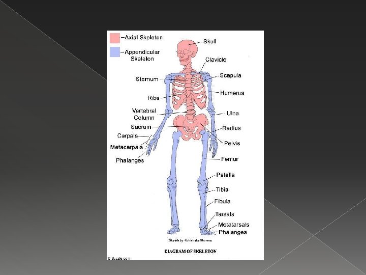
The Appendicular Skeleton � Includes: limb bones 2. bones that connect limbs to axial skeleton 1. › › shoulder girdle pelvic girdle
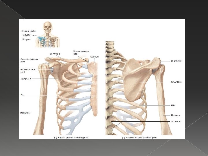
The Pectoral Girdle (Shoulder) � 2 pectoral girdles � attach bones of upper limbs to axial skeleton � each: 1 clavicle � 1 scapula
Pectoral Girdle � does not form complete belt-like bony structure � anteriorly: clavicles attached to sternum � laterally: clavicles attach to scapulae � posteriorly: › scapula attach to vertebral column via muscle attahments
Clavicle � S-shaped, (medial ½ convex anteriorly, lateral ½ concave anteriorly) slender bone � lies horizontally across anterior thorax superior to 1 st rib
Clavicles � Functions: anchor muscles 2. hold upper limbs and scapula out laterally away from the narrow superior part of thoracic cage 1.
Clavicle � medial end = sternal end is rounded & articulates with the manubrium @ sternoclavicular joint
Clavicle � lateral end = acromial end is flat � articulates with acromion of the scapula to form acromialclavicular joint
Clavicle � last bone to stop growing � 1 of most frequently fx’d bones (2 curves) usually from fall on outstretched arm � or see compression fx in auto accidents from shoulder strap which can cause damage to median n. (between clavicle & 2 nd rib)
Scapula � aka shoulder blade, angel bone � large, triangular, flat bone � in superior part of posterior thorax between levels of 2 nd & 7 th ribs � spine: prominent ridge that runs diagonally across posterior surface
� lateral edge: acromion a flattened expanded process, easily felt as hi pt of shoulder (tailors use it as landmark to measure length of arm) � glenoid cavity: inferior to acromion, smooth, shallow depression that accepts head of humerus in shoulder joint
Scapula
Upper Limb � 1. 2. 3. 4. 5. 6. 6 parts: Humerus Ulna Radius Carpals Metacarpals Phalanges � � � Joints: Shoulder Elbow Wrist Hand
Humerus � longest & largest bone of upper limb � articulates proximally with scapula & distally with ulna & radius � head: rounded proximal end �articulates with glenoid cavity of scapula to form glenohumeral joint
Humerus
Humerus � distal end: � capitulum: rounded knob on lateral aspect that articulates with head of radius � trochlea: medial to capitulum, spool-shaped, articulates with ulna
Humerus
Forearm (Antebrachium) � 2 parallel bones: Ulna & Radius � articulate: › proximally with humerus elbow › distally with carpal bones wrist › with each other along their length radioulnar joint
Ulna � medial aspect of forearm › (in anatomical position: pinky finger side) � longer than radius � proximal end: olecranon (prominence in elbow) � distal end: head, styloid process (posterior)
Ulna
Radius lateral aspect of forearm � proximal end: head of radius: articulates with capitulum � distal end: styloid process (palpable proximal to thumb) �
Radius
Hand � includes: 1. Carpals › 2. Metacarpals › 3. wrist palm Phalanges › digits
Carpals proximal to the hand, distal to radius & ulna � 8 small bones joined by ligaments � articulations w/each other called intercarpal joints �
Carpal Tunnel
Metacarpals
Phalanges � 14 bones of the digits (each hand) � #’d I to V beginning with thumb � thumb is the pollex has only 2 phalanges, other digits have 3 � joints between phalanges called interphalangeal joints
Phalanges
Pelvic (Hip) Girdle � attaches lower limbs to axial skeleton › transmitting full weight of trunk to lower limbs � supports visceral organs of pelvic cavity � attachment to axial skeleton (compared to shoulder girdle) stronger via strongest ligaments in body
Pelvic Girdle � 2 hip bones (os coxa) which unite anteriorly at pubic symphysis and posteriorly with the sacrum @ sacroiliac joint
Pelvic Girdle Functions: � provides sturdy support for vertebral column � connects lower limb to axial skeleton �
Newborn Pelvis 3 bones on each side: 1. Ilium � › superior Pubis 2. › anterior & inferior Ischium � posterior & inferior 3.
Ilium � largest of the 3 hip bones � distinguishing features: 1. Iliac Crest � along superior surface (hands akimbo resting on them) 2. Sacroiliac Joint (SI Joint) � between sacrum and ilium
Ilium
Ischium � ramus of ischium fuses with pubis � distinguishing features: Ischial Tuberosity � what you feel when someone sits on your lap 1.
Ischium
Pubis � Pubic Symphysis › anterior joint between the 2 hip bones
Acetabulum � point of fusion of all 3 pelvic bones � a deep hemispherical socket
True Pelvis/ False Pelvis � Pelvic Brim: line that distinguishes between true & false pelvis
Male Pelvis � generally male bone heavier & stronger & have larger surface marker (because larger muscles attach) � Pelvis: › › deeper false pelvis, smaller, narrower pelvic brim heart-shaped acetabulum larger, faces posterior obturator foramen round
Female Pelvis � generally bones lighter & thinner � Pelvis: › › false pelvis shallow, widers pelvic brim larger, more oval acetabulum smaller & faces anterior obturator foramen oval
Male or Female?
Male or Female?
Lower Limb � carries the weight of the entire erect body � so bones are thicker & stronger than comparable bones in upper limb
Lower Limb � 30 bones in each: � 1 femur � 1 patella � 1 tibia � 1 fibula � 7 tarsals � 5 metatarsals � 14 phalanges
Femur � longest, heaviest, & strongest bone in the body � proximally articulates with the acetabulum to form hip joint › Head of the Femur: “ball” part of joint �small, central depression: fovea capitis › Greater Trochanter �prominence felt & seen @ side of hip
Femur: Proximal End
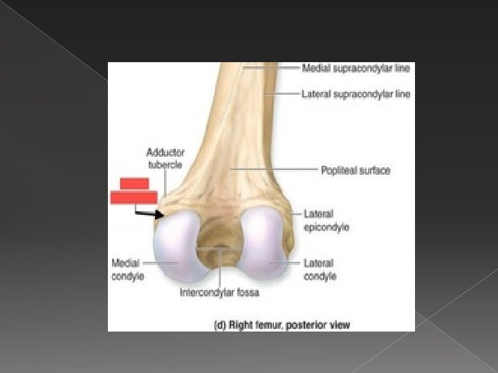
Femur: Distal End � broadens lateral & medial condyles › articulation points with tibia � each flanked superiorly: lateral & medial epicondyles › sites of muscle attachments
Femur � distally articulates with: › Patella › Tibia
Patella (kneecap) � small, triangular, sesamoid bone � develops in tendon of quadriceps femoris muscle � Parts: � Base: broad, superior end � Apex: pointed, inferior end
Patella
Tibia “shin bone” larger, medial, weight-bearing bone of lower leg proximally articulates with femur & fibula distally articulates with fibula & tarsals
Tibia � medial malleolus forms prominence that is palpable & visible on medial ankle
Fibula parallel & lateral to the tibia & considerably smaller � head of fibula on proximal end � lateral malleolus at distal end �
Tibia & Fibula � Interosseous membrane between tibia and fibula: is less flexible but more stable than radius and ulna
Foot � Functions: supports body weight 2. acts like a lever to propel body forward as we walk or run 1.
Tarsals � 7 bones: � 1 calcaneous: heel bone, largest of the tarsals
Metatarsals � 5 bones between tarsals & phalanges � #’d I to V from medial lateral
Phalanges 14 bones that make up the 5 digits � #’d I to V medial to lateral � Hallux: great or big toe has 2 large heavy phalanges �
Arches of the Foot � 2 arches in foot: 1. allows the foot to support weight of body by distributing weight over the soft & hard tissues 2. provide leverage while walking fully developed by age 12 - 13
Arches of the Foot � 2 longitudinal arches (medial & lateral � 1 transverse arch