APOPTOSIS DR SANJIV KUMAR ASSTT PROFESSOR DEPTT OF


 Activates")


 PATHWAY Initiated by engagement of cell surface death receptors Type 1")
PATHWAY Withdrawl of growth factors or cell stress Increased permeability of mitochondrial membrane")
METHOD BYPASS MECHANISM FOR CELLS THAT REFUSE SUICIDE VIA EXTRINSIC OR INTRINSIC")


- Slides: 27
APOPTOSIS DR. SANJIV KUMAR ASSTT. PROFESSOR, DEPTT. OF PATHOLOGY BVC, BASU, PATNA
CONTENTS INTRODUCTION EXAMPLES BIOCHEMICAL FEATURES MORPHOLOGICAL FEATURES MECHANISM
INTRODUCTION A PATHWAY OF CELL DEATH INDUCED BY A TIGHTLY REGULATED SUICIDAL PROGRAMME WORD “PTOSIS” MEANSSHEDDING OFF OR FALL OFF. Active form of cell death Requires energy Mediated by Caspases (a family of proteins) Does NOT elicit inflammation like typical necrosis Acts at the individual cell level or small clusters Balance to mitosis
Often called Programmed cell death which helps in: Deletion of un-needed cells during embryogenesis Normal involution Regression of hyperplasia /tumour Removal of viruses Immunity
Caspases KEY PLAYER What is this Cysteine dependent aspartate directed Proteases
EXAMPLES APOPTOSIS In which situation PHYSIOLOGICAL PATHOLOGICAL
PHYSIOLOGIC SITUATIONS DEATH BY APOPTOSIS IS A NORMAL PHENOMENON THAT SERVES TO ELIMINATE CELLS THAT ARE NO LONGER NEEDED Embryogenesis - Implantation - Organogenesis Hormone dependent involution Cell deletion in proliferating pools Death of senile cells Elimination of self reactive lymphocytes Cell death induced by cytotoxic T-cells
PATHOLOGIC SITUATIONS v Cell death following an injury v Cell injury in some viral diseases - Viral hepatitis v Pathogenic atrophy in some organs - Duct obstruction v Cell death in some tumors
BIOCHEMICAL FEATURES
BIOCHEMICAL FEATURES Apoptotic cells usually exhibit a distinctive constellation of biochemical modifications that underlie the structural changes in cells Protein cleavage Activation of endonucleases Phagocytic recognition
PROTEIN CLEAVAGE Occurs via activation of several members of cysteine protease family (caspases) Activates several proteases which microfilaments like laminin, actin etc. Break up nuclear scaffold & cytoskeleton acts on
DNA BREAKDOWN Apoptotic cells exhibit characteristic breakdown of DNA into 50 -300 kb pieces – There is intra-nucleosomal cleavage into multiple 180200 bp fragments by CA 2+ & Mg 2+ dependent endonucleases Resembles ladders electrophoresis) on a gel (on agarose gel
PHAGOCYTIC RECOGNITION Apoptotic cells express phosphatidylserine in outer layer of the membrane - Allows for early recognition of apoptotic cells by macrophages resulting in phagocytosis without the release of proinflammatory cellular components
MORPHOLOGICAL FEATURES
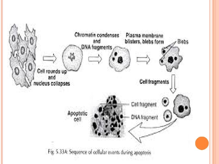
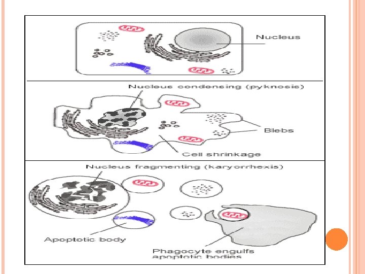
MORPHOLOGICAL FEATURES Ø Cells shrink & round up Ø Cells become more dense Ø Cells detach from neighbors Ø Chromatin becomes very dense & separates into homogeneous masses Ø Cells will undergo budding & blebbing -Buds break off to become apoptotic bodies Ø Phagocytosis by surrounding cells Ø Removal of apopotic bodies
MECHANISM
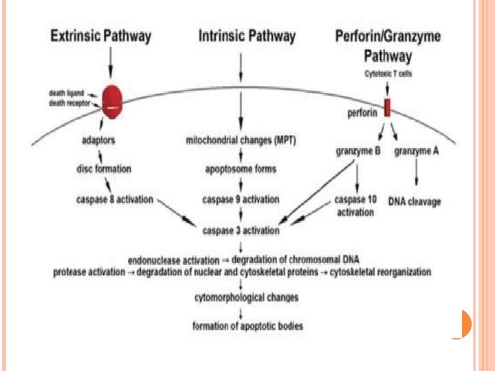
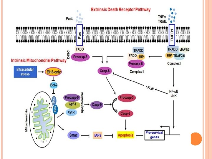
EXTRINSIC (DEATH RECEPTOR) PATHWAY Initiated by engagement of cell surface death receptors Type 1 TNF receptor (TNFR 1), Fas (aka CD 95) Fas is cross-linked by a ligand (Fas. L) 3 or more Fas. L molecules come together & their cytoplasmic death domains from a binding site for FADD then binds pro-caspase 8 (10 in humans) which cleave each other to become active Initiation of caspase cascade APOPTOSIS
INTRINSIC (MITOCHONDRIAL)PATHWAY Withdrawl of growth factors or cell stress Increased permeability of mitochondrial membrane Result of increased mitochondrial permeability & release of pro-apoptotic molecules into the cytoplasm Leakage of cytochrome C Cytochrome C binds to Apaf-1 in the cytoplasm Caspase 9 is activated Caspase cascade APOPTOSIS
CYTOTOXIC T-CELL (BY-PASS)METHOD BYPASS MECHANISM FOR CELLS THAT REFUSE SUICIDE VIA EXTRINSIC OR INTRINSIC • Cytotoxic T-lymphocytes recognize foreign Ag present on infected cell membranes • On recognition, CTLs release perforins (allows entry of granzyme B, a serine protease ) • Gransyme B cleaves proteins at aspartate residues & activates several caspases • Most commonly seen with viral infections
HISTOPATHOLOGY
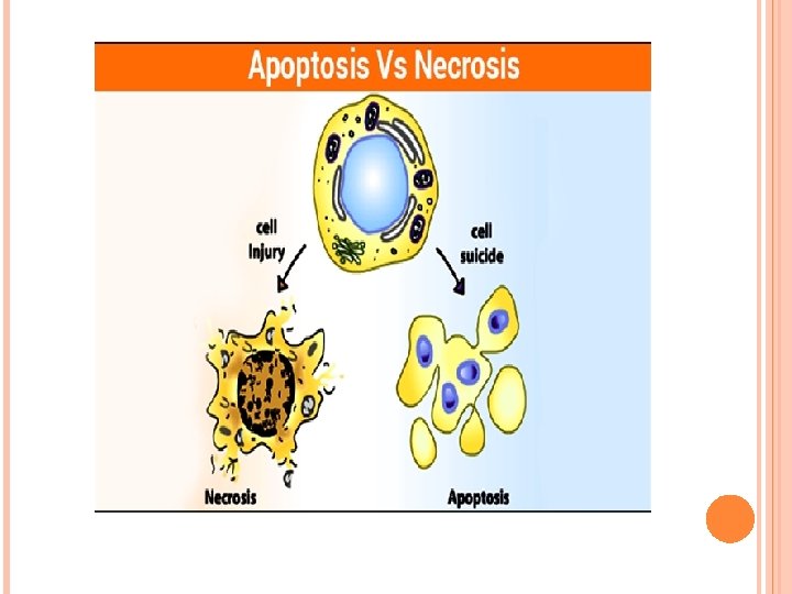
DON’T CONFUSE WITH Necrosis: death of cells & tissues while the body is whole (still living) - Note it is an inflammatory response - Some cells & tissues are dead. Autolysis: also called self-digestion - destruction of tissues by certain substances (such as enzymes) that are produced within the organism
THANK YOU