APOPTOSIS Definition Apoptosis or programmed cell death is


 , start")


 TOO")
 Neuronal dysfunction or")


- Slides: 16
APOPTOSIS
Definition Apoptosis or programmed cell death, is coordinated collapse of cell, protein degradation , DNA fragmentation followed by rapid engulfment by neighboring cells. Apoptosis is gene directed active form of cell death Between 50 and 70 billion cells die each day due to apoptosis in the average human adult.
Why should a cell commit suicide? 1. Apoptosis is needed for proper development Examples: The formation of the fingers and toes of the fetus and the formation of the proper connections between neurons in the brain 2. Apoptosis is needed to destroy cells as defense mechanism Examples: Cells infected with viruses or with DNA damage or cancer cells 3. In aging , senescence and tissue degeneration
APOPTOSIS vs NECROSIS In contrast to necrosis, which is a form of traumatic cell death that results from acute cellular injury, apoptosis is a highly regulated and controlled process that confers advantages during an organism's lifecycle.
Necrosis vs. Apoptosis Necrosis • • • Passive death External stimulus Cellular swelling Membranes are broken ATP is depleted Cell lyses, eliciting an inflammatory reaction • DNA fragmentation is random, or smeared • In vivo, whole areas of the tissue are affected Apoptosis • • • Active death Internal or external stimulus Cellular condensation (shrink) Membranes remain intact Requires ATP Cell is phagocytosed, no tissue reaction • Ladder-like DNA fragmentation • In vivo, individual cells appear affected
NECROSIS Vs APOPTOSIS Wilde, 1999
Molecular mechanism of apoptosis q Because apoptosis cannot stop once it has begun, it is a highly regulated process. q Apoptosis can be initiated through one of two pathways. q In the intrinsic pathway the cell kills itself because it senses cell stress, while in the extrinsic pathway the cell kills itself because of signals from other cells. q Both pathways induce cell death by activating caspases, which are proteases, or enzymes that degrade proteins. q The two pathways both activate initiator caspases, which then activate executioner caspases, which then kill the cell by degrading proteins indiscriminately.
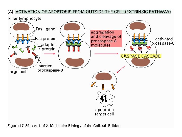
A. Extrinsic pathway - Initiated by death receptors (FAS or TNF receptors) , start with ligand binding to death receptors and then activate pro caspase 8 to yield caspase 8 (active) that triggers effector caspases, (caspase 3, 7). - Activation of death receptors may also activate intrinsic pathway. - TNF or Fas. L are expressed by activated T immune cells to begin death programme to other cells.
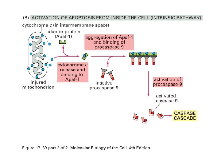
B. Intrinsic Pathway - Usually initiated by cellular stress (UV, cytotoxic drugs, reactive oxygen species) as all leads to change in mitochondrial membrane permeability (MMP) - Regulated by Bcl-2 family member of proteins – regulate the release of pro-apoptotic factors from mitochondria. Pro apoptotic : e. g. Bax , Bad Anti apoptotic : e. g. Bcl-2 - Battle between the ratio of pro and anti levels to determine cell’s response to apoptotic stimuli such as ROS (reactive oxygen species), radiation etc.
Apoptosis: Pathways “Extrinsic Pathway” Death Ligands Death Receptors “Intrinsic Pathway” DNA damage & p 53 Mitochondria/ Cytochrome C Initiator Caspase 8 Effector Caspase 3 Initiator Caspase 9 APOPTPSIS
APOPTOSIS: Role in Disease TOO MUCH: Tissue atrophy Neurodegeneration AIDs (lymphocyte induced depletion) TOO LITTLE: Hyperplasia Cancer Autoimmune diseases (reactive lymphocytes not removed)
APOPTOSIS: Role in Disease Neurodegeneration Neurons are post-mitotic (cannot replace themselves) Neuronal dysfunction or damage results in loss of synapses and neurons that leads to : PARKINSON'S DISEASE ALZHEIMER'S DISEASE
APOPTOSIS: Role in Disease Cancer Apoptosis eliminates damaged cells (damage => mutations => cancer Tumor suppressor p 53 controls apoptosis responses to damage Most cancer cells are defective in apoptotic response (damaged, mutant cells (P 53 mutant) survive) High levels of anti-apoptotic proteins or Low levels of pro-apoptotic proteins ===> CANCER
USEFUL LINKS • https: //www. youtube. com/watch? v=7 WRk Y 8 q_F 3 k • https: //www. youtube. com/watch? v=Syv. OP Xeg 4 ig • http: //www. mcqlearn. com/biology/g 9/apopt osis-and-necrosis-mcqs. php