APARATO AUDITIVO 1 OIDO EXTERNO 2 OIDO MEDIO


 Trago* Anti-Trago*")


 Zona vascular Fibras Meridionales Fibras Radiales")


 arco cricoides M Cricoaritenoideo post (N")


 COSTO- PLEURAL 1 VERTEBRO-PLEURA (D 1) COSTO-VERTEBRO-PLEURAL (C")


 Dr M Monzo mmonzo@ub. edu")


- Slides: 60
APARATO AUDITIVO • 1 OIDO EXTERNO • 2 OIDO MEDIO • 3 OIDO INTERNO Dr M Monzo mmonzo@ub. edu
1 OIDO EXTERNO Adaptación al medio Estructuras Limites Longitud Significado morfológico Dr M Monzo mmonzo@ub. edu
PABELLON AUDITIVO Relieves Constitución Vascularización Hélix Anti-hélix Canal del Hélix (fosa escafoidea) Trago* Anti-Trago* Lóbulo auricular Tej Cartilaginoso Tej Adiposo Tej Muscular Linfaticos Art Auricular pots Art Temporal Superf Inervación Vens Auricular pots Vens Temporal Superf Anteriores Posteriores Inferiores Motora , F Sensitiva N auricular M N auricular T Dr M Monzo mmonzo@ub. edu
CONDUCTO AUDITIVO EXTERNO • Limites • Constitución Relaciones ** Vascularización Cara externa Cara Interna Pared Anterior Pared Posterior Pared Superior Pared Inferior Linfaticos Art Auricular pots Art Temporal Superf Vens Auricular pots Vens Temporal Superf Significado Clínico Inervación Anteriores Posteriores Inferiores Ramas: Auriculo T. Facial Vago Dr M Monzo mmonzo@ub. edu
MEMBRANA TIMPANICA** • FORMA • DIMENSIONES • ESTRUCTURAS ANATOMICAS • CARAS • CONSTITUCION • DISPOSICION Vascularización Cara Exter. Art auricular prof (rama maxilar) Cara Inter. Art auricular post Vena: Cara Exter. Drenan Y E Cara Inter. Drenan Seno trasnsverso Dr M Monzo mmonzo@ub. edu
2 OIDO MEDIO • CONTENIDO • PAREDES • TROMPA CONTENIDO HUESOS Martillo Yunque Estribo MUSCULOS Martillo Estribo LIGAMENTOS Martillo Yunque Estribo Dr M Monzo mmonzo@ub. edu
2 OIDO MEDIO HUESOS MARTILLO YUNQUE ESTRIBO Cabeza Mango Apof Lateral Apof Anterior Cuerpo Rama Corta Rama Larga Apof Lenticular Cabeza Cuello Rama Ant Rama Post Platina MUSCULO Martillo (Timpánico) Estribo (Estapédio) MUSCULOS O I INERV ACCION Trompa Porción cartilaginosa Mango Martillo Rama Mandibular Trigémino Amortg Vibraciones Memb timp Pirámide Cuello estribo Facial Amortg Vibraciones Estribo Dr M Monzo mmonzo@ub. edu
2 OIDO MEDIO VASCULARIZACION INERVACION Art Estilomastoideda Art Timpánica Art Meningea media Art Faríngea Art Carótida Interna Venas: Plexos pterigoideos Plexos faríngeos Vena meningea media Golfo yugular interna Motora (Fibras del facial y trigemino) Sensitiva (Nerv Jacobson) Dr M Monzo mmonzo@ub. edu
2 OIDO MEDIO LIGAMENTOS MARTILLO YUNQUE ESTRIBO Ligamentos Maleolar Sup Maleolar Inf Ligamento Sup Yunque Ligamento Cabeza PAREDES (Caja Timpánica)** • Dimensiones • Anterior • Posterior • Externa • Interna • Superior • Inferior Dr M Monzo mmonzo@ub. edu
TROMPA AUDITIVA / TROMPA DE EUSTAQUIO • LIMITES • DIMENSIONES • PORCIONES • RELACIONES VASCULARIZACION: Ramas Art faringea des (R C EX) Ramas Art Meningea med ( RMax Inf) VENAS : Drenan plexo venoso pterig INERVACION: Ramas plexo timpánico (facial glosofaringeo) Dr M Monzo mmonzo@ub. edu
LA VISION 1 OJO O GLOBO OCULAR 2 ESTRUCTURAS ANEXAS Dr M Monzo mmonzo@ub. edu
LA VISION PARED CONTENIDO CAPA EXTERNA / ESCLEOTICA/ FIBROSA CAPA MEDIA / UVEA/COROIDES/ VASCULAR CAPA INTERNA / RETINA/ NERVIOSA CRISTALINO HUMOR ACUSOSA HUMOR VITREO Dr M Monzo mmonzo@ub. edu
LA VISION CAPA EXTERNA / ESCLEOTICA/ FIBROSA SUPERFICIE EXTERNA SUPERFICIE INTERNA Orificios posteriores Orificios Ecuatoriales Orificios Anteriores Lamina Fusca Dr M Monzo mmonzo@ub. edu
LA VISION CAPA MEDIA/ UVEA/COROIDES/VASCULAR • COROIDES PROPIA • ZONA CILIAR • IRIS ZONA CILIAR IRIS COROIDES P Dr M Monzo mmonzo@ub. edu
COROIDES PROPIA Capa externa / Art Ciliares largas post Capa media /Venas vorticosas Capa profunda/Art Ciliares cortas post LA VISION Dr M Monzo mmonzo@ub. edu
ZONA CILIAR LA VISION Zona muscular (Mus ciliar) Zona vascular Fibras Meridionales Fibras Radiales Fibras circulares (Mus Rouget) Procesos ciliares Orbiculo ciliar Ora Serrata Zona muscular Zona vascular Dr M Monzo mmonzo@ub. edu
LA VISION IRIS Cara Anterior /Mus constr /Miosis Cara Posterior/Mus dilatador/ Midriasis Dr M Monzo mmonzo@ub. edu
LA VISION CONTENIDO CAMARAS CRISTALINO HUMOR ACUSOSA HUMOR VITREO Anterior Posterior Dr M Monzo mmonzo@ub. edu
LA VISION FONDO DE OJO Venulas de la retina Punto ciego Mácula /fovea central Dr M Monzo mmonzo@ub. edu
LA VISION 2 ESTRUCTURAS ANEXAS • Grasa orbitaria • Conjuntiva • Parpados • Aparato Lacrimal GRASA ORBITARIA Dr M Monzo mmonzo@ub. edu
LA VISION 2 ESTRUCTURAS ANEXA • Conjuntiva Dr M Monzo mmonzo@ub. edu
LA VISION 2 ESTRUCTURAS ANEXA • Parpados Vascularizacion: Ramas oftalmica/ Art pal Sup Inervacion: Ramas facial Dr M Monzo mmonzo@ub. edu
LA VISION 2 ESTRUCTURAS ANEXA • Aparato Lacrimal Dr M Monzo mmonzo@ub. edu
APARATO RESPIRATORIO Dr M Monzo mmonzo@ub. edu
APARATO RESPIRATORIO VIAS RESPIRATORIAS SUP Fosas Nasales VIAS RESPIRATORIAS INF Laringe Traquea Bronquios Pulmones Dr M Monzo mmonzo@ub. edu
FOSAS NASALES Dr M Monzo mmonzo@ub. edu
FOSAS NASALES MEATO INFERIOR MEATO MEDIO MEATO SUPERIOR Dr M Monzo mmonzo@ub. edu
FOSAS NASALES Art Etmoidea/R Oftal/R CI Art Esfenopalatina/R Max In/ R CE Art Facial /R CE Ven Facial Pteriogomaxilar VASCULARIZACION LINFATICOS INERVACION Sub-maxilares Retro-faringeos Yugo-carotideos Ramas sensitivas ganglio esfeno-palatino Dr M Monzo mmonzo@ub. edu
LARINGE • LIMITES • CONSTITUCION Cartilagos articulares Ligamentos Musculos Mucosa Dr M Monzo mmonzo@ub. edu
LARINGE Dr M Monzo mmonzo@ub. edu
LARINGE NOMBRE O M Cricotiroideo (N laringeo Sup) arco cricoides M Cricoaritenoideo post (N R) lamina cart Cricoides (Sup dorsal) M Cricoaritenoideo lat (NR) arco cricoides I lamina cartilago tiroides FUNCION tensor cuerdas v Apof mus Aritenoides Expande glotis Apof mus Aritenoides Cierra glotis M Aritenoideo transv cart aritenoides (NR) M Aritenoideo oblicuo cart aritenoides (NR) M Vocal (NR) cart Tiroides Cart ariteoindes opuesto Apof mus opuesta Cierra glotis Apof vocal Tensor lig vocal M Tiroaritenoideo Apof Mus Dr M Monzo Estrecha glotis cart Tiroides Ensancha glotis mmonzo@ub. edu
LARINGE Dr M Monzo mmonzo@ub. edu
LARINGE Dr M Monzo mmonzo@ub. edu
LARINGE Dr M Monzo mmonzo@ub. edu
LARINGE Art Laringea craneal / R Tiroidea CR / R CE VASCULARIZA CION INERVACION Art Laringea caudal/ R Tiroidea CD/ R Art SClv Rama Interna Ner Laringeo Cra Rama Externa Ner Laringeo Caudal / Recurrente G Supragloticos/ YI LINFATICOS G Infragloticos/YI Dr M Monzo mmonzo@ub. edu
TRAQUEA Dr M Monzo mmonzo@ub. edu
TRAQUEA ARTERIAS Tiroideas Sup/Inf Art Bronquiales Art Diafragmaticas Sup VENAS ------ Vena Acigos Recurrente LINFATICOS Ramas plexo simpático y pulmonar Dr M Monzo mmonzo@ub. edu
PULMONES • COLORACION • PESO • FORMA Dr M Monzo mmonzo@ub. edu
PULMONES Dr M Monzo mmonzo@ub. edu
PULMONES Ap Post 1 AP 1 2 2 Post 3 Ant 6 Sup 4 Ling S 4 lat 5 med 10 9 B lat B Post 8 B Ant 5 Ling I 8 B Ant 9 B Lat 10 B Post Dr M Monzo mmonzo@ub. edu
PULMONES 7 7 Dr M Monzo mmonzo@ub. edu
PULMONES PD PI Dr M Monzo mmonzo@ub. edu
BRONQUIOS Dr M Monzo mmonzo@ub. edu
PLEURAS PARIETAL COSTAL MEDIASTINICA DIAFRAGMATICA CERVICAL/CUPULA PLEURAL VISCERAL SENOS COSDIAFRAGMATICO COSTFRENICO Dr M Monzo mmonzo@ub. edu
1 2 LIGAMENTOS PULMONARES (CUPULA PLEURAL) COSTO- PLEURAL 1 VERTEBRO-PLEURA (D 1) COSTO-VERTEBRO-PLEURAL (C 7) Dr M Monzo mmonzo@ub. edu
MEDIASTINO SUPERIOR D 4 MEDIASTINO INFERIOR ANTERIOR MEDIO POSTERIOR D 8 Dr M Monzo mmonzo@ub. edu
TORAX Bronq D AORTA AR P VCS AU D OR AU I VE VERT Dr M Monzo mmonzo@ub. edu
TORAX (D 4) Dr M Monzo mmonzo@ub. edu
Imágenes New England Journal of Medicine
A 10 -year-old boy has a brown right eye and a blue left eye, as well as mild ptosis and miosis of his left eye Gesundheit, B. et al. N Engl J Med 2005; 353: 1502
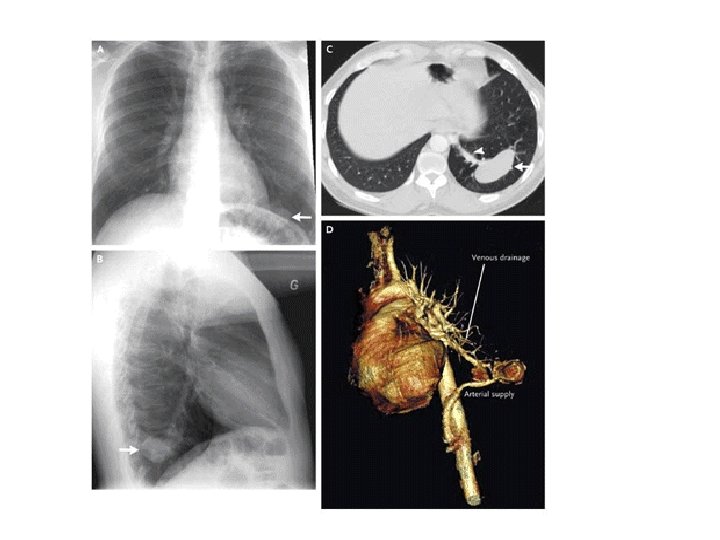
A healthy 48 -year-old man without prior pulmonary symptoms underwent routine chest radiography for minor thoracic pain. The frontal and lateral images (Panels A and B) showed a well-defined, lobulated mass (arrows) in the left lower lobe of the lung. A subsequent contrast-enhanced multislice computed tomographic (CT) scan (Panel C) showed the homogeneous mass (arrow), with an adjacent large feeding vessel (arrowhead). Three-dimensional reconstruction (Panel D) showed the arterial supply of this lesion, with an aberrant origin in the celiac trunk, and normal venous drainage through the pulmonary veins. These findings are diagnostic of intralobar pulmonary sequestration, which is characterized by anomalous pulmonary tissue that is typically localized in the left lower lobe and supplied by the systemic circulation. The CT volume-rendering reconstruction is particularly useful in demonstrating the arterial and venous pattern of the malformation, obviating the need for an invasive imaging technique.