Aortic Valve Stenosis High Gradient Vs Low Gradient


- Slides: 36
Aortic Valve Stenosis High Gradient Vs. Low Gradient Hetal Gandhi MD FACC Advocate Heart Institute
Introduction Natural History Pathophysiology and symptoms Echocardiographic Low measurement flow gradient AS Asymptomatic Management Severe AS
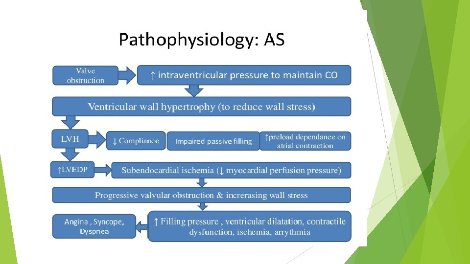
Natural History of Aortic Stenosis Most common valvular heart disease and third most prevalent form of cardiovascular disease in western world Average rate of increase in mean pressure gradient is 4 to 7 mmhg per year Valve area decline at average rate of 0. 1 cm 2/year, but some patients have little or no progression and other progress more rapidly Aortic jet velocity is increase by an average of 0. 3 m/s per year Patients with mild disease (< 3. 0 m/s jet velocity) less likely to develop symptoms over 5 years
A substantial number of patients with severe aortic stenosis are not yet symptomatic and risk of sudden cardiac death is low <1%/year but once symptomatic increase mortality 50% at 2 year AHA/ACC 2014 guideline
Natural History of AS without treatment Brawanwald E. aortic stenosis 1968 circulation
Aortic valve stenosis and low LVEF
Heart Failure reduces the life expectancy <2 years Angina and Syncope reduces life expectancy between 2 -5 years
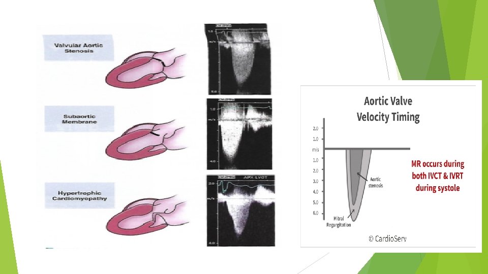
Causes of Aortic stenosis Supravalvular Subvalular: Valvular Tunnel, Discrete : Congenital (1 -30 years old) Bicuspid (40 -60 years old) Rheumatic (40 -60 years old) Senile degenerative ( >65 years old)
Aortic Valvular Morphology
Two Different Patterns of Low-Flow, Low-Gradient AS NORMAL-LVEF NORMAL-FLOW HIGH-GRADIENT 50 -70% «PARADOXICAL» LOW-FLOW LOW-GRADIENT 10 -25% LOW-LVEF «CLASSICAL» LOW-FLOW LOW-GRADIENT 5 -10%
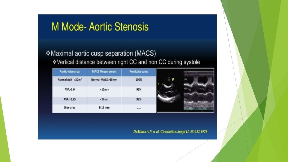
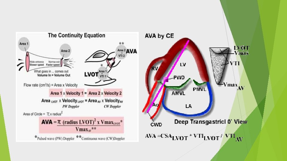
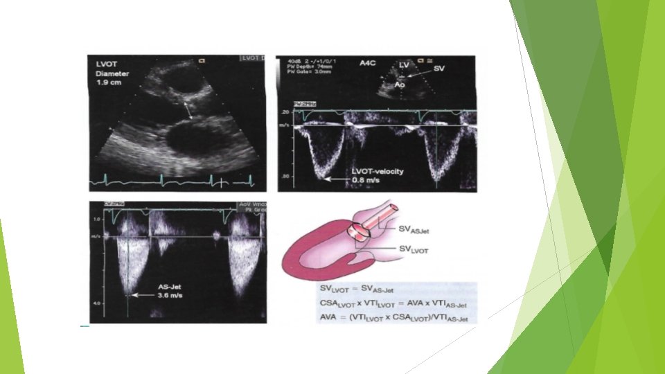
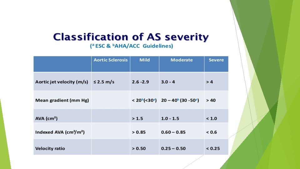
Echo approach to Aortic Stenosis Valve anatomy 2 d views, LVOT diameter Exclude LV outflow trach obstruction Stenosis severity: a. Peak velocity b. Mean pressure gradient c. Continuity equation valve area Degree of coexisting aortic regurgitation Left ventricular hypertrophy and LVEF
Calcification affect LOVT measurement
Pitfalls in Echo measurement Outflow tract diameter images Intercept angle between aortic stenosis jet and ultrasound beam Identification of flow signal origin ( AS vs. MR. vs HOCM) Beat to Beat variability ( AF, PVCs) Respiratory motion affect acoustic window
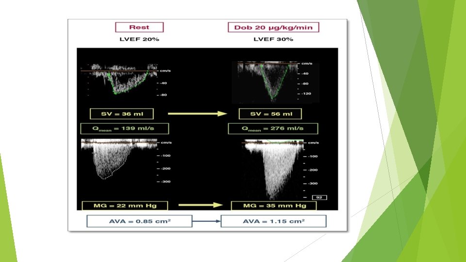
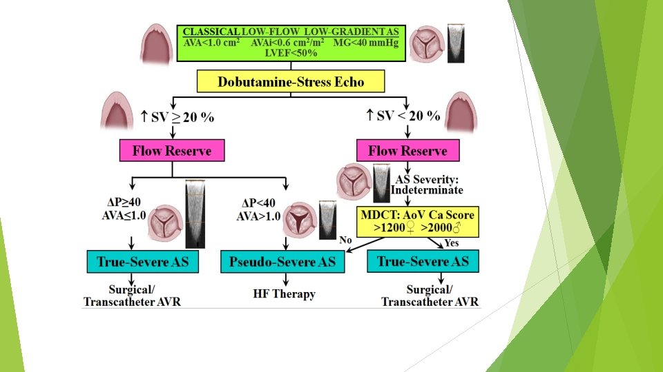
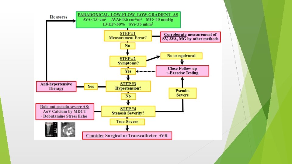
Low gradient low flow AS To differentiated low gradient is due to true aortic stenosis vs. pseudo aortic stenosis from Low LVEF related It can be also seen in patient with normal LVEF (about 5 -7% cases) Criteria: (i) AVA <1. 0 cm 2 (ii) mean aortic transvalvular pressure gradient <40 mm. Hg (iii) LV ejection fraction <50% (iv) SVi <35 m. L/m 2. Low-dose Dobutamin Echocardiogram can help distinguish between pseudo severe AS vs. true severe AS.
Dobutamine Echocardiography
Images at Baseline and after Dobutamine infusion
Low gradient AS with normal LVEF Most challenging Valve area <1. 0 cm 2, mean gradient <40 mm. HG, LVEF is normal or above 50% Transvalvular flow SVi <35 ml/m 2 Usually seen in elderly, with small LV cavity, more hypertrophy, HTN Dobutamin echo may be helpful Calcium score CT for aortic valve
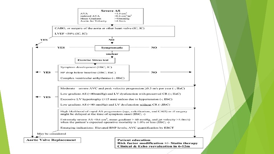
Asymptomatic Severe Aortic Valve Stenosis The management and the clinical decision making in asymptomatic patients with aortic stenosis are challenging. An “aggressive” management, including early aortic valve replacement, is debated in these patients. Optimal timing for surgery remains controversial due to the lack of prospective data on the determinants of aortic stenosis progression Exercise stress testing with or without imaging is strictly contraindicated in symptomatic patients with severe aortic stenosis. Exercise stress test is recommended by current guidelines in asymptomatic patients and may provide incremental prognostic value. Indeed, the development of symptoms during exercise or an abnormal blood pressure response are associated with poor outcome and should be considered as an indication for surgery Exercise stress echocardiography may also improve the risk stratification and identify asymptomatic patients at higher risk of a cardiac event. During exercise, an increase >18 to 20 mm Hg in mean pressure gradient, absence of improvement in left ventricular ejection fraction (i. e. , absence of contractile reserve), and/or a systolic pulmonary arterial pressure >60 mm Hg (i. e. , exercise pulmonary hypertension) are suggestive signs of advanced stages of the disease and impaired prognosis.
Stress Echocardiogram in AS
Summary AVR is class I indication in patient with symptomatic severe aortic stenosis, improve survival significantly Decision about timing of AVR in asymptomatic severe AS patients require careful assessment of risk and benefit ratio of AVR vs. watchful waiting Exercise testing/stress echo in asymptomatic Severe AS patients provide for risk stratification and has prognostic valve Need more prospective trials to assess Global Strain- LGS to detect early LV systolic dysfunction Careful assessment of echocardiographic measurement help to differentiate between low flow gradient from true vs. pseudo AS
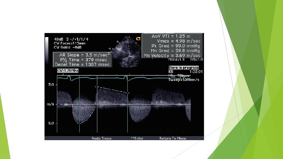
CASE A 64 -year-old man is referred for systolic murmur and abnormal echocardiogram. He has a history of hypertension. He does not smoke, is physically active (walks briskly for three miles per day), and consumes a healthy diet. He has no symptoms. Medications include aspirin, high-intensity statin (atorvastatin), and angiotensin converting enzyme inhibitor. Blood pressure is 120/80 mm. Hg and heart rate is 65 beats per minute. Echocardiogram: Normal biventricular systolic function, ejection fraction 60 -65%, with mild concentric LVH Normal biatrial size Heavily calcified aortic valve, unable to discern valve morphology; Vmax 5 m/s, peak gradient 99 mm. Hg, mean gradient 60 mm. Hg, calculated aortic valve area 0. 5 m 2. (Figure 1) Pulmonary artery systolic pressure is 28 mm. Hg based on an RA pressure of 5 mm. Hg.
Which of the following is not correct? A. Refer to surgical aortic valve replacement B. Refer to Transcatheter aortic valve replacement C. Treadmill exercise testing D. Watchful waiting
Which of the following is not correct? A. Refer to surgical aortic valve replacement B. Refer to Transcatheter aortic valve replacement C. Treadmill exercise testing D. Watchful waiting
A patient with severe AS and CHF has mild mitral valve thickening and moderate MR. Your advice to the surgeon is: A. Aortic valve replacement and mitral valve repair B. Aortic valve replacement only C. Aortic and mitral valve replacement D. Aortic valve replacement; re-evaluate MR by TEE after coming off bypass.
A patient with severe AS and CHF has mild mitral valve thickening and moderate MR. Your advice to the surgeon is: A. Aortic valve replacement and mitral valve repair B. Aortic valve replacement only C. Aortic and mitral valve replacement D. Aortic valve replacement; re-evaluate MR by TEE after coming off bypass.