AORTIC DISSECTION Dr M SOFI MD FRCP London
; FRCPEdin; FRCSEdin")

, which starts from the left")


")


 • Altered mental status")


 is a relatively good test in the diagnosis of aortic")
 is currently the gold standard test, sensitivity of 98% and a")


- Slides: 25
AORTIC DISSECTION Dr. M. SOFI MD; FRCP (London); FRCPEdin; FRCSEdin
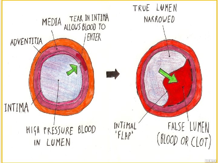
AORTIC DISSECTION Aortic dissection is defined as separation of the layers within the aortic wall. The primary event in aortic dissection is a tear in the aortic intima. • Degeneration of the aortic media, or cystic medial necrosis, is felt to be a prerequisite for the development of non-traumatic aortic dissection. • Blood passes into the aortic media through the tear, separating the intima from the surrounding media and/or adventitia, and creating a false lumen. • Mortality is still high despite advances in diagnostic and therapeutic modalities
Dissection of the descending part of the aorta (3), which starts from the left subclavian artery and extends to the abdominal aorta (4). The ascending aorta (1) and aortic arch (2) are not involved.
Classification Type I – Originates in ascending aorta, propagates at least to the aortic arch and often beyond it distally. It is most often seen in patients less than 65 years of age and is the most lethal form of the disease. Type II – Originates in ascending aorta and is confined to the ascending aorta. Type III – Originates in descending aorta, rarely extends proximally but will extend distally. It most often occurs in elderly patients with atherosclerosis and hypertension. Percentage 60% 10 -15% 25 – 30 % Type De. Bakey III Stanford A (Proximal) Stanford B Distal
Pathophysiology • Aorta is made up of three layers, the intima, the media, and the adventitia. • The intima is in direct contact with blood and consists of a layer of endothelial cells on a basement membrane; • Media contains connective and muscle tissue • Adventitia, comprising connective tissue outer layer • Blood penetrates the intima and enters the media layer. The high pressure rips the tissue of the media apart along the laminated plane splitting the inner 2/3 and the outer 1/3 of the media apart. • This can propagate along the length of the aorta for a variable distance forward or backwards
Pathophysiology: The aortic dissections originate with an intimal tear in: 1. Ascending aorta (65%) 2. Aortic arch (10%) 3. Descending thoracic aorta (20%) 4. No evidence of tear (13%) Blood penetrates the intima and enters the media layer.
A U T O P S Y H E A R T Dissection media as well outer media
Pathophysiology • The blood traveling • The initiating event is a through the media, tear in the intimal lining creating a false lumen of the aorta. separating from the true • High pressure blood lumen is a layer of intimal enters the media at the tissue. point of the tear. • This tissue is known as the • The force of the blood intimal flap. causes the tear to extend. • May extend proximally or • The majority of dissection are in distally or both. – ascending aorta (65%) – aortic arch (10%), – descending thoracic aorta (20%).
Signs and symptoms Aortic dissection can be rapidly fatal, with many patients dying before presentation to the emergency department or before diagnosis is made in the ED. • Sudden onset of severe chest pain that often has a tearing or ripping quality • Chest pain may be mild • Anterior chest pain: Usually associated with anterior arch or aortic root dissection • Neck or jaw pain: Aortic arch involvement and extension into the great vessels • Tearing or ripping intrascapular pain: indicate dissection involving the descending aorta • No pain in about 10% of patients • Syncope
Signs and symptoms • CVA symptoms: hemianesthesia, and hemiparesis, hemiplegia) • Altered mental status • Numbness and tingling, pain, or weakness in the extremities • Horner syndrome (ptosis, miosis, anhidrosis) • Dyspnea • Hemoptysis • Dysphagia • Flank pain (with renal artery involvement • Abdominal pain (with abdominal aorta involvement) • Fever • Anxiety and premonitions of death
Possible physical examination findings include: • Hypertension/Hypotension • Inter-arm blood pressure differential greater than 20 mm Hg • Signs of aortic regurgitation (bounding pulses, wide pulse pressure, diastolic murmurs) • Cardiac tamponade (muffled heart sounds, hypotension, pulsus paradoxus, jugular venous distention, Kussmaul sign) • Neurologic deficits (e. g. , syncope, altered mental status) • Peripheral paresthesias • Horner syndrome • New diastolic murmur • Asymmetrical pulses (e. g. , carotid, brachial, femoral) • Progression or development of bruits
Laboratory findings include the following: • Leukocytosis: Stress state • Decreases in hemoglobin and hematocrit values: Leaking or rupture of the dissection • Elevation of the BUN and creatinine levels: Renal artery involvement or prerenal azotemia • Elevation of the cardiac enzymes, myoglobin, and troponin I and T levels: Myocardial ischemia from coronary artery involvement • LDH: Hemolysis in false lumen • Smooth muscle myosin heavy-chain assay: levels in the first 24 hours are 90% sensitive and 97% specific • FDP elevation: • FDP of 12. 6 μg/m. L or higher suggests possible aortic dissection with a patent false lumen • FDP level of 5. 6 μg/m. L or higher suggests possibility of dissection with complete thrombosis of false lumen
Causes • Marfan syndrome is • Aortic dissection is often noted in 5– 9%. Individuals associated with Marfan syndrome hypertension tend to have aneurysms of • Chest trauma. 72 to 80% the aorta and are more of individuals have a prone to proximal previous history of dissections of the aorta. hypertension. • A bicuspid aortic valve (a type of congenital heart disease involving the aortic valve) is found in 7– 14%. • Risk is not associated with the degree of stenosis of the valve.
Diagnosis The diagnosis of acute aortic dissection requires a high index of suspicion and involves the following: • History and physical examination • Imaging studies • Chest X-Ray • CT with contrast • MRI • Aortoraphy • Electrocardiography • Complete blood count, serum chemistry studies, cardiac marker assays
Imaging studies Chest radiography: CT with contrast: • Initial imaging • The definitive test in technique if it is most patients with readily available at the suspicion of aortic bedside dissection • Widening of the • Useful only in mediastinum is the hemodynamically stable classic finding patients • Hemothorax may be • Findings help determine evident if the whether hypothermic dissection has circulatory arrest is ruptured necessary for surgery
Chest X-Ray • Mediastinum widening: CXR has moderate sensitivity in an ascending aortic dissection. • Pleural effusions may be seen on CXR. Commonly in descending aortic dissections. Typically in left hemi-thorax. • Obliteration of the aortic knob, depression of the left mainstream bronchus, loss of the para-tracheal stripe, and tracheal deviation. • About 12 to 20% of individuals presenting with an aortic dissection have a "normal" chest x-ray Chest radiograph demonstrating widened mediastinum in a patient with aortic dissection.
Calcium sign: Aortic dissection The calcium sign on CXR suggests aortic dissection. It is the separation of the intimal calcification from the outer aortic soft tissue border by 10 mm
The transesophageal echocardiogram (TEE) is a relatively good test in the diagnosis of aortic dissection, with a sensitivity of up to 98% and a specificity of up to 97%. It has become the preferred imaging modality for suspected aortic dissection. An echocardiogram displaying the true lumen and false lumen of an aortic dissection. In the image to the left, the intimal flap can be seen separating the two lumens. In the image to the right, color flow during ventricular systole suggests that the upper lumen is the true lumen
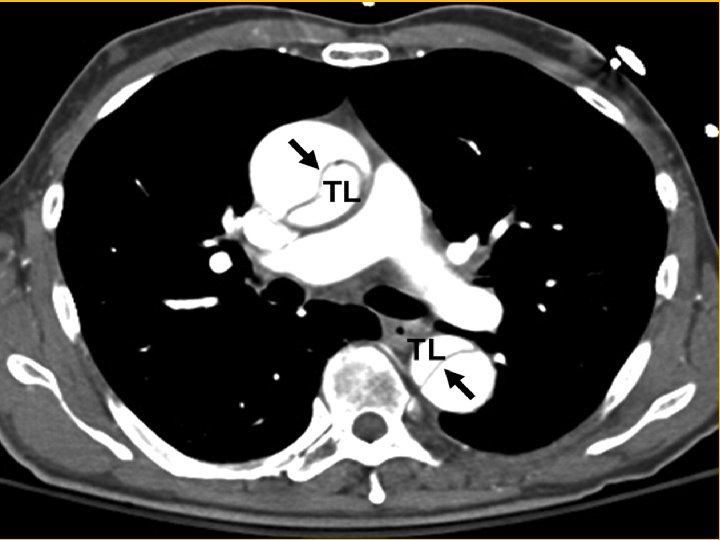
v v (MRI) is currently the gold standard test, sensitivity of 98% and a specificity of 98%. An MRI, allowing the physician to determine the: v v Location of the intimal tear Involvement of branch vessels Locate any secondary tears. Detect and quantitate the degree of aortic insufficiency. MRI of an aortic dissection 1 Aorta descends with dissection 2 Aorta isthmus
Management: Medical • Beta blockers are first line • Calcium channel blockers treatment for acute and can be used if there is a chronic contraindication to the use of beta blockers. • In acute dissection, rapidly acting, titratable parenteral • The calcium channel agents (such as esmolol, blockers typically used or labetalol) are verapamil and diltiazem, for their combined • Vasodilators such vasodilator and as sodium nitroprusside negative inotropic effects. for ongoing hypertension, but they should never be • Pain management: used alone. Narcotics and opiates are the preferred agents
Surgical Treatment Indications for the surgical treatment include: • Acute proximal aortic dissection • Acute distal aortic dissection with complications. Complications include: – Risk of a vital organ damage – Rupture of the aorta – Retrograde dissection ascending aorta • Marfan syndrome • Ehlers-Danlos Syndrome In surgical treatment, the area of the aorta with the intimal tear is usually resected and replaced with a Dacron graft. • Resect the most severely damaged segments of the aorta • Obliterate blood into false lumen • Endovascular repair is emerging as the preferred treatment for descending aortic dissection.
Epidemiology & Prognosis • Of all people with aortic • In ascending aortic dissection, 40% die almost dissection, if there is a straight away and do not decision that surgery is not reach hospital. appropriate, 75% die within 2 weeks. • Of the remainder, 1% die every hour, making • With aggressive treatment prompt diagnosis and 30 -day survival for thoracic treatment a priority. dissections may be as high as 90% • Even after diagnosis, 5– 20% die during surgery or in the immediate postoperative period.
THANK YOU FOR YOUR ATTENTION