AORTIC ARCH ANOMALIES Dr Santhosh Narayanan Topic outline


")


 Aorticopulmonary septal defect and intact ventricular septum, TGA with IVS")


- Slides: 92
AORTIC ARCH ANOMALIES Dr. Santhosh Narayanan
Topic outline • • Embryology Anatomy Classification Individual anomalies
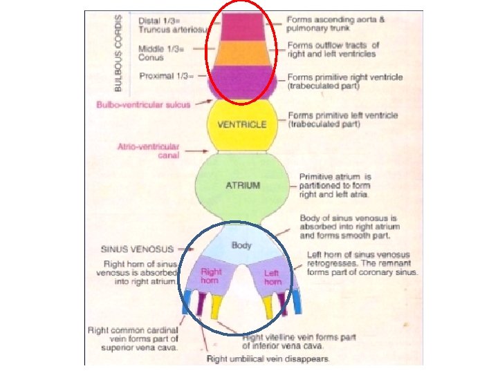
Embryology-Sequence of Events • Day I 8 - Cardiac precursor cells seen in the form of blood islands • Day 20 - First intraembryonic blood vessels • Day 21 - Folding, heart tube formation, looping • Day 22 – heart starts to beat • Day 28 – embryonic circulation established
The Cardiac Neural crest
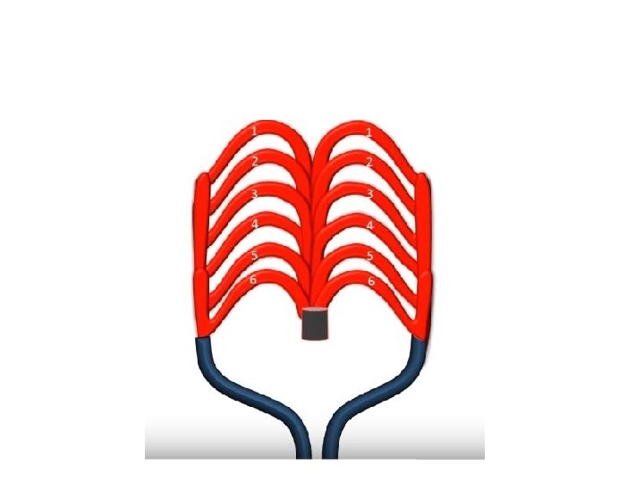
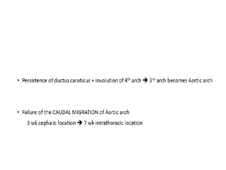
Normal aortic arch development Rathke's model
1 st arch remnant-Maxillary Art. 2 nd arch remnant-Stapedial Art.
Intersegmental Arteries Intersegmental artery Development Cervical All except 7 th merge into vertebral artery 7 th cervical intersegmental Subclavian artery Thoracic Intercostal artery Lumbar All except 5 th -becomes lumbar arteries 5 th Lumbar Common iliac artery Sacral Lateral sacral artery
Arch sidedness
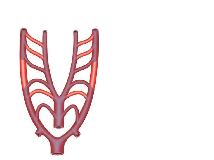
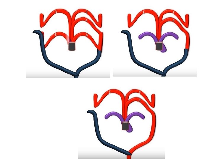
Edward's hypothetical double aortic arch model
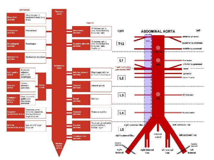
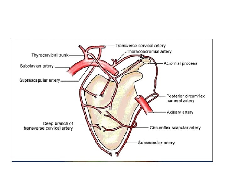
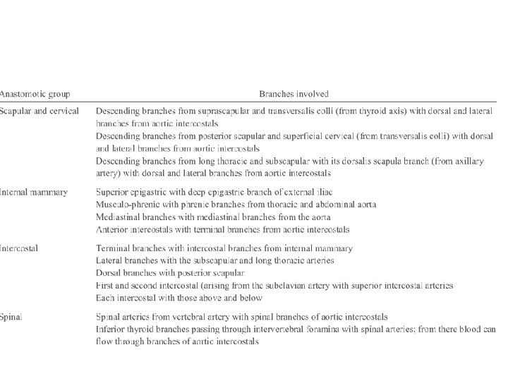
Anatomy • Branches • Anastomoses
History • Hunault in 1735 - pathologic description of anomalous right subclavian artery. • Double aortic arch-Hunault 1735 • First report of clinical syndrome of vascular compression. Wolman 1939 • Kommerel in 1936 –xray findings of anamolous right sca • First division of a vascular ring -Gross in 1945 • First successful repair of interrupted aortic arch-Merrill et al. in 1957
Stewert's Anatomical Classification • 1. Abnormal branching - of Lt Arch • 2. Abnormal arch position -Rt Arch, Cervical Arch • 3. Supernumary arches - DAA, Persistent 5 th Arch • 4. Interrupted aortic arch • 5. Anomalous origin of PA branches
Freedom's clinical classification • Vascular rings • Non ring vascular compression of the trachea, bronchi, or esophagus • Noncompressive arch malformations • Ductal-dependent arch anomalies including interrupted aortic arches • Isolated subclavian, carotid, or innominate arteries • Genetic syndromes and associated abnormalities.
Clinical Presentations Aortic Arch Configuration Clinical Presentation 1. LEFT AORTIC ARCH A. Branching variants Asymptomatic B. Aberrant Rt Subclavian Tracheoesophageal compression(Rare) C. Isolation of Rt Subclavian steal 2. RIGHT AORTIC ARCH A. Mirror image Symptomatic with other associated CHD B. Aberrant Rt Subclavian Tracheoesophageal compression(Rare) C. Isolation of Rt Subclavian steal 3. DOUBLE AORTIC ARCH Tracheoesophageal compression 4. CERVICAL AORTIC ARCH Neck mass, pressure symptoms
Branching variants
Separate origin of vertebral artery
Bovine arch variant in humans
True bovine arch -Uncommon
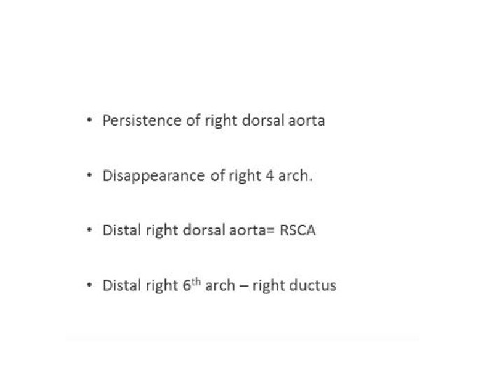
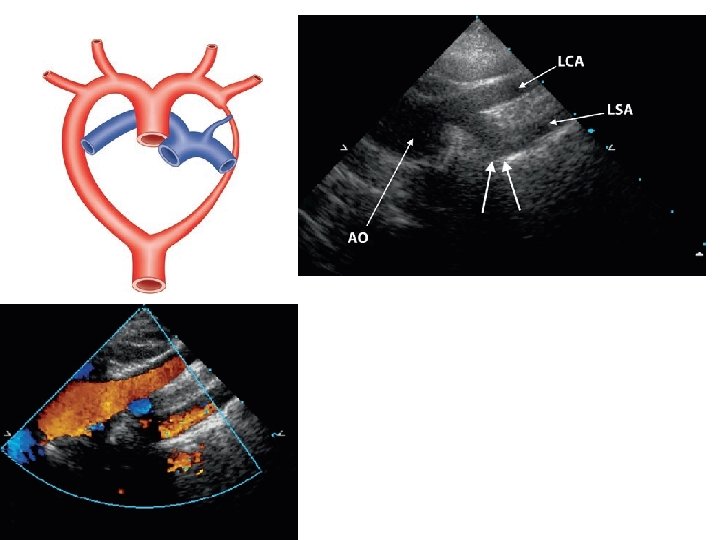
Left aortic arch with aberrant right subclavian(ARSA)
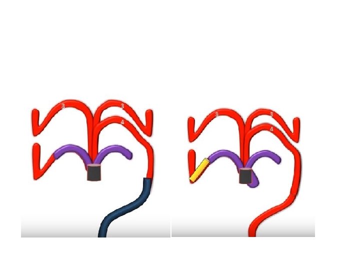
Embryology
Left arch with diverticulum
Edward's model
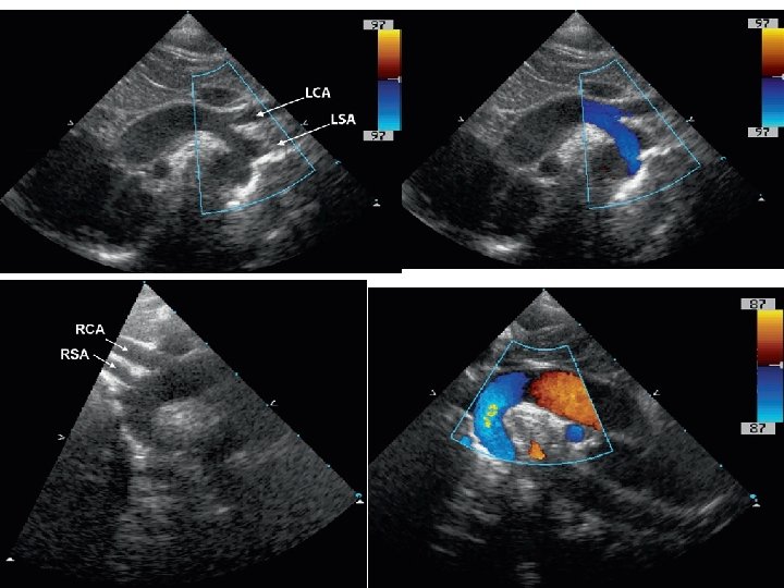
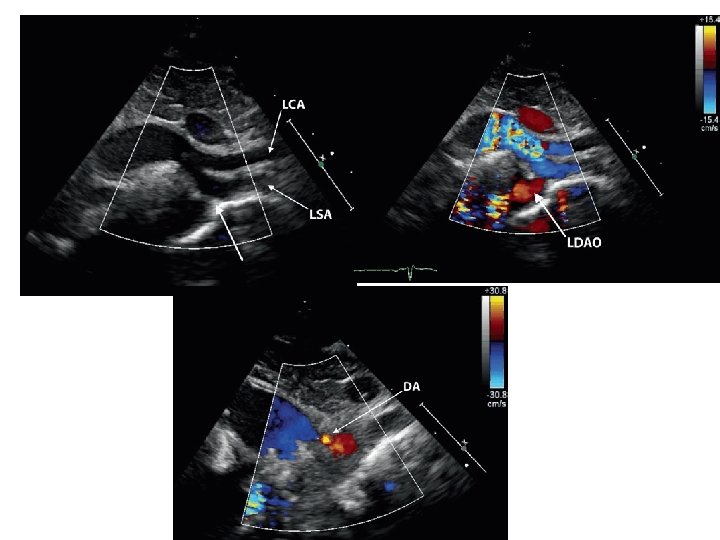
Left arch with isolation of right subclavian artery
Left arch with isolation of right subclavian artery • Rare • SCA arises from ductus arteriosus • If PDA closes, retrograde flow from vertebral artery via circle of willis • Subclavian steal • Treatment - Re implantation of SCA to aorta
Right Aortic Arch • Associated with CHD -mainly conotruncal anomalies TOF -13 -34% Truncus arteriosus -50% max incidence TGA VSD PS, Pulm. atresia, DORV • 4 Major patterns • 1. Mirror image branching • 2. Retroesophageal LSCA • 3. Retroesophageal Diverticulum • 4. Circumflex aorta with rt arch
• Rt aortic arch is rare with CCTGA LVOT Obstruction-cong AS HLHS
Rt Arch with mirror image branching
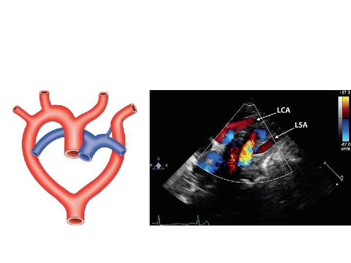
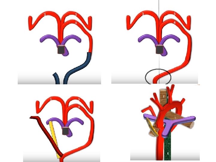
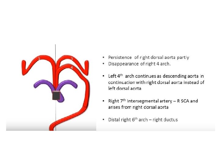
Rt arch with retroesophageal LSCA
Rt arch with retroesophageal LSCA
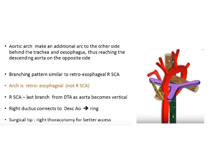
Rt Arch with diverticulum
Circumflex aorta with Lt Arch
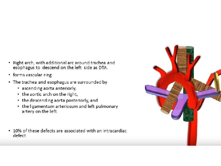
Circumflex aorta with Rt arch
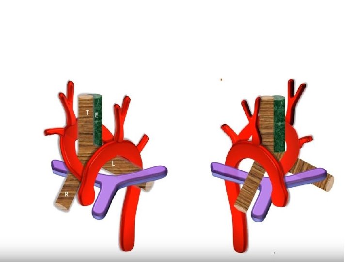
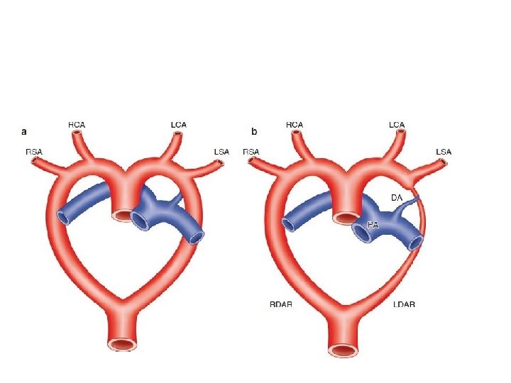
Double aortic arch • Both R and L arches persist • Most common of the vascular rings(55%) • Types (1) Balanced - both arches dominant(equal size) (2) Right Arch dominant and Left partially atretic (3) Left Arch dominant and right partially atretic • Not usually a/w other cong. heart defects • Trisomy 21, 18, TOF, VSD -Only in 10 %
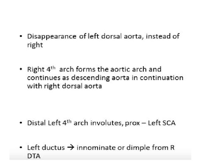
Embryology
Double aortic arch with Type A of IAA
DAA with Type A IAA Similar to RT arch with mirror image branch and left DTA Indistinguishable by imaging
DAA with Type B IAA
Cervical aortic arch
Cervical arch-different types
Cervical aortic arch
Persistent 5 th Arch
Persistent fifth arch
Embryology
Persistent fifth arch with IAA
Interrupted aortic arch
Celoria and patten classification
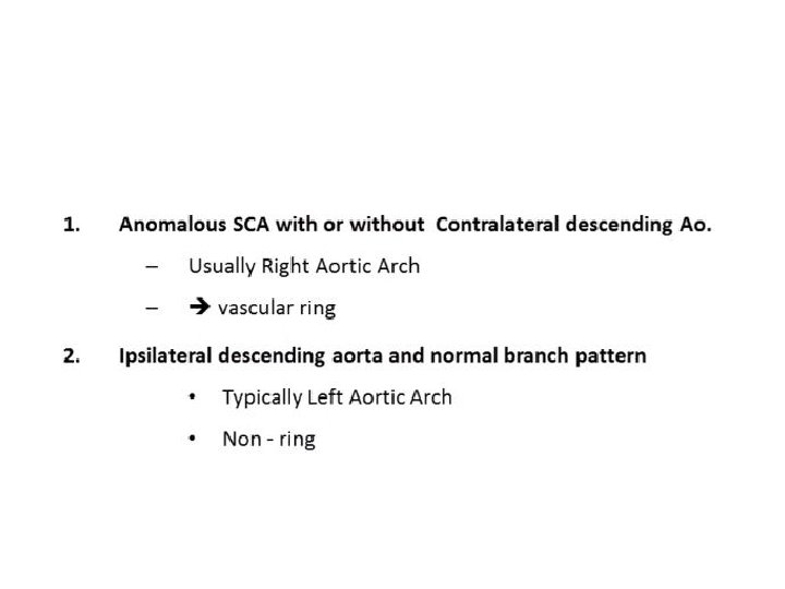
3 Subcategories in each • Without retroesophageal or isolated SCA • With retroesophageal SCA • With isolated SCA
Type A (30 -44%) Aorticopulmonary septal defect and intact ventricular septum, TGA with IVS Type B (51 -70%) • More common • a/w conotruncal anomaly with normally aligned great arteries • Large malalignment-type VSD • Posterior displacement of the infundibular septum and subaortic obstruction Type C Very rare
Type B and Digeorge syndrome • 43% were found to have type B interruption • 68% of interrupted arch patients had Di. George syndrome • Truncus arteriosus - comparable figures were 34% and 33%, respectively
• Presentation Duct dependent obstructive lesion CHF after PDA closure Pulse discrepancy depends on branching pattern Absence of all limb pulses in type B Differential cyanosis rare if Large VSD+
Treatment • PGE 1 • Surgical -Staged repair -Single stage repair LVOT Obstruction Aberrant RSCA
Other anomalies of aortic arch system • 1. Anomalous origin of pulmonary artery from ascending aorta • 2. Anomalous origin of LPA from RPA • 3. Innominate artery compression of trachea
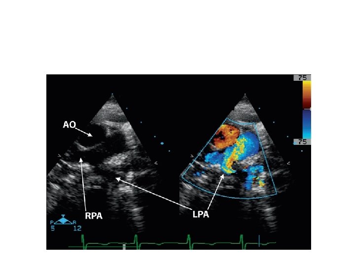
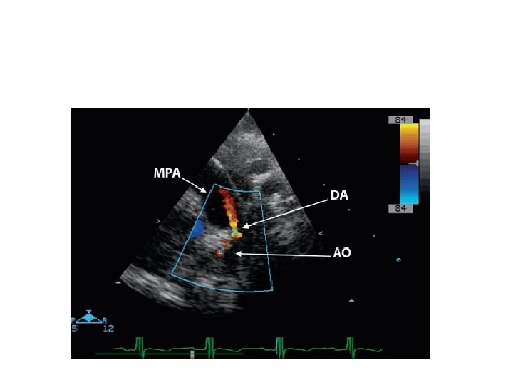
PA from Asc. Aorta • MPA arises from heart • RPA or LPA arises from ascending aorta • RPA-more common -82%
• Anomalous RPA -abnormal migration -aortopulmonary septation anomalies, IAA • Anomalous LPA - Failure to join TA sac -RAA and TOF • Differential pulmonary blood flow • CCF in infancy f/b development of PVR
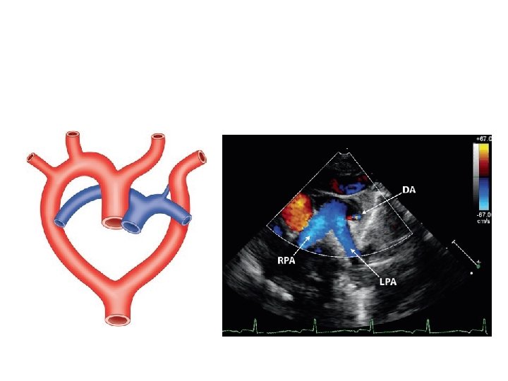
Pulmonary artery sling
• LPA arises as a branch of RPA • LPA courses in a position cephalad to right mainstem bronchus; between trachea & esophagus • Forms a "sling" and partially surrounds lower trachea • a/w tracheal stenosis • ONLY condition where a major vessel passes b/w trachea & esophagus
• Severe respiratory distress and stridor • Symptomatic patients should be evaluated by bronchoscopy at the time of surgical repair frequent association of complete cartilaginous rings • Surgical approach - division of the left pulmonary artery from the right and reanastomosis in front of the trachea
Innominate artery compressing trachea
MCQ 1 • Which of the arches present as pulsatile neck mass • • 1. double aortic arch 2. Persistent 5 th arch 3. Circumflex rt arch 4. Cervical arch
MCQ 2 • Commonest type of IAA is
MCQ 3 • All of the following can be associated with right aortic arch except 1. TOF 2. TGA 3. HLHS 4. DORV
MCQ 4 • Which of the following is true • 1. In circumflex rt arch -arch is on right and DTA is on left • 2. Persistent fifth arch is characterised by two lumens on opposite side • 3. In DAA with IAA, Right arch is more commonly atretic • 4. Right arch with mirror image branch is not always associated with Congenital heart disease
THANK YOU