Antimicrobial Photodynamic Therapy as an Adjunct to Non

Antimicrobial Photodynamic Therapy as an Adjunct to Non. Surgical Treatment of Deep Periodontal Pockets : A Clinical Study Dr. Farrukh Faraz Assoc. Prof. Maulana Azad Institute of Dental Sciences. New Delhi (India)

Historical perspective Use of light as therapy dates back to ancient Egyptian, Indian & Greek civilizations. Danish physician Niels Finsen invented Finsen Lamp. John Toth of Cooper Medical devices, acknowledged the “ photodynamic Chemical Effect” of therapy using early clinical Argon Lasers.

INTRODUCTION q PATHOGENIC BACTERIA ARE THE MAIN CAUSE OF TREATMENT FAILURE IN DENTISTRY! q THE BIOFILM PROVIDES A SAFE HABITAT FOR THEM. q QUORUM SENSING ENABLES BACTERIA TO COMMUNICATE WITH ONE ANOTHER AND COORDINATE THEIR ACTIVITIES. q THE MORE MATURE THE BIOFILM, THE MORE RESISTANT THE BACTERIA WITHIN IT BECOME.

Rationale of Conventional Treatment for Chronic Periodontitis Decreased Bacterial load Supra- and Sub-gingival debridement Disease Progression stops
 § Removes plaque and calculus (sub-and supragingival)")
Treatment options Mechanical debridement (closed / open) § Removes plaque and calculus (sub-and supragingival) § Has no influence on the germ spectrum, can cause hard tissue damage, can be painful Rinsing solutions § Can remove superficial or unattached bacteria § Are rinsed out from the sulcus, can cause staining & taste dysfunction Antibiotics § Side Effects § Interaction with other medicines § Risk of Resistance

Adjunctive modalities • Local and systemic antibiotics. • Laser assisted pocket therapy • Antimicrobial Photodynamic Therapy

Local Antibiotic s Insufficient concentration of the drug in GCF Systemic Antibiotics Disturbance of intestinal microflora Antibiotic resistance Plaque Biofilm confers resistance
, also known as photo radiation therapy, phototherapy or photo")
Photodynamic Therapy Photodynamic therapy (PDT), also known as photo radiation therapy, phototherapy or photo chemotherapy is a new treatment modality that has been developing rapidly within various medical specialties since the 1960 s.

Photodynamic Therapy It has been defined as “the light induced athermal inactivation of cells, microorganisms or molecules. ” REDUCES PATHOGENIC BACTERIA BY > 99% STOPS INFLAMMATION/INFECTION

Mode of Action of a. PDT By using a colorant, the bacteria are: • stained • sensitized • destroyed after irradiation with light of a suitable wavelength and energy density
 which absorbs light can be preferentially taken up by")
PHOTODYNAMIC THERAPY An agent (photosensitizer) which absorbs light can be preferentially taken up by bacteria It is subsequently activated by light of appropriate wavelength, in the presence of oxygen, Photo activation by light of 660 nm Generation of singlet oxygen and free radicals Cytotoxic to microorganisms Dye left for 3 minutes Injection of dye into the pocket

The chemical/physical process is carried out in 3 steps……

Step 1: Staining the microorganisms Diffusion-determining step Migration and attachment of the dye molecules on the wall of microorganisms Process parameters are viscosity, p. H-value, temperature, charge, time, structure of the plaque etc.

Step 2: Exposure and activation of the photosensitiser Laser light at 660 nm Excited stained molecules Absorptionn bacterium Energy-controlled step Determined by physical-optical properties With excitation of the sensitizer molecules From singlet state to triplet state Process parameters are optical, electronic/ chemical states, p. H-value, time etc.

Step 3: Oxygen radical formation & destruction of the microorganisms Local oxygen bacterium Singlet oxygen Exposure and activation of photosensitiser leads to the build-up of singlet -oxygen radicals and oxidative destruction of membrane lipids and enzymes

In principle, all molecules are affected by oxygen radicals. Unsaturated fatty acids in the bacterial membranes are particularly susceptible to damage by oxygen radicals, as these do not have the body’s own enzyme defences as is the case with healthy cells. It is thus specifically the pathogenic bacteria that are destroyed by antimicrobial photodynamic therapy. Healthy cells are at no risk!

3 D Pocket/Endo Probe: For infected Pocket/canals For three dimensional, even exposure • With overload protection through flexible design Micro-lens for even light distribution • Angled for optimum access to all areas, even distal • Sterile disposable product to prevent spread of bacteria • Power density > 60 m. W/cm 2 3 D Pocket / Endo Probe

3 D Pocket/Endo Probe - Indications Periodontal Pocket Periimplantitis Alveolar ostitis Endodontics

What is achieved by the photobiological effect? Inflammation is suppressed Wounds heal faster Pain is reduced

How does the photobiological effect work? The mitochondria in the cells are stimulated. Inhibiting influences on the metabolism and the energy supply are reduced.

Why does the photobiological effect work? Radiation with blue to red laser light (wavelength 380 nm to 700 nm) stimulates synthesis from ADP to ATP through oxidative phosphorylation.

Simplify your therapy • No eye protection needed • No warning signs • … no other preventive measures necessary!!! => therapy is delegable!!!

AIM To evaluate the additional benefit of antimicrobial photodynamic therapy, if any in the non-surgical treatment of deep periodontal pockets.

MATERIALS AND METHODS Prospective, split mouth, controlled clinical trial. 30 adult patients (21 M+9 F)

SAMPLE SELECTION Inclusion criteria • Adults, with age above 18 years. • Presence of at least 2 teeth in different quadrants with probing depth ≥ 6 mm, and bleeding on probing.

SAMPLE SELECTION Exclusion criteria • Systemic conditions which can modify the outcome of treatment, like uncontrolled diabetes mellitus. • Any acute systemic disease/ life threatening disease. • Pregnant or lactating mothers. • Any history of smoking or smokeless tobacco use. • History of antibiotic use in last 1 month. • Undergoing long term treatment with immunosuppressant, NSAIDs, phenytoin, anticoagulants or calcium channel blockers. • Presence of carious lesion or restoration on the root surface

MATERIALS The Photodynamic laser system includes…. 1. A battery-operated diode laser (HELBOs minilaser 2075 F dent, , Bredent Medical, Germany). • Wavelength of 660 nm • Power density of 75 m. W/cm 2 2. A fiber optic applicator (HELBO 3 D Pocket Probe, Bredent Medical, Germany).
")
MATERIALS 3. Photosensitizer- Phenothiazine chloride 10 mg/ml (HELBO Blue, Bredent Medical, Germany)
 • Modified Sulcular Bleeding Index")
CLINICAL PARAMETERS • Plaque Index ( Silness & Loe-1964) • Modified Sulcular Bleeding Index ( Mombelli- 1987) • Probing Pocket Depth • Clinical Attachment Level

STUDY METHODOLOGY Selection of patients based on inclusion criteria and after informed consent (n= 30) Recording of baseline clinical parameters : m. SBI, PD, CAL 2 sites with deepest probing depth selected, and stents were fabricated Full mouth SRP Flip of Coin Test site Control site Photodynamic therapy & Oral Hygiene instructions Follow up at 1 month & 3 months : recording of clinical parameters Statistical Analysis

Steps in PDT Photosensitizer dye injected in the pocket and left for 3 minutes Pocket irrigated with saline Diode laser applied for 10 seconds X 6 sites

FOLLOW UP …. after 1 week to reinforce oral hygiene instructions. & After 1 month & 3 months of therapy to record Clinical parameters Modified Sulcular Bleeding Index ( Mombelli- 1987) Plaque Index ( Silness & Loe-1964) Probing Pocket Depth Clinical Attachment Level

COMPARISON OF MEAN VALUES OF PARAMETERS AT BASELINE Parameters SRP+a. PD p- value T Mean values of Parameters at Baseline 10 9 8 7 6 5 4 3 2 1 0 8. 46 8. 7 7. 53 7. 9 1 1. 09 1. 17 1. 28 PI Time Interval m. SBI PD CAL SRP+PDT PI 1± 0. 45 1. 09± 0. 47 0. 07 m. SBI 1. 17± 0. 38 1. 28± 0. 41 0. 06 PD (mm) 7. 53± 1. 31 7. 90± 1. 71 0. 26 CAL (mm) 8. 46± 1. 81 8. 7± 1. 91 0. 27

Comparison of mean values of PI at Baseline, 1 month and 3 months Baseline 1 month 3 months 1. 2 Mean values of PI 1 1 SRP+a. PDT p-value 1± 0. 45 1. 09± 0. 47 0. 07 0. 57± 0. 40 0. 54± 0. 41 0. 25 0. 46± 0. 17 0. 47± 0. 18 0. 32 1. 09 0. 8 0. 57 0. 54 0. 6 0. 47 0. 4 0. 2 0 Baseline Time Interval 1 month 3 months SRP+PDT

Comparison of mean values of m. SBI at Baseline, 1 month and 3 months Baseline 1 month 3 months 1. 4 Mean values of m. SBI 1. 2 1. 17 SRP+a. PDT p-value 1. 17± 0. 38 1. 28± 0. 41 0. 06 0. 63± 0. 40 0. 43± 0. 42 0. 02 0. 46± 0. 27 0. 31± 0. 30 0. 03 1. 28 1 0. 8 0. 63 0. 6 0. 43 0. 46 0. 31 0. 4 0. 2 0 Baseline Time Interval 1 month 3 months SRP+PDT

Comparison of mean change in m. SBI SRP+a. PDT p-value 1 month 0. 54± 0. 47 0. 85± 0. 41 0. 002 3 months 0. 73± 0. 42 0. 97± 0. 45 0. 015 1. 2 0. 97 Reduction in m. SBI 1 0. 85 0. 73 0. 8 0. 6 0. 54 0. 2 0 1 month Time Interval 3 months SRP+PDT

Comparison of mean values of PD at Baseline, 1 month and 3 months Baseline 1 month 3 months 9 Mean values of PD (mm) 8 7. 53 SRP+a. PDT p-value 7. 53± 1. 31 7. 90± 1. 71 0. 26 6. 17± 1. 20 6. 27± 1. 44 0. 64 5. 37± 0. 85 5. 43± 1. 36 0. 77 7. 9 7 6. 17 6. 27 5. 37 5. 43 6 5 4 3 2 1 0 Baseline Time Interval 1 month 3 months SRP+PDT

Comparison of mean change in PD SRP+a. PDT p-value 1 month 1. 3± 0. 95 1. 77± 0. 86 0. 01 3 months 2. 13± 1. 22 2. 37± 1. 10 0. 47 2. 37 2. 5 2. 13 Reduction in PD (mm) 2 1. 5 1. 77 1. 3 1 0. 5 0 1 month Time Interval 3 months SRP+PDT

Comparison of mean values of CAL at Baseline, 1 month and 3 months 10 9 SRP+a. PDT p-value Baseline 8. 47± 1. 81 8. 70± 1. 91 0. 54 1 month 7. 53± 1. 52 7. 43± 1. 43 0. 74 3 months 6. 73± 1. 31 6. 67± 1. 45 0. 83 8. 47 8. 7 7. 53 7. 43 Mean values of CAL (mm) 8 7 6. 73 6. 67 6 5 4 3 2 1 0 Baseline Time Interval 1 month 3 months SRP+PDT

Comparison of mean change in CAL SRP+a. PDT p-value 1 month 0. 97± 0. 96 1. 27± 1. 11 0. 12 3 months 1. 73± 1. 26 2. 10± 1. 35 0. 18 2. 5 2. 1 Improvement in CAL (mm) 2 1. 73 1. 5 1 1. 27 0. 97 0. 5 0 1 month Time Interval 3 months SRP+PDT

DISCUSSIO N Deep periodontal pockets were selected as they are difficult to be debrided completely by SRP alone. (5 mm) Antimicrobial PDT was done after haemostasis was achieved as heavy bleeding after SRP may prematurely flush the photosensitizer out of the pockets

DISCUSSIO N At the baseline no statistically significant difference between two groups in terms of PI, m. SBI, PD and CAL was recorded. All these parameters improved in both treatment groups after 1 month and 3 months post-treatment interval as observed in many previous studies.

DISCUSSIO N Larger reduction in m. SBI in SRP+a. PDT group, statistically significant at both 1 month and 3 months post treatment interval. a. PDT has a positive effect on patient care, mainly due to the considerably faster resolution of overt inflammation in the gingival tissues, as a result of bacterial load reduction and inactivation of bacterial virulence factors and cytokines when the methylene blue is irradiated with laser.

DISCUSSIO N Inter- group difference in reduction of PD at 1 month was statistically significant, in favour of SRP+a. PDT group. Plausible explanation being additional shrinkage in inflamed pocket wall after reduction in microbial load achieved by photodynamic therapy.

DISCUSSION Incomplete debridement in deep pockets might have prevented significant reduction in PD.

Advantages of a. PDT Photodynamic therapy damages only the pathogenic bacteria sparing the host cells. Unsaturated fatty acids in the bacterial membranes are particularly susceptible to damage by oxygen radicals, as these do not have the body’s own enzyme defences as is the case with healthy cells.

Advantages of a. PDT It significantly reduces inflammation without antibiotics and without surgical intervention while providing maximum therapeutic safety. PDT is safe for both human tissue and dental materials. Because it utilizes cold laser light for activation of the sensitizing solution, there is no risk of thermal damage to patient tissues, prosthetics or implants.

Other Advantages Antimicrobial PDT has other advantages as well, like: 1. Broad spectrum of antimicrobial effect 2. No development of resistance 3. Easy application 4. No repetition required until BOP reappears.

Conclusion It would be worthwhile to include larger sample and follow the outcomes for longer periods to assess the outcome in terms of CAL gain.
 plays an important adjunctive role in improvement of")
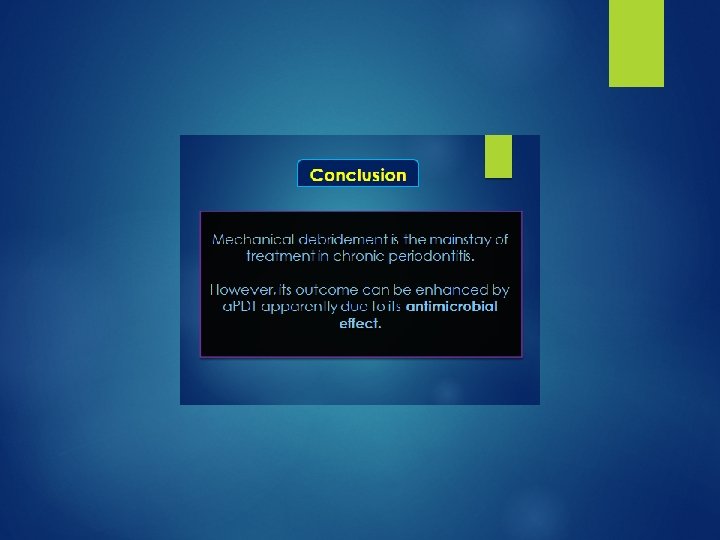
Conclusion Antimicrobial Photodynamic Therapy (a. PDT) plays an important adjunctive role in improvement of bleeding on probing obtained through Scaling and Root Planing (SRP) in deep periodontal pockets.


“ Thank You ”

 improved significantly in both groups, however, inter-group difference")
DISCUSSIO N Clinical Attachment Level (CAL) improved significantly in both groups, however, inter-group difference was not significant either at 1 month or 3 months confirming results of study by Balata et al (2013) They had evaluated the parameters in deep periodontal pockets (PD ≥ 7 mm), similar to present study.

Comparison of mean change in PI SRP+a. PDT p-value 1 month 0. 43± 0. 50 0. 48± 0. 60 3 months 0. 47± 0. 53 0. 62± 0. 47 0. 59 0. 12 0. 7 0. 62 Reduction in PI 0. 6 0. 5 0. 43 0. 48 0. 47 0. 4 0. 3 0. 2 0. 1 0 1 month Time Interval 3 months SRP+PDT

Conclusion Antimicrobial PDT seems to have little role in improvement of Clinical Attachment Level.

DISCUSSION Bactericidal activity of a. PDT explains the additional improvement in bleeding scores

Effect on Biofilms reduce the effectiveness of PDT , but not as much as reported in cases of antibiotics. Fontana et al. (2009) • 63% killing of bacteria in suspension, while • 32% killing in biofilms made of the samples.
- Slides: 60