ANTIGEN ANTIBODY REACTIONS AgAb reactions occur continuously in

ANTIGEN – ANTIBODY REACTIONS

Ag-Ab reactions occur continuously in vivo within the host, forming the basis of humoral immunity Given appropriate conditions & necessary reagents, Ag-Ab reactions may occur in vitro, in the laboratory which are termed as “Serological Reactions”

Uses of Serological Reactions Diagnosis of infectious diseases by demonstration of either Ag or Ab in body fluids Help in confirmation or give additional information of a disease Help in evaluation of different stages of a disease, the prognosis & also efficacy of treatment Help in identification of various pathogens (serotyping)

General properties & terms used The reaction between an Ag & its corresponding Ab is specific, i. e, the Ab combines only with the Ag that was responsible for its formation. This ‘specificity’ is not absolute as closely related Ag’s may react with the same Ab’s, resulting in what are called ‘cross reactions’. ‘Specificity’ or a ‘Specific test’ – refers to the ability of a test to detect only those reactions which occur between homologous Ags and Abs and no other.

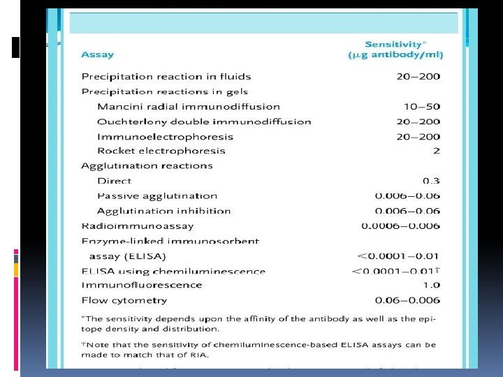
General properties & terms used ‘Sensitivity’ or a ‘Sensitive test’ – refers to the ability of a test to detect even very minute quantities of Ags or Abs ‘Affinity’ -> refers to the intensity of attraction between the Ags and Abs ‘Avidity’ -> refers to the strength of the bond after the formation of Ag-Ab complex

General properties & terms used ‘Qualitative test’ -> is one which detects the presence of Ags or Abs ‘Quantitative test’ -> is one which measures the amount of Ag or Ab present ‘Titre’ -> the highest dilution of the sample which shows a detectable reaction

Stages in Ag-Ab reactions Primary stage: The initial interaction which results in the binding between the Ag and the Ab. Binding is due to non covalent binding forces such as – Hydrogen bonds, Ionic bonds, Van. Der. Waal’s forces. There is no visible effect of this primary stage. This stage is reversible
 events occur due to the")
Secondary stage: Follows the primary stage wherein visible (demonstrable) events occur due to the Ag binding with the Ab. Demonstrable events may include -> - Precipitation - Agglutination - Lysis of cells - Killing of live antigens - Immobilization of motile organisms - Enhancement of phagocytosis

Mechanism of Ag-Ab reactions Marrack’s Lattice Hypothesis: 1. Multivalent Ags combine with bivalent Abs in varying proportions depending on the amount of each present, in the reaction mixture 2. A visible reaction such as precipitation/agglutination occurs only when a large lattice (network) of alternating Ag & Ab molecules is formed 3. This phenomenon occurs only when the Ags and Abs are in equivalent proportions in the reaction mixture –> “Zone of Equivalence”
,")
Marrack’s Lattice Hypothesis: 4. If Ab molecules greatly outnumber the Ag molecules (Ab excess), then only few valencies of the Ab molecules are bound by Ag – resulting in little or no visible effect as no lattice is formed -> “Prozone phenomenon” 5. If Ab molecules are too few in number when compared to the Ag molecules, again, there is no lattice formed – resulting in little or no visible effect (precipitation /agglutination) -> “Zone of Antigen excess”

Marrack’s Lattice Hypothesis
 Precipitation reactions Agglutination reactions Complement Fixation tests Neutralization tests")
Types of Ag-Ab Reactions (Tests) Precipitation reactions Agglutination reactions Complement Fixation tests Neutralization tests Immunofluorescence tests Enzyme Immunoassays Radioimmunoassay Western Blotting techniques

Precipitation Reactions Definition: When a soluble Ag reacts with its corresponding Ab in optimal proportions at a suitable temperature & p. H and in the presence of electrolytes, the Ag-Ab complexes form an insoluble precipitate. If instead of sedimenting, the precipitate remains suspended as floccules, the reaction is called ‘flocculation’. Mechanism: As explained by Marrack’s Lattice Hypothesis

Precipitation Reactions Factors affecting precipitation reactions: Nature of Ags and Abs Proportion of Ags & Abs present (Zone phenomenon) Temperature & p. H Presence of electrolytes Proper mixing of reagents General applications of precipitation reactions: Highly sensitive, used to detect Ags/Abs (as little as 1 µg of protein) Forensic applications – detection of blood, seminal stains, etc

Precipitation Reactions Precipitation in Solutions Immunodiffusion Immunoelectrophoresis Single ID Double ID In 1 Dimension Precipitation in Gels Electroimmunodiffusion Rocket Electrophoresis in 2 Dimensions Countercurrent IE

Precipitation in Solutions 1. Slide Precipitation/Flocculation test: A drop each of the Ag & Ab are mixed on a slide, resulting in flocculation Eg: VDRL Test for diagnosis of Syphilis 2. Tube Precipitation Tests: Ring precipitation tests: - Ags and Abs are mixed in capillary tubes and a ring of precipitation will appear at the interphase of the two solutions Eg: CRP test, Ascoli’s Thermoprecipitin test (for Anthrax), Lancefield’s grouping of Streptococcus Tube precipitation tests: - Serial dilutions of Ab/Ag are prepared in test tubes & equal volumes of corresponding Ag/Ab is added and then incubated, resulting in precipitation

Precipitation in Gels Precipitation reactions can be conducted in semisolid media prepared from Agarose or Polyacrylamide gels. In gels, Ag & Ab diffuse towards each other and where they meet in optimal proportions, they form a line of precipitate which is clearly visible in the clear gel.

Precipitation in Gels Advantages: The line or band of pptn. Is stable and can be stained & preserved for long durations. As each Ag-Ab reaction gives rise to an individual line of pptn. , the number of Ag’s in a mixture can be resolved. Pptn. In gels also indicate identity, cross reactions, and nonidentity between different Ags.

Types of Precipitation in Gels IMMUNODIFFUSION: When Ags & Abs diffuse passively towards each other in gels to form lines of pptn. Single Gel Diffusion -> When only the Ag or Ab diffuses passively and the corresponding Ab or Ag is incorporated in the gel layer. 1. Single gel diffusion in one dimension 2. Single gel diffusion in two dimensions – Also known as “Radial Immunodiffusion”.

Radial Immunodiffusion The Ab is incorporated into the gel on a slide and the Ag is added onto wells cut into the gel. Ag diffuses out of the well into the gel and reacts with the Ab present, forming a ring of pptn. around the well. Uses – Estimation of Ig classes & Complement system proteins Detection of Abs to Influenza viruses

Radial Immunodiffusion

Double Gel Diffusion -> When both Ags & Abs diffuse passively towards each other in the gel and form lines of pptn where they meet each other in optimal proportions. 1. Double diffusion in one dimension 2. Double diffusion in two dimensions – Also known as the “Ouchterlony procedure”

Ouchterlony procedure

Ouchterlony procedure Here, wells are cut in agarose layers taken on petridishes. Antiserum is added to the central well and different Ags are added to surrounding wells. If two adjacent Ags are identical, the lines of precipitation formed will fuse but if they are unrelated, the lines will cross each other. If Ags are cross reacting or partially related to each other, one line of precipitation might fuse with the other, forming a ‘spur’.

Uses of Double diffusion in two dimensions -> In detecting identity of different Ag preparations Detection of Ag/Ab in unknown preparations To detect Abs against fungal Ags such as H. capsulatum, C. immitis To detect production of Diphtheria toxin production by C. diphtheriae (Elek’s Gel Pptn test)

IMMUNOELECTROPHORESIS In this type of precipitation in gel, the Ags & Abs are first separated (resolved) from a mixture by the help of electrophoretic current. They then diffuse passively towards each other and where they meet each other, they will form lines of pptn. Uses: - To detect presence or absence of serum proteins - For the detection of unusual serum proteins (myeloma proteins) - To detect changes in globulin levels in various diseases

IMMUNOELECTROPHORESIS

ELECTROIMMUNODIFFUSION The process of Ag – Ab reactions and the formation of precipitation lines may be greatly enhanced/speeded up by electrophoretically driving both the Ags & Abs towards each other in gel. Types: One dimensional single electroimmunodiffusion (Rocket immunoelectrophoresis) One Dimensional Double Electroimmunodiffusion (Counter Current Immunoelectrophoresis
 Antiserum (Abs) to particular Ags is incorporated into")
One dimensional single electroimmunodiffusion (Rocket immunoelectrophoresis) Antiserum (Abs) to particular Ags is incorporated into the agarose layer so that it does not migrate. Ags are placed in a well and resolved (separated) with the help of electrophoretic current. Now, the separated Ags diffuse towards the Ab in the gel, and form lines of pptn, which resemble spikes or rockets. Use: To quantitate serum Ig & Complement components

Rocket immunoelectrophoresis
 In this method, both Ags & Abs")
One Dimensional Double Electroimmunodiffusion (Counter Current Immunoelectrophoresis) In this method, both Ags & Abs are electrophoretically driven towards each other in gel with resultant precipitation at a point intermediate between their origins. Advantages: - Rapid tests; Highly sensitive. Uses: 1. To detect HBs Ag in serum 2. To detect Capsular Ags of C. neoformans, H. influenzae in CSF

Counter Current Immunoelectrophoresis

Agglutination Reactions Definition: When particulate Ags combine with their specific Abs at optimal proportions in the presence of electrolytes and a suitable p. H, the Ag-Ab complexes formed will get clumped to form agglutinates – the reaction is known as Agglutination reaction. Mechanism of Agglutination: Same as pptn reacs, as explained by the Marrack’s Lattice Hypothesis. The Zone phenomenon (Prozone) occurs even in Aggln reacs. If incomplete Abs or Blocking Abs are present, they will react with the Ag but this will not result in Agglutination.

Types of Agglutination reactions & their applications Agglutination tests are highly sensitive in detecting Abs and therefore, they are usually used in the detection of Abs to various infectious agents in the patients clinical samples such as serum, CSF, etc. 1. Direct (active) agglutination tests 2. Indirect/passive agglutination tests 3. Reverse passive agglutination tests 4. Agglutination inhibition tests
 AGGLUTINATION TESTS Slide Agglutination tests: - A drop each of the")
A. DIRECT (ACTIVE) AGGLUTINATION TESTS Slide Agglutination tests: - A drop each of the Ag & Ab is mixed on a slide, resulting in agglutination. Uses: - Bacterial slide agglutination tests for identification of bacteria - Blood Grouping (using RBC’s – haemagglutination test)
 containing the")
• Tube Agglutination tests: Serial dilutions of the clinical sample (serum) containing the Abs are made in test tubes and equal volumes of Ag suspensions are added to these tubes. These tubes are now incubated in a water bath or incubator at a suitable temperature for an appropriate period of time after which agglutination is observed in the tubes. Because of the serial dilutions, these tests provide information regarding the amount of Abs present and not just their presence – called the ‘Quantitative tests’

Examples of tube agglutination tests : - 1. Bacterial agglutination tests –> - Widal test for serodiagnosis of Enteric Fever - Brucella agglutination test for diagnosis of Brucellosis - Weil-Felix agglutination test for Rickettsial fevers

2. Hemagglutination tests –> Here, different species of RBC’s are used as Ags to detect Abs in Patient’s serum Paul Bunnel test which uses sheep RBC as Ag, is used to diagnose Infectious Mononucleosis Cold agglutination test which uses human ‘O’ blood group RBC as Ag and where the incubation temperature is 4 o C – used in diagnosis of Primary Atypical Pneumonia (Cold Agglutinins – Ig. M Abs capable of agglutinating human RBC between 0 – 4 O C, found in Prim. Atyp. Pneum. , Malaria, Trypanosomiasis & Acquired Hemolytic Anemia)

Coomb’s test Principle -> A test used for the detection of incomplete Anti Rh Abs which have the ability to react with Rh +ve RBC’s but which do not have the ability to agglutinate them. When such Anti Rh Ab coated RBC’s are treated with Antiglobulin (Ab to Human Gamma Globulin, Coomb’s serum), the antiglobulin reacts with the incomplete Abs coating the RBC’s bringing about agglutination.

Coomb’s test Types -> Direct – This test is used to diagnose Hemolytic disease of the newborn which occurs due to Rh incompatibility. Here, sensitization of the fetal Rh+ve RBC’s occurs in vivo, in the womb, by the mother’s incomplete anti Rh Abs. Indirect – This test is used to detect Anti. Rh Abs in the mother’s serum. Here, Rh+ve RBC’s are mixed with the mothers serum so that if Anti Rh Abs are present, they will bind to the RBC’s. In the 2 nd step, Coomb’s serum is added which brings about agglutination of the RBC’s.

Coomb’s test

B. INDIRECT/PASSIVE AGGLUTINATION TESTS When soluble Ags are attached onto the surfaces of Carrier particles thereby converting them into particulate Ags, which then react with their corresponding Abs leading to agglutination. Here, the carrier particles are passively agglutinated as they themselves are not involved in the Ag-Ab reaction. Thus, precipitation reactions may be converted into agglutination reactions by converting soluble Ags into particulate Ags.

Carrier particles used -> Latex beads, Bentonite beads, Carbon particles, etc. When RBC’s are used, the reactions are called - ‘Passive Haemagglutination tests’ Egs: - Latex agglutination tests -> ASO test (Rheumatic fever), RA test (Rheumatoid Arthritis), CRP test - Passive hemaggln tests -> TPHA (Syphilis) Rose -Waaler’s test (R A) - Using Carbon Particles -> RPR test (Syphilis)

Passive Agglutination Test

C. REVERSE PASSIVE AGGLUTINATION TESTS Principle -> Abs are attached to carrier particles instead of Ags and this complex is used to detect Ags in biological fluids Egs -> Using RBC’s (Reverse passive hemaggln) – HBs. Ag detection in serum Using latex beads (Reverse passive latex aggln) – HBs. Ag detection in serum Co-agglutination test – Here, killed & stabilized Staphylococcus aureus ‘Cowan strain I’ cells are used as carrier particles.

Co-agglutination test Reason for using Staph aureus cells is because the cell wall of this bacterium contains ‘Protein A’ which has high affinity for the Fc portion of the Ig molecule. Therefore, Ab molecules bind to these cells with great affinity/avidity through their Fc portion and this reagent can be used to detect Ags in biological fluids.

Co-agglutination test

Co-agglutination test Egs : - Detection of capsular Ags of -> meningococcus, pneumococcus & cryptococcus in CSF - Detection of Pneumococcal Ag in sputum - Detection of ‘Vi’ Ag of Salmonella in urine - Lancefield’s grouping of Streptococcus

D. AGGLUTINATION INHIBITION TESTS Principle -> Agglutination of particles such as RBC’s by agglutinating Ags is inhibited by Abs present in the clinical sample such as serum. ‘Haemagglutination inhibition tests’ are used routinely in virology, to detect Abs against various hemagglutinating viruses such as - Arboviruses, Influenza viruses, Measles virus & Rubella virus.

Procedure -> The Patients serum is mixed with RBCs and the virus particles against which Abs are to be detected in the serum and incubated. The virus would cause haemagglutination of the RBCs. But if Abs were present, they would bind to the virus and neutralize them. Therefore, if no haemagglutination is observed, test is positive, Abs present. If haemagglutination is observed, then test is negative, Abs absent.

Agglutination inhibition test
 Definition: - A serological test which is used to detect")
Complement Fixation Tests (CFT) Definition: - A serological test which is used to detect and quantitate either Ags or Abs in serum samples through activation of the Complement system. Principle: - Whenever Ag – Ab reacs take place in the presence of Complement (C), the C gets activated/fixed by the Ag-Ab complex, resulting in various biological effects of activation of C such as – lysis of bacterial, tumor or RBC’s, - enhancement of phagocytosis, - tissue damage in hypersensitivity reactions, etc “The ability of C to lyse RBC’s when activated by an Ag-Ab reaction is used in the CFT’s”

Reagents used -> 1. The ‘Ag’ against which the Abs are to be detected 2. Patient’s serum – containing the Abs which are to be detected 3. Complement – source is the Guinea pig serum which is rich in Complement proteins 4. Indicator (Hemolytic) system – A readymade Ag-Ab complex, the Ag being sheep RBC and the Ab being anti sheep RBC ab’s (antisheep RBC Abs are also known as ‘AMBOCEPTOR’ and they are raised in rabbits)

Procedure -> Test is performed in two stages. 1 st step – To patient’s serum is added the Ag & C. This mixture is incubated for 1 hr at 370 C. 2 nd step – After incubation, the indicator/hemolytic system is added to the above mixture and incubated again for 1 hr at 370 C. After the incubation period, the mixture is observed for hemolysis, if any.
 Explanation: -")
Observation -> Absence of Hemolysis – indicates a positive test (Abs present) Explanation: - If patient’s serum had contained Abs, they would have reacted with the added Ag to form Ag-Ab complexes which in turn, would have activated/utilized all the C present. In the 2 nd step when the hemolytic system was added, as there was no free C present, there would be no hemolysis of the sheep RBC. Thus, absence of hemolysis – indicated presence of Abs in patient’s serum.
 Explanation: - If patient’s")
Hemolysis present – indicates a negative test (Abs absent) Explanation: - If patient’s serum had not contained any Abs, no Ag-Ab reaction would have occurred and the C present would not have been utilized after the completion of the 1 st step. In the 2 nd step when the hemolytic system was added, which was a readymade Ag-Ab complex, this would have activated the free C still present after the first step, resulting in hemolysis of the sheep RBC’s of the hemolytic system. Thus presence of hemolysis – indicated absence of Abs in the patients serum.

Complement Fixation Test

Applications of CFT -> Used in diagnosis of various Viral, Parasitic, Fungal & Bacterial infections Formerly used in the diagnosis of Syphilis – the Wasserman’s Complement Fixation Test

Neutralization Tests Definition -> Tests which detect the presence of Abs by their ability to neutralize the biological effects of microbes or their products such as toxins or enzymes. Types -> 1. Virus neutralization tests 2. Toxin neutralization tests

1. Virus neutralization test Principle -> Abs in patient’s serum are detected by their ability to neutralize the invasiveness or infectivity of viruses. Procedure -> A viral suspension (against which Abs are to be detected) is mixed with patient’s serum & incubated. Then the virus in this mixture is inoculated into cell cultures / embryonated eggs / experimental animals and observed for signs of growth of the virus (CPE in cell cultures/disease or death of embryo/animal).

Observation -> Viral growth -> indicates absence of neutralizing Abs in pt’s serum, test negative. No viral growth -> indicates that neutralizing Abs present in pt’s serum had neutralized the infectivity of the virus, test positive. Uses -> Used in diagnosis of most viral infections

2. Toxin Neutralization tests Principle -> Abs in patients serum are detected by their ability to neutralize the toxic effects of various toxins either in vivo or in vitro. In vivo tests: Schick test – to detect susceptibility to diphtheria Dick test – to detect susceptibility to scarlet fever Schultz-Charlton reac – for scarlet fever Toxin-antitoxin tests in animals – for determination or standardization of toxins/antitoxins In vitro: - Neutralization of Streptolysin O by ASO in pt’s serum

Radio-immunoassay These tests may be used to detect & quantitate Ags that can be radiolabelled. Principle -> It is based on the competetion for specific Abs between a known concentration of radiolabelled Ag & the unlabelled Ag whose presence and concentration in the clinical sample is to be detected. After Ag – Ab reac has occurred, the Ag-Ab complexes are separated from the mixture & the radioactivity present in them is measured.

The strength of radio activity is compared with a standard curve prepared for that Ag. This will provide us an idea about the presence and concentration of the unlabelled Ag present in the clinical sample. Applications -> Assay of drugs & hormones in serum Radioallergosorbent test (RAST) – for detection of Ig E Advantages -> Highly sensitive (upto picogram concentrations) Disadvantages -> Expensive, hazardous, specialized personnel and equipment required.

Immunofluorescence tests Definition -> Tests which detect either Ags or Abs in pt’s serum using fluorescent dye labelled Abs or Ags, respectively. Principle -> Fluorescent dyes (fluorochromes) are bound (tagged) to Ags or Abs and these are then made to react with their respective Abs or Ags in the clinical sample. This preparation is illuminated by UV light in a fluorescent microscope, so that the combination of labelled Ag/Ab to its respective Ab/Ag in the sample may be observed due to fluorescence of the tagged dye.

Egs of Fluorescent dyes -> Fluorescin isothiocyanate Lissamine-Rhodamine Types of Immunofluorescent tests -> Direct Immunofluorescence tests (Fluorescent Ab tests) : - Principle : Ags in clinical samples are detected using fluorochrome labelled Abs
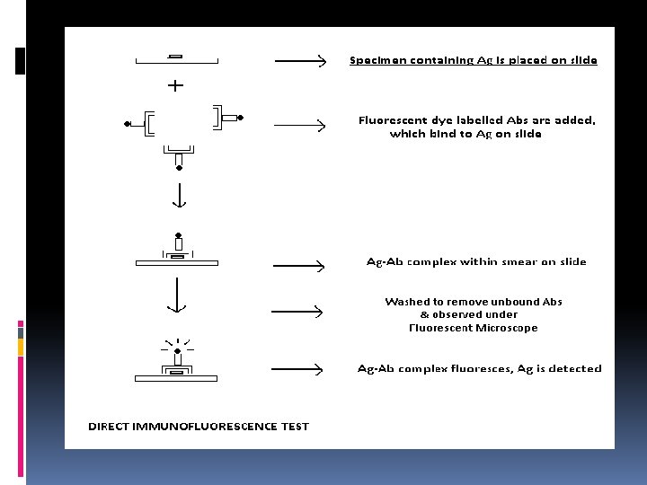
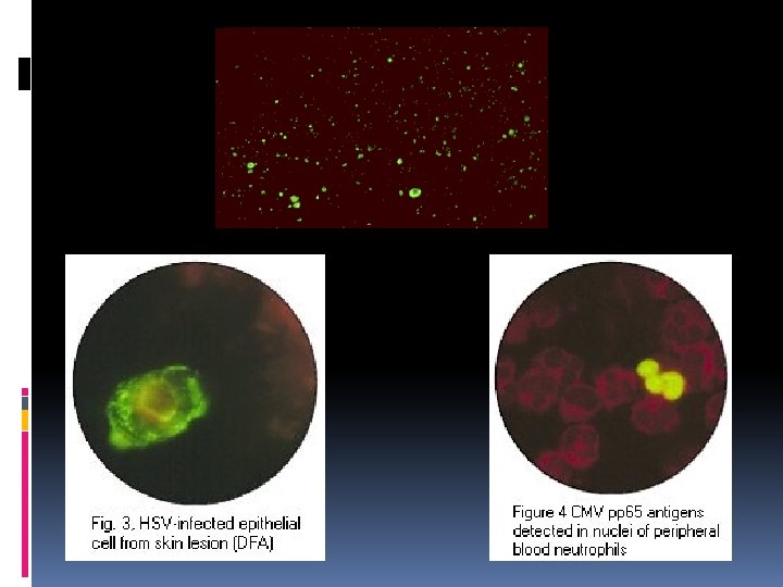

Applications: - Detection of Viral Ags in samples (rabies Ag in corneal impression smears, HSV, CMV Ag detection, etc) - Detection of Bacterial Ags (T. pallidum in serous exudate, chlamydial Ag in urethral exudate, etc) - Detection of Parasite Ags (Cryptosporidia, Pneumocystis) Advantages: - Rapid diagnosis; Diagnosis in acute stage of infection; Diagnosis where culture is not possible

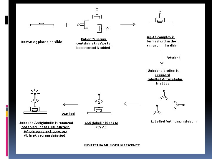
• Indirect Immunfluorescence Tests -> Principle: Abs against various infectious agents present in the clinical sample (serum, CSF, etc) are first made to react with known Ags. This Ag-Ab complex formed, is detected with the help of fluorochrome labelled Antiglobulin. Applications: Abs against bacteria -> Treponemal Abs, Rickettsial Abs against viruses -> HSV, EBV, Rubella Abs against Parasites -> Toxoplasma

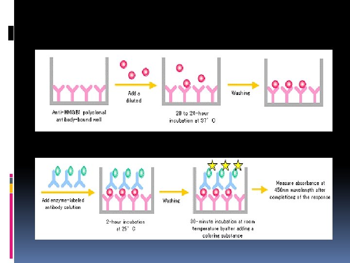
ENZYME IMMUNOASSAYS Principle: These serological tests use Enzyme-Substrate reactions to demonstrate the specific combination of Ags and their Abs, thereby helping in the demonstration of either Ags or Abs Such enzyme-substrate systems consist of -> An enzyme which is linked to either Ag or Ab A substrate which is added after the Ag-Ab reaction. This substrate is broken down by the enzyme molecules bound to either the Ag or Ab in the Ag-Ab complex resulting in a change of colour. The intensity of colour change gives an indication of the amount of bound Ag or Ab whose presence and concentration was to be detected.

Types of Enzyme Immunoassays Homogenous Assays -> Used for the detection & assay of: - Drugs of abuse - Antibiotics - Hormones These tests are competitive, and there is no separation of the Ag-Ab complexes from free Ags & Abs. Also, there is no solid phase used.
 Definition: A type of Enzyme Immunoassay where either Ags or Abs")
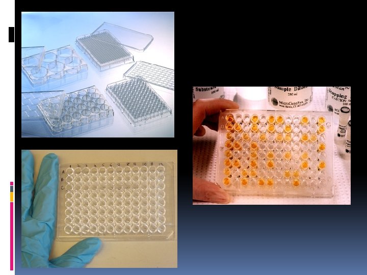
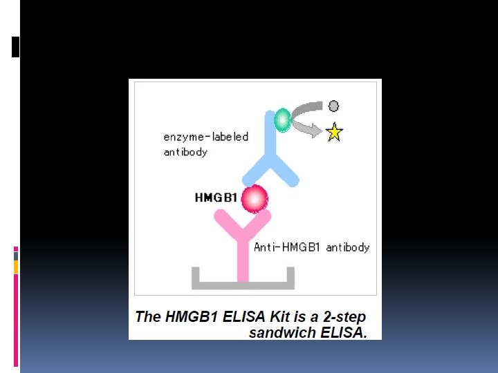
Heterogeneous Assays (ELISA) Definition: A type of Enzyme Immunoassay where either Ags or Abs are bound to a solid phase and the process involves separation of the Ag-Ab complex from free Ags or Abs. Enzyme Linked Immuno Sorbent Assays (ELISA) can be used to detect both Ags & Abs.

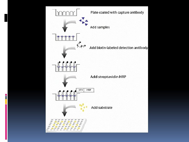
Steps involved: The known Ag/Ab are bound to a solid phase Solid phase is – usually polystyrene (microwells, tubes, beads) Abs used – highly specific monoclonal Abs Enzyme systems used – - Alkaline phosphatase : Paranitrophenyl phosphate - Horse Radish Peroxidase : O-phenylene diamine dihydrochloride Change in colour of reaction mixture – spectrophotometer

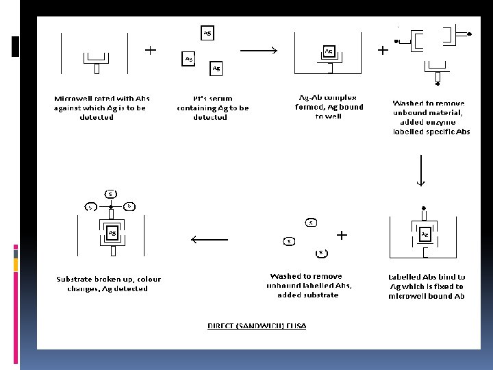
DETECTION OF ANTIGENS Ags are detected by “Sandwich ELISA” technique or Direct ELISA As Ag to be detected will be sandwiched between two molecules of Ab, the name ‘Sandwich ELISA’ is used. Applications of Direct/Sandwich ELISA: Detection of viral Ags – Rotavirus in Stool samples Detection of bacterial Ags – Chlamydial Ag in Urethral exudates

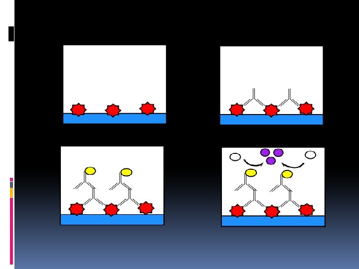
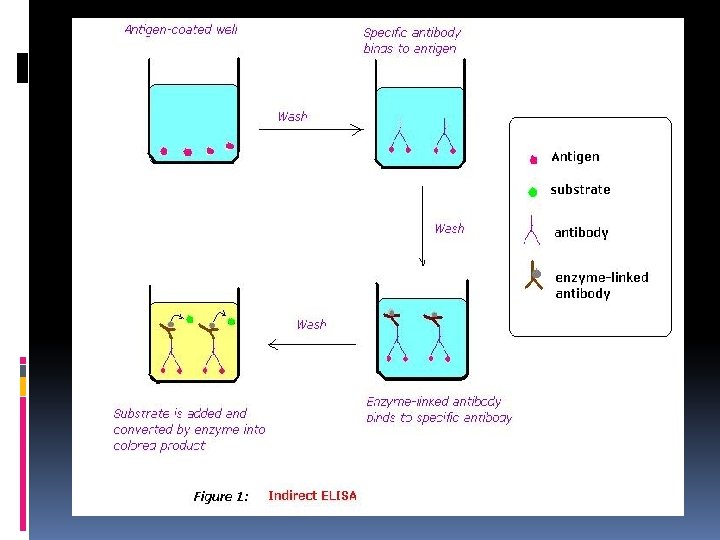
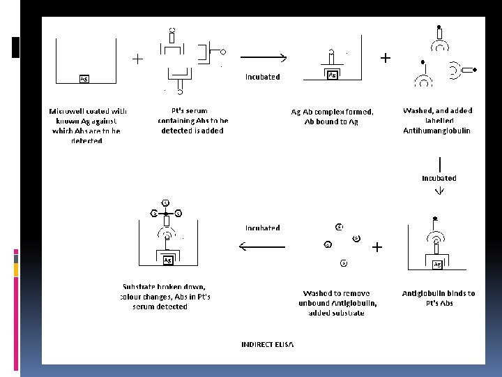
DETECTION OF ANTIBODIES Also referred to as ‘Indirect ELISA’ Antibodies in clinical samples are detected using enzyme labelled Antiglobulin. Used in serodiagnosis of several viral, parasitic and bacterial infections. Viral infections -> HIV, HBV Bacterial infections -> Tuberculosis, Chlamydial infections Parasitic -> Toxoplasmosis Advantages: - Reproducible, sensitive, inexpensive

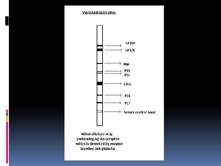
IMMUNOBLOTTING TECHNIQUES Also called ‘Western Blotting’ A highly specific serological technique which involves the detection of Abs against multiple Ags of a single pathogen Procedure: - Separation by SDS-PAGE of different Ags from a crude mixture prepared from an infectious agent - Ags after separation, are transferred to a ‘Nitrocellulose’ membrane (blotting) - Each Ag will be present as a separate band

- Patient’s serum containing Abs is applied over the membrane strip containing the separated Ags - Abs bind to different Ags - This complex is detected using enzyme labelled Antihuman globulin Applications: Used as a confirmatory test for HIV infection & Lyme disease
- Slides: 89