Antibody structure and Humoral Immunity Dr Eman Albataineh

Antibody structure and Humoral Immunity Dr. Eman Albataineh, Associate Prof. Immunology College of Medicine, Mu’tah university Immunology, 2 nd year students

– When blood clot, the remaining fluid called serum which include antibody and serology is any study include serum and antibody detection – 3 g of antibody produced daily and most of them is IGA in GIT and RT secretions – Whereas, In serum, the most distributed antibody is IGG

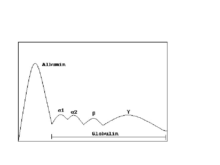
Antibody structure • it is difficult to use normal human blood to study antibody structure as it has variable types or clones of antibodies (different variable regions) so that hypridoma technique that can produce one type of antibody was used • This technique is; stimulation B cells with certain antigen to be antibody producing cells then fuse these cells with cancerous cell plastocytoma (the fused complex called hypridoma) by this we make these B cells to proliferate and continuously producing one type of antibodies called monoclonal antibodies against that antigen • imunoglobulins can be detected in human serum using protein electrophoresis, they are in the band of ϒ globulin

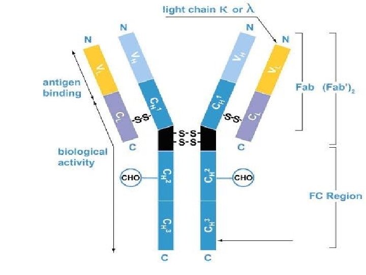
General structure • 4 polypeptide chains, 2 identical heavy chains and 2 identical light chains combined by di-sulphide bonds • Heavy chain constitute of one variable and 3 -4 constant domains depending on the class of immunoglobulin • Constant domains Differ with in heavy chains only between 5 classes of Ig. M, Ig. D, Ig. E, Ig. G, Ig. A • Light chain have one variable and one constant domains and the constant is between the 2 types Kappa and lambda (κ, λ) both of which can associate with the 5 types of heavy chains.


• The N-terminal part; 2 identical antigen binding sites. Each is formed by one variable domain of light and one of heavy domains • Variable domain contribute to variability in antigen receptors with different antigenic specificities • The carboxy terminal consist only constant of heavy chain (Fc part). • Heavy chain determine – the type of antibody – and do the functional effect of Ab; – bind to Fc receptors on innate cells

Antigen binding site • Much of the variation between Igs located in 3 hot spots of hypervariable regions ( HVR 1, 2, 3) or called CDR 1, 2, 3 found in variable region. Because they determine the complementary fit of antibody to antigen, they are referred to as complementarily determining regions or CDR (i. e. , CDR 1, CDR 2, and CDR 3). • antigen combining sites consist of 3 HVR from light and 3 HVR from heavy chains • The most variable is CDR 3 of heavy chain on antibody and BCR and on beta chain on TCR • Each HVR is about 10 amino acid residues in length. The hypervariable regions make up about 15– 20% of a variable domain, with the remaining 80– 85% being made up of four framework regions • Intervening sequences between the CDRs have restricted variability and show little difference in amino acid sequence between chains. These invariant segments make up the framework residues

Hypervariable regions

follow • In human adult there about 107 -9 different antibodies with different antigen combining sites • The constant domains of the heavy chain are called depending on class of antibody (Cϒ for IGG, Cδ for IGD, Cε for IGE, Cμ for IGM and Cα for IGA), little difference in structure • Constant L chains (2 classes)- one or the other not both Lambda (λ) [40% in humans] and Kappa (κ) [60% in humans]

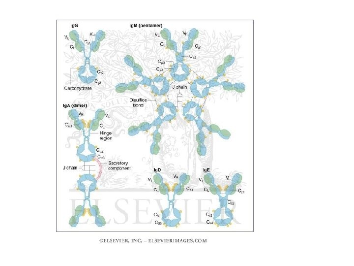
Further additions on the structure • Antibodies also demonstrate segmental flexibility, which means that the two Fab portions can move relative to one another on antigen binding. The angle varies from 60 to 180 degrees. This flexible region where the arms meet the stem of the Y is called the hinge region and is located between the CH 1 and CH 2 domains. Only Ig. G, Ig. A, and Ig. D antibody molecules have hinge regions • Ig. M and Ig. A also have a polypeptide called the joining ( J) chain, which is disulfide- linked to the tail of the antibody and stabilizes the multimeric structure. • Secretory part in IGA

• How many molecules can a single antibody molecule bind (i. e how many combining sites does it have, called valency ( in IGM they are 10 sites whereas in IGA are 4 and 2 in IGG, IGE and IGD) • What is the strength of binding of the epitope to single combining site on the antibody molecule called affinity whereas the combining strength of all combining sites to the epitopes on surface of same antigen called avidity

Generation of antibody fragments • Papain enzyme digest the antibody in the n terminal side of the disulphide bonds result in 2 fab and one Fc • Pepsin in the c-terminal side of the bonds and result in f(ab)2 and smaller fc fragments (pfc)

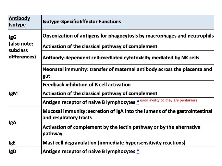
Classes and subclasses • Abs can be classified as isotypes or allotypes or as idiotypes. Her we will use the first system • 5 classes or isotypes; IGG, IGM, IGA, IGE and IGD • IGG into 4 subclasses, IGG 1, 2, 3, 4. IGA into IGA 1, IGA 2 while no subclasses in IGGE, IGM and IGD. all of these classes and subclasses found in every person • Antibody isotypes differ in their chemical (charge, size, and solubility) and serologic (in vitro reactions with antigens) properties,

• . isotypes show functional differences in constant regions of their heavy chains; whereas structures of constant regions are nearly identical except change in 1 amino acid may occur kappa constant chain and gamma constant chain (GM and KM allotypes respectively) the types of allotypes depend on races. • Idiotypic determinants The structure formed by the CDR is known as the idiotope. They are unique to immunoglobulins of particular antigenic specificity. These determinants classify antibodies into idiotypes.


IGM • Ig. M, primary response to antigens, is largest antibody, it is pentamer that makes up about 8% • The H chain have 1 v and 4 c chains • The five monomeric Ig. M molecules are arranged radially, the Fab fragments pointing outward and the Fc fragments pointing to the center of the circle • Because of its many antigen-binding sites, Ig. M can quickly clump antigen (agglutinate) • Ig. M acts as one of the main receptors on the surface of mature B cells, along with Ig. D. When Ig. M is a surface receptor, it is in its monomeric form. • the CH 1 and CH 3 domains are where the J chain binds. The CH 2 domain is equivalent to the hinge regions • The membrane form of Ig. M is made up of additional transmembrane segment • Function; -complement activation • Indirect opsonization • Antigen clumping • In IGA deficiency IGM can appear in secretions linked to secretory piece

Natural antibodies • TI antigens also contribute to the generation of natural antibodies, mainly IGM, Most natural antibodies are low-affinity anticarbohydrate antibodies, postulated to be produced by peritoneal B-1 cells stimulated by bacteria that colonize the gastrointestinal tract and by marginal zone B cells in the spleen.

Natural antibodies

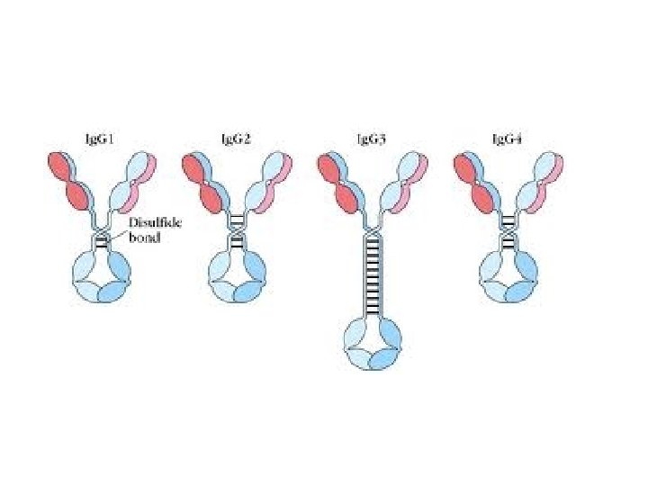
IGG • • • Ig. G, induced by protein antigens, constitutes about 80% (12. 5 mg/ml) of the antibody in serum. The four polypeptide chains are covalently held together by disulfide bonds. Human Ig. G consists of four subclasses (isotypes), which are numbered in order of their serum concentrations (Ig. G 1, Ig. G 2, Ig. G 3, and Ig. G 4). The four subclasses have 90 to 95% identity with each other. The H chain is made up of four domains, one in the V portion and three in the C portion of the chain. The chief distinguishing characteristic among the four Ig. G subclasses is the pattern of interchain linkages in the hinge region. Produced particularly in secondary immune response It’s presence indicate previous exposure and the higher the titer the higher the protection is. It activate the classical complement pathway via Cγ 2 domain bind to complement IGG and IGG 3 interact with the 3 Fc receptors expressed on various cells. Function; – FcγR 1 and 2 and 3 on phagocytes help in phagocytosis, low affinity FcγR 3 on NK help in extracellular killing, (ADCC) – Complement activation – Opsonization – IGG cross the placenta to give babies their immunity – Do neutralization of toxins

• A higher than normal Ig. G antibody level can suggest an Ig. G monoclonal gammopathy, such as multiple myeloma — a cancer of the blood and bone marrow • A lower than normal Ig. G antibody level may suggest some types of leukemia or nephrotic syndrome, which often results in kidney damage.

Complement activator Binds to Fc receptor on phagocytic cells Name Percent Crosses placenta easily Ig. G 1 66% yes (1. 47)† second-highest high affinity Ig. G 2 23% no (0. 8)† third-highest extremely low affinity Ig. G 3 7% yes (1. 17)† highest high affinity Ig. G 4 4% yes (1. 15)† no intermediate affinity †: Quota cord/maternity concentrations blood. Based on data from a Japanese study on 228 mothers.
 of the antibody")
IGA • Human Ig. A constitutes only 13% (2. 1 mg/ml) of the antibody in human serum, but it is the predominant class of antibody in extravascular secretions. The Ig. A present in secretions (tears, saliva, nasal secretions, bronchial and digestive tract mucus, and mammary gland secretions) is secretory Ig. A. • The J chain is synthesized by plasma cells and attaches to Ig. A (or Ig. M) either before or at the time of secretion. The J chain attaches to the carboxyl-terminal of either the a or the m chain. • IGA may be monomeric in serum or dimeric in secretions • The H chain is made up of one V domain and three C domains. Ig. A 1 is the most prevalent form in serum, but Ig. A 2 is slightly more prevalent in secretions. • Another difference between Ig. A allotypes is the size of their hinge regions. • Function; -Bind neutrophils through FCαR 1 and mediate phagocytosis – although most of its protection result from direct neutralization of toxins in gut and RT – Do agglutination of antigens gaining access via mucosa – They secreted in breast milk help in infant immunity

IGD • Ig. D constitutes less than 1% of the antibody in human serum. • Ig. D is an antibody whose function remains unknown, even though it is one of the main receptors on mature B cells and may regulate cell activation. • The d-chain C region is divided into three domains • The hinge region of Ig. D longer than any other antibody class

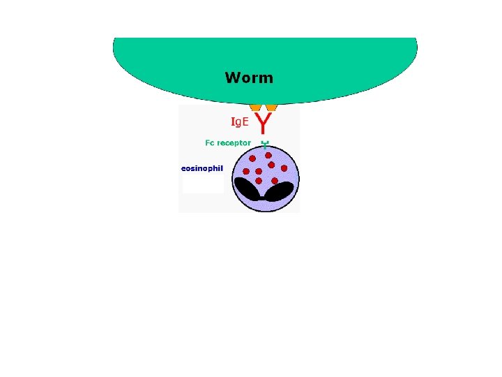
IGE • Human Ig. E makes up less than 0. 003% of the antibody in serum. • Ig. E binds through its high affinity FcεR 1 part to mast cells or basophils. On later exposure to the same antigen, mast cells and basophils bind antigen with Ig. E and trigger allergic reactions. • Ig. E protects against parasites by binding low affinity Fc εR 1 on eosinophils and then releasing mediators (ADCC). • FC εR 2 on B cells • Like the m chain, the e chain contains four C-region domains.

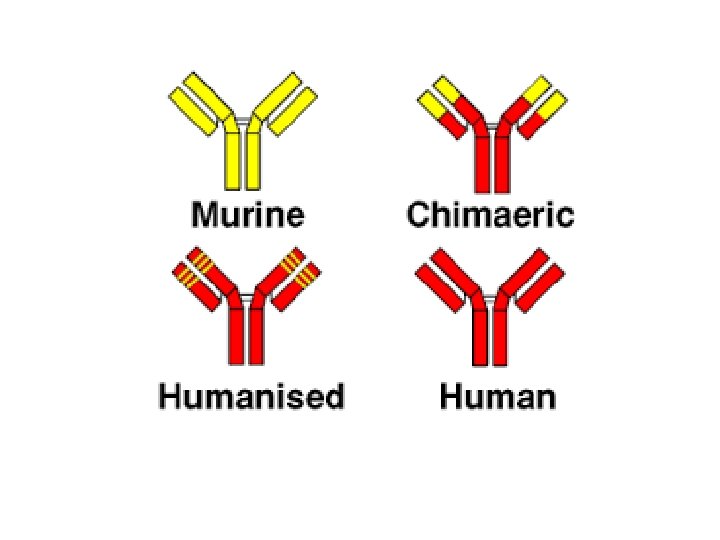
Monoclonal antibodies • Are pure antibodies with single antigen specificity produced from one B cell clone by hypridoma technique • Uses; – Diagnostic uses • Identification and separation of microbe antigens; identification of autoimmune disease, level of vaccination, diagnose immune complex disease, diagnose pregnancy – Therapeutic uses; • • • antitumor therapy alone or with cytotoxic agents (magic bullet), Immunosuppressive; anti-CD 3 (T cell) in graft rejection Neutralize drug toxicity; digitalis Anti RH incompatibility (hemolytic disease of newborn) Passive immunotherapy to protect against vericella zoster and CMV

FC receptors
 are expressed on")
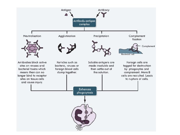
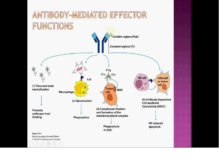
Fc receptors and antibody functions • Immunoglobulin Fc receptors (Fc. Rs) are expressed on all hematopoietic cells • Binding of antigen-antibody to Fc. R activates effector cells, leading to 1 - Opsonization of microbe (coating to make it obvious) using IGG or IGM. Then phagocytosis • 2 types • Direct opsonization by IGG • Indirect opsonization by IGM + complement 2 - and antibody-dependent cellular cytotoxicity (ADCC). By NK

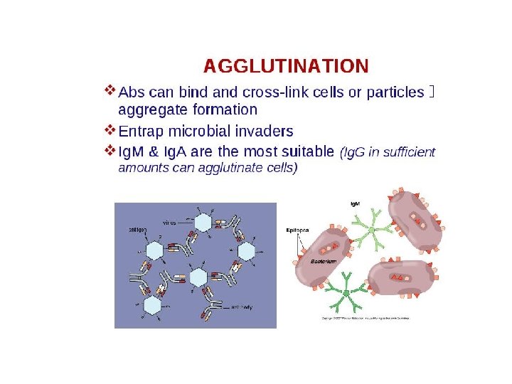
Fc receptors and antibody functions 3 - Mast cell degranulation in allergy 4 - Extracellular killing by eosinophils 5 - complement activation and cell lysis, IGG, IGM, IGA 6 - Neutralization; Antibodies against microbes and microbial toxins block the binding sites of these toxins and viruses so un able to bind cellular receptors (IGG) 7 - Precipitation and agglutination (IGM and IGG)


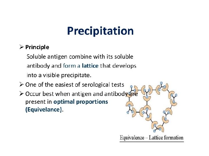
Serology • In serology; Precipitation, Precipitation reactions are based on the interaction of antibodies and antigens. They are based on two soluble reactants that come together to make one insoluble product, the precipitate which appear as line between 2 solutions • In serology, Agglutination; Agglutination is the visible expression of the aggregation of antigens and antibodies. Agglutination reactions apply to particulate test antigens is cell bound ( on RBC or artificially fixed on particles as. The endpoint of the test is the observation of clumps resulting from that antigenantibody complex formation.

• Fc receptors have been described for all classes of immunoglobulins: – – – FcγR and neonatal Fc. R (Fc. Rn) for Ig. G, FcεR for Ig. E, FcαR for Ig. A, FcδR for Ig. D, and FcμR for Ig. M. • Of these receptors, leucocyte FcγR and FcεR are characterized most extensively. • Structurally, all known Fc receptors belong to the immunoglobulin superfamily, except for Fc. Rn and FcεRII,

• Among them, FcγRI and FcεRI are highaffinity Fc receptors with dissociation constants ranging high. • All other Ig. G receptors, such as FcγRII and FcγRIII, are low-affinity receptors with dissociation constants are low.

FcγR • In addition to the affinity variations among the receptors, each Fcγ receptor displays distinct Ig. G subtype specificities; for example, – FcγRIII and FcγRI binds Ig. G 1 and Ig. G 3 better than Ig. G 2 and Ig. G 4. • Structural knowledge regarding antibody recognition by Fc receptors is critically important to antibody-mediated immune therapeutics.

FcγR • FcγRII A, B, and C. These isoforms have similar extracellular domains and ligand specificities but differ in cytoplasmic tail structure, cell distribution, and functions • FcγRI and FcγRII A and C, do opsonization function • FcγRIIB deliver inhibitory signals to B lymphocytes and IVIG (intravenous immunoglobulin) may use this mechanism to treat B cell cancer • FcγR 1 and 2 and 3 on phagocytes help in phagocytosis,

 cells and eosinophils bind to antibody-coated cells by Fc")
• Natural killer (NK) cells and eosinophils bind to antibody-coated cells by Fc receptors and destroy these cells. This process is called antibody-dependent cell mediated cytotoxicity (ADCC). • It was first described as a function of NK cells, which use their Fc receptor, low affinity FcγRIIIA (CD 16), to bind to antibody-coated cells. • After binding the receptors, NK secrete cytokines such as IFN-γ as well as to discharge the contents of their granules, which mediate the killing functions • anti-CD 20 antibody kill B cell–derived tumor cells by NK cells by ADCC

ADCC by NK cells

FcεR • FcεRI on eosinophils, function to mediate the killing and expulsion of some helminthic Parasites carrying Ig. E by ADCC • Killing molecules are secreted out side from eosinophils as major basic protein • Binding of FcεRI on Mast cell with an allergen mediate a rapid release of mediators ( Histamine) that may induce bronchoconstriction and increased local motility, contributing to the formation of hypersensitivity reaction 1

Effecter phase

• Some data suggest the existence of Fc. Rs for Ig. M and Ig. D on leukocytes, although structural entities for such receptors have not been well defined. • In addition, two epithelial cell Fc. Rs have been well characterized: – Fc. Rn which mediates both Ig. G transport across the placenta and Ig. G uptake by neonatal intestinal epithelium, – and the p. Ig. R (poly Ig receptor, also known as secretory component) which transports Ig. A into mucosal secretions.

Neonatal Fc receptors • Neonatal mammals are protected from infection by maternally produced antibodies transported across the placenta into the fetal circulation and by antibodies in ingested milk transported across the gut epithelium of newborns by a specialized process known as transcytosis. • Transport of maternal Ig. G across the placenta and across the neonatal intestinal epithelium is mediated by an Ig. G-specific Fc receptor called the neonatal Fc receptor (Fc. Rn). • The Fc. Rn is unique among Fc receptors in that it resembles a class I major histocompatibility complex (MHC)
- Slides: 52