Anthoceros Division Bryophyta Anthocerotopsida Class Anthocerotales Order Family

Anthoceros Division: Bryophyta Anthocerotopsida Class: Anthocerotales Order: Family: Anthocerotaceae Anthoceros Genus:

Vegetative Structure: External Features It occurs in moist, shaded habitats in subtropical and warm temperate regions. The gametophyte are dorsiventral and are often rosette-like. On the ventral surface, many smooth walled rhizoids are present which help in fixation.

Scales and tuberculate rhizoids are absent. Some species of Anthoceros are unisexual and others are bisexual. The antheridia and archegonia are sunken on the dorsal surface of the gametophyte. Numerous sporophytes may develop on the same gametophyte.

Vegetative Structure: External Features

Vegetative Structure: Internal Features Thallus is not differentiated into photosynthetic zone and storage zone. All cells are green and contain chloroplasts. Chloroplasts are associated with pyrenoids which is a unique feature of Anthocerotales. There are no air chambers or pores. Schizogenous cavities filled with mucilage are present. These are often inhabited by Nostoc (cyanobacteria), which supply nitrogen through nitrogen fixation to their host plants.

Vegetative Structure: Internal Features

Vegetative Reproduction Fragmentation: occurs through progressive death and decay of posterior portion of thallus. Tubers: develop at the end of growing seasons. Under unfavorable conditions, they perennate and grow in the next growing season. Gemmae: borne on short stalks on dorsal surface and along margins of thalli. They detach and germinate to produce new plants. Persistent growing apices

Sexual Reproduction It is oogamous type. Male sex organs are antheridia and female archegonia. Many species are monoecious while some are dioecious. Formation of sex organs are dependent on specific photoperiods. Anthoceros being a short day plant, sex organs develop in winters. Antheridia develop much earlier to archegonia.

Antheridia Occurs on dorsal surface of thallus in an acropetal succession inside closed cavities called antheridial chambers. It is differentiated into a long stalk and clubshaped body. The stalk is multicellular, slender and consisted of 4 vertical rows of elongated cells. The club shaped body of antheridium has single layered sterile jacket enclosing a mass of androcytes which metamorphose into biflagellated curved anthrozoids.
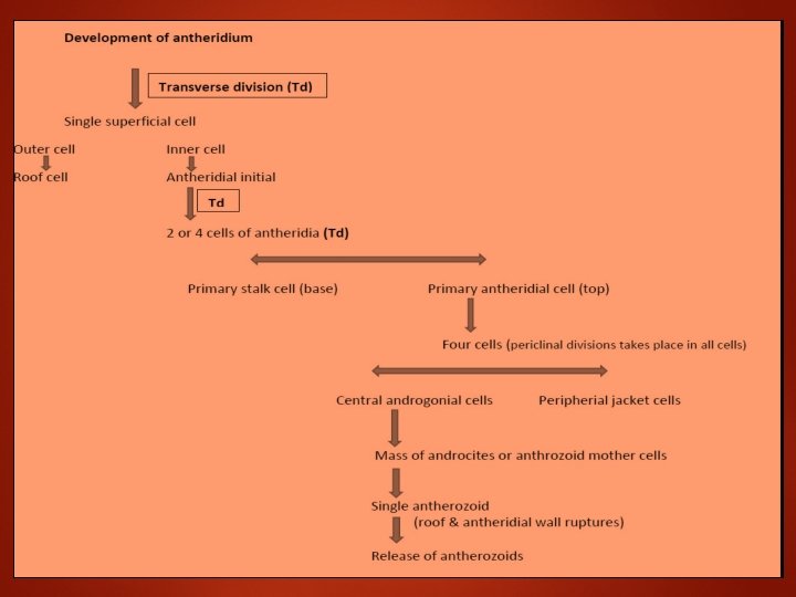

Development of Antheridium

Archegonia These are embedded on dorsal surface of thallus in an acropetal succession near growing point. The archegonial chambers are absent. It is flask shaped consisted of neck and venter. It contains 4 -6 neck canal cells, a venter cell and an egg. At maturity, the neck canal cells disinitegrate and become mucilagenous. There are no jacket cells covering the Archegonia. Cover cells or Lid cells are found at the tip of Archegonium.

Development of Archegonium

Fertilization The anthrozoids swim in water film. Some of these reach mature archegonium. The mature archegonium is characterized by mucilaginous mound at its apex. The antherozoids pass through mucilage and enter its wide open canal. Single antherozoid fuses with egg to form a diploid zygote.

Fertilization

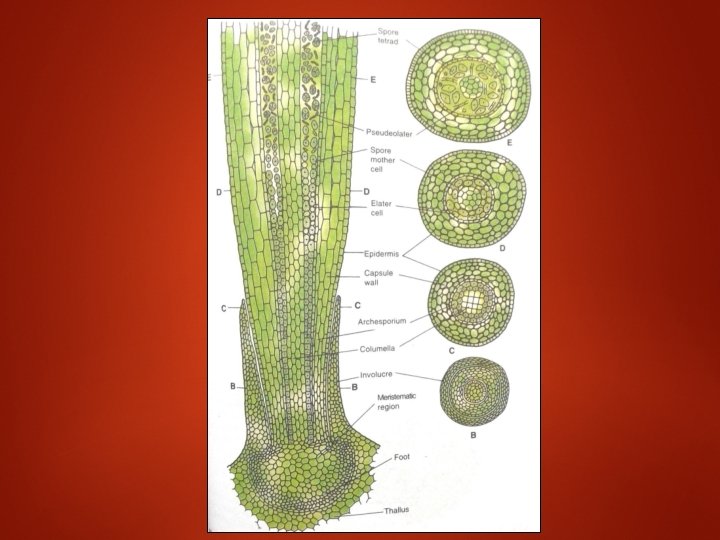
Sporophyte/Sporogonium The sporophyte is differentiated into 3 distinct regions: Foot: It is basal, bulbous parenchymatous structure found deeply embedded in the gametophyte. It helps in attaching the sporophyte to gametophyte and in absorption of water and nutrients from it. Intermediate or intercalary zone: A narrow zone of meristematic cells located between the basal foot and the upper capsule. These cells help in the continuous growth of the sporophyte. Capsule: It is the fertile, major and conspicuous part of the sporophyte which is long and cylindrical. It is green when young, but turns grey or brown on maturity.
 Columella: It is central solid")
The capsule is composed of the following structures: a) Columella: It is central solid core of sterile cells, consisting of 16 vertical rows of cells. It extends from the base to almost to the tip of the capsule. b) Sporogenous tissue: It is the mass of fertile spore-forming cells surrounding the columella, like a dome. At the base of the capsule, it is single layered and called archesporium. It becomes 2 -4 layered and develops into diploid spore mother cells upwards. Towards the tip of the capsule, the spore mother cells divide by meiosis and produce haploid spores.

• Capsule wall: It is the outer wall of the capsule which is 4 -6 layers in thickness. The outermost layer is called epidermis which is interrupted by stomata. The inner layers consist of chlorenchymatous cells and are photosynthetic.

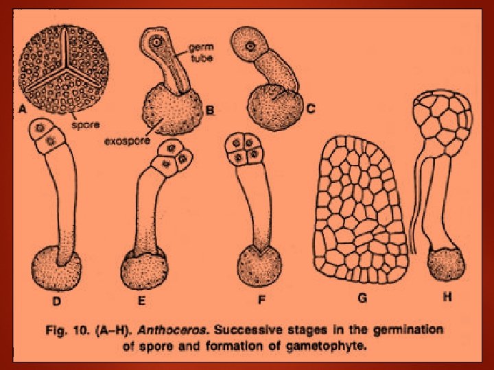
Structure of Spore: • The spores are haploid, uninucleate, semicircular with a conspicuous triradiate mark. Each spore remains surrounded by two wall layers. • The outermost layer is thick ornamented and is known as exospore. It varies in colour from dark brown to black. • The inner layer is thin and is known as endospore. Wall layers enclose colourless plastids, oil globules and food material.

Germination of spore and formation of young gametophyte Under favourable conditions the spores germinate immediately. At the time of germination spore absorbs water and swells up Exospore ruptures at the triradiate mark and endospore comes out in the form of a tube. It is called germ tube.

• Contents migrate into the germinal tube where the colourless plastids turn green. Two successive transverse walls are laid down at the tip of a the germinal tube resulting in the formation of three celled filament. The upper cell divides by a vertical division followed by similar vertical division in the lower cell (quadrant stage).

Life Cycle

THANK YOU
- Slides: 25