ANTERIOR COMMUNICATING ANEURYSMS DR M ABBASZADEH NEUROSURGEON RAZI

ANTERIOR COMMUNICATING ANEURYSMS DR M. ABBASZADEH NEUROSURGEON RAZI HOSPITAL

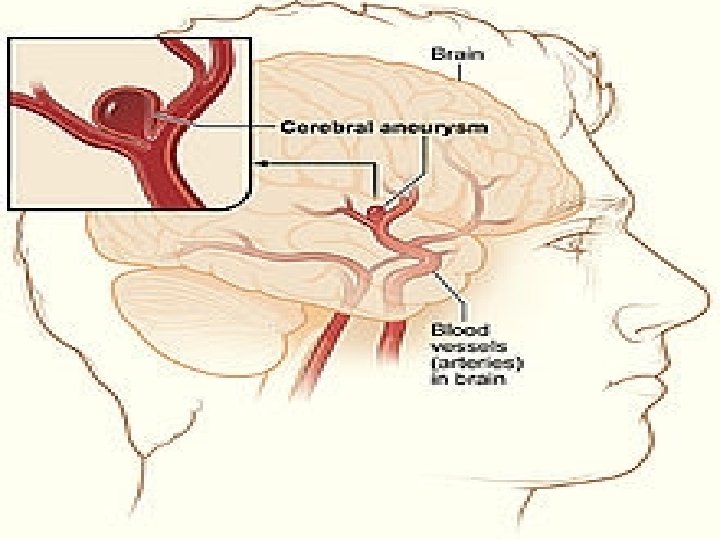
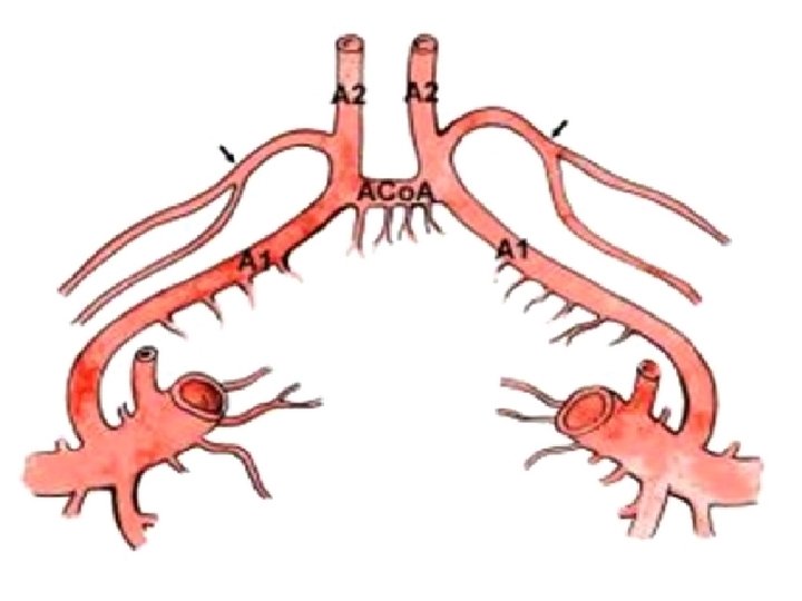
EPIDEMYOLOGY � � � Acom aneurysm is the most common site for cerebral aneurysms. 30% to 39% of cerebral aneuryms are located in A 1 segment-Acom-distal ACA region. Highest rate of rebleeding among anterior cerebral circulation arteries.

This particular aneurysm has a wide variety of complexities and technical difficulties due to variations in parent artery anatomy, aneurysm projection, and clinical presentation.

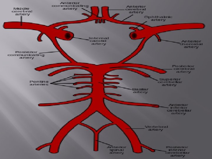
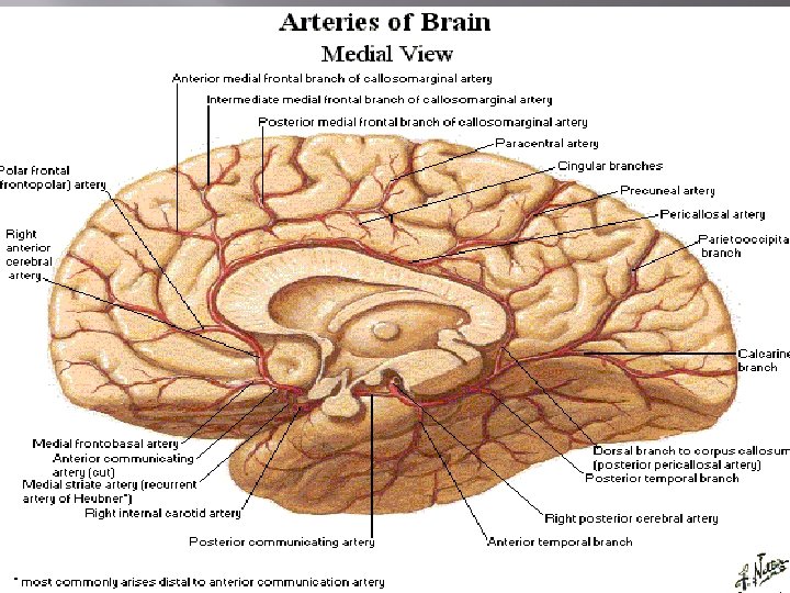
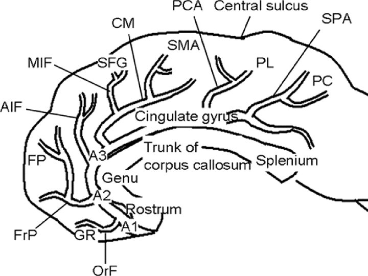
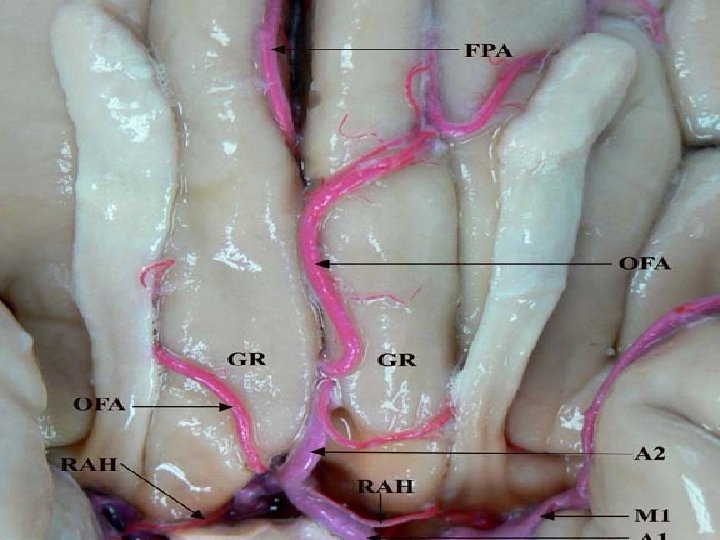
ANATOMY ACo. A comlex is adjacent to : � 11 Cerebral arteries � Hypothalamus � Opic apparatus � Cognitive-emotional centers of basal frontal lobes Arteies emanating from ACo. A complex supplies : � Basal ganglia � Internal capsule � Sensory-motor cortex
")
BRANCHES OF ANTERIOR CEREBRAL ARTETY � � � � Reccurent artery of heubner (2) Orbitofrontal artery (2) Frontopolar artery (2) Perforant arteries A 1 segments (2) A 2 segments (2) A 3 A 4

CLINICAL PRESENTATION � Sudden onset severe headache � Obtundation Coma Visual field defect (giant aneurysms) Endocrine dysfunction due to mass effect on hypothalamus (giant aneurysms) Hydrocephalus due to obstruction of monro foramen Cognitive dysfunction Memory impairment Seizure � � � �
 CT angiography MRA Four vessels angiograghy (DSA)")
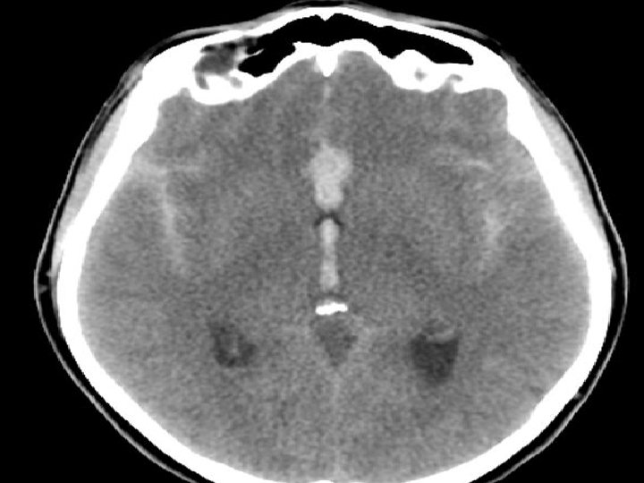
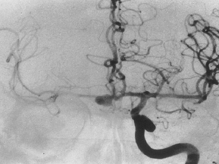
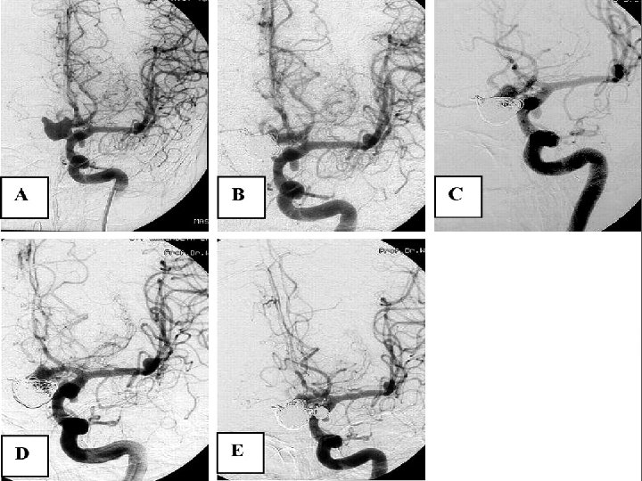
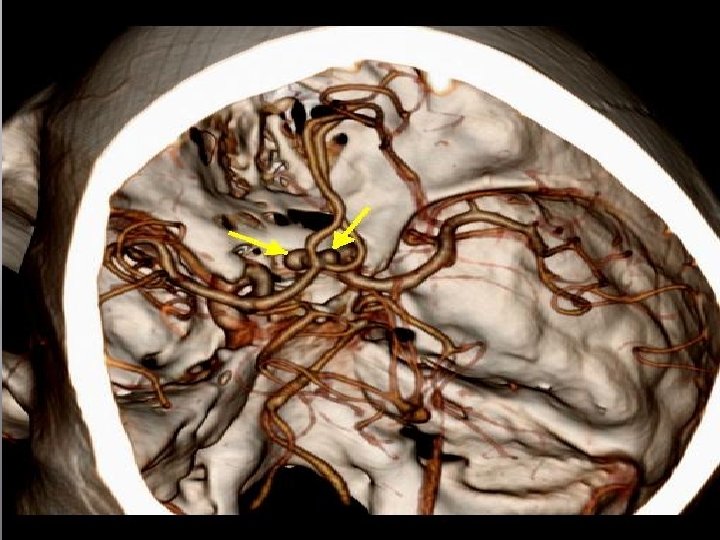
DIAGNOSTIC IMAGING � � CT scan (noncontrast) CT angiography MRA Four vessels angiograghy (DSA) : gold standard but With high false-negative results

PREOPERATIVE MANAGMENT � � � After a ruptured aneurysm is diagnosed it should be secured within 48 to 72 hours during this period efforts are made to minimize the risk of rerupture. Most important is systolic blood pressure control under 140 mm hg Hyrocephalus is present in approximately one fourth of patients and resolves with ventriculostomy insertion Intubation and mechanichal ventilation in poor-grade patients Ca channel blocker, AED, laxatives,

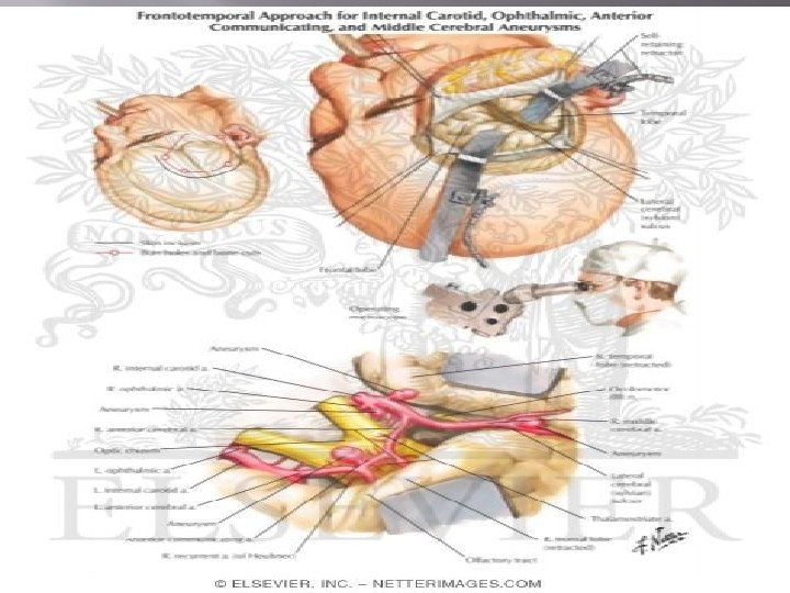
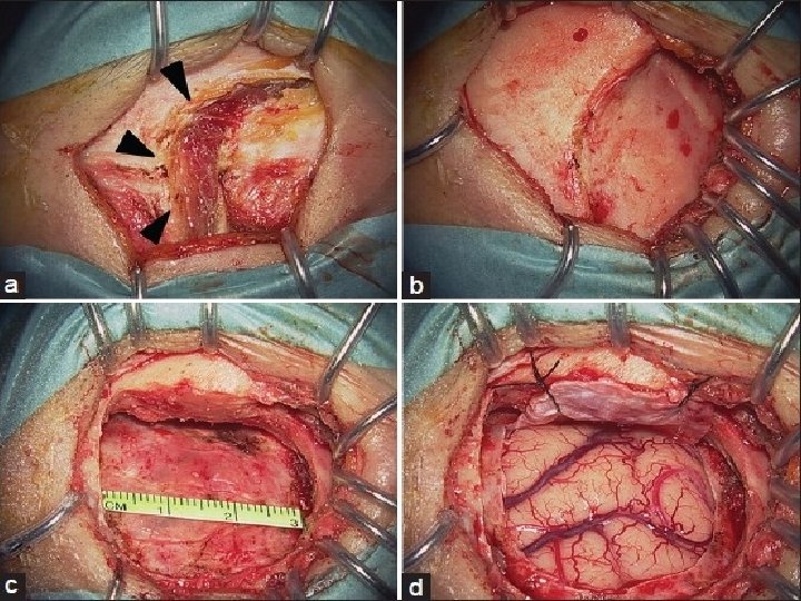
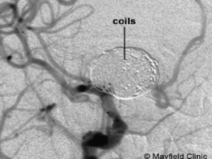
MANAGEMENT � Microsurgical clipping � Endovascular coiling

- Slides: 23